Abstract
Purpose
To compare the postoperative results of 2.2 mm microcoaxial cataract surgery (MCCS) and conventional 3.0 mm cataract surgery (CCS).
Methods
This study included 30 patients (40 eyes): 17 patients (21 eyes) had microcoaxial cataract surgery using a 2.2 mm microcorneal incision, and 13 patients (19 eyes) had conventional cataract surgery using a 3.0 mm corneal incision. In these two groups, we measured phacoemulsification time, total phacoemulsification percentage, and effective phacoemulsification time (EPT) during cataract surgery. We compared corneal thickness, endothelial cell count, uncorrected visual acuity, best‐ corrected visual acuity, and surgically, induced astigmatism (SIA) by corneal topography between the two groups preoperatively and at 1 day, 1 week, 1 month, and 2 months postoperatively.
Results
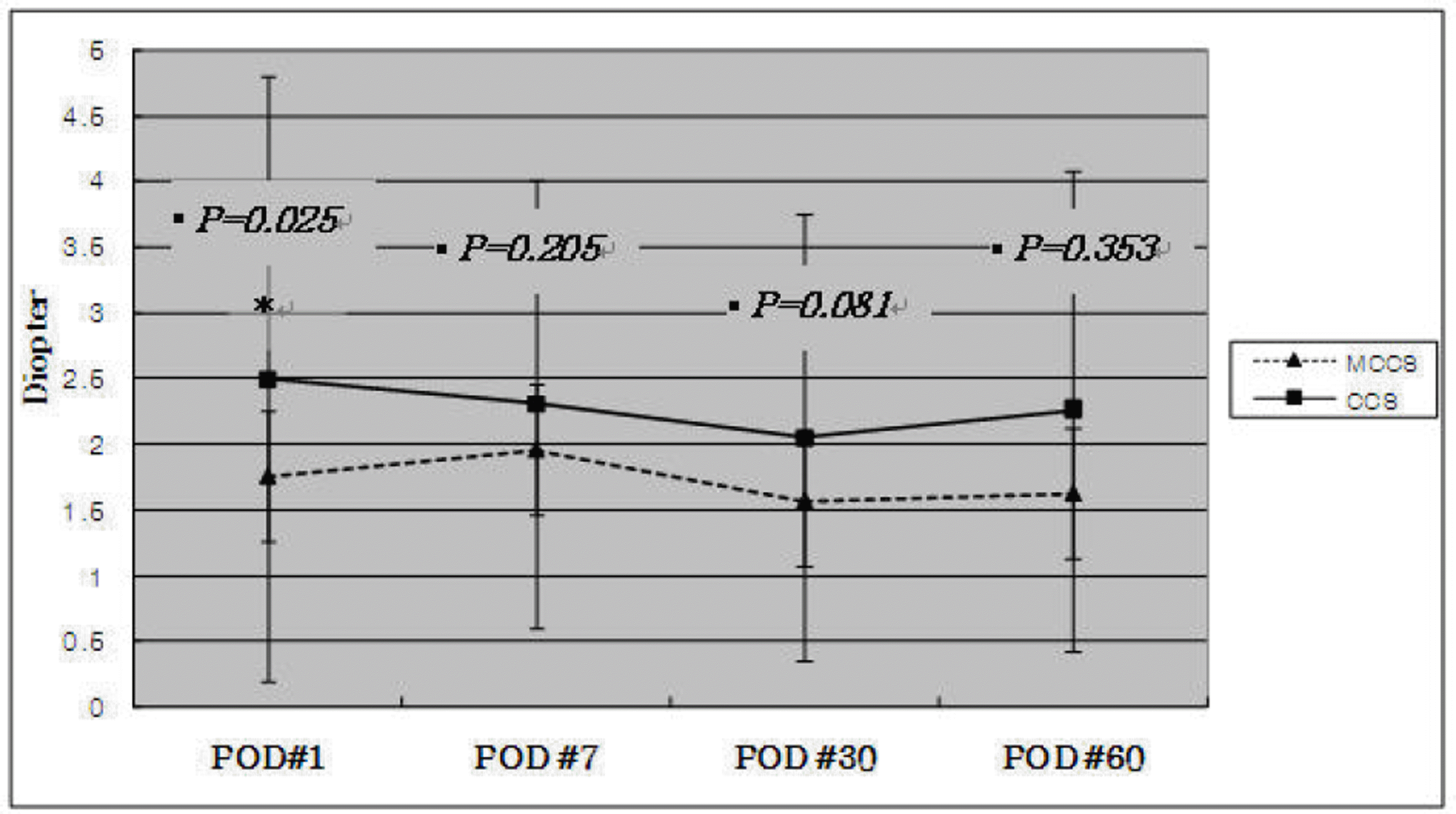
There were significantly lower SIA ( p=0.025) at postoperative day 1 in MCCS, and there was no significant difference after that. No difference in phacoemulsification time, total phacoemulsification percentage, EPT, corneal thickness, postoperative uncorrected visual acuity or corrected visual acuity was noted between MCCS and CCS.
References
1. Kellman CD. Phacoemulsification and aspiration; a new technique of cataract removal; a preliminary report. Am J Ophthalmol. 1967; 64:23–35.
2. Crema AS, Walsh A, Tamane Y, Nose W.Comparative study of coaxial phacoemulsification and microincision cataract surgery. One-year follow-up. J Cataract Refract Surg. 2007; 33:1014–8.
3. Ku CH, Kim HJ, Joo CK. The comparison of astigmatism according to the incision size in small incision cataract surgery. J Korean Ophthalmol Soc. 2005; 46:416–21.
4. Jee DH, Lee PY, Joo CK. The comparison of astigmatism according to the incision size in cataract operation. J Korean Ophthalmol Soc. 2003; 44:594–8.
5. Vasavada V, Vasavada V, Raj SM, Vasavada AR. Intraoperative performance and postoperative outcomes of microcoaxial phacoemulsification: Observational study. J Cataract Refract Surg. 2007; 33:1019–24.
6. Osher RH. Miacocoaxial phacoemulsification Part 2: clinical study. J Cataract Refract Surg. 2007; 33:408–12.
7. Cavallini GM, Campi L, Masini C. . Bimanual microphacoemulsification vs coaxial miniphacoemulsification: Prospective study. J Cataract Refract Surg. 2007; 33:387–92.
8. Fine IH, Packer M, Hoffman RS. Power modulations in new phacoemulsification technology: improved outcomes. J Cataract Refract Surg. 2004; 30:1014–9.
9. Cravy TV. Calculation of the change in corneal astigmatism following cataract extraction. Ophthalmic Surg. 1979; 10:38–49.
10. Huang FC, Teng SH. Compression of surgically induced astigmatism after sutureless temporal clear corneal and slceral frown incision. J Cataract Refract Surg. 1998; 24:477–81.
11. Lee DS, Joo CK. Effect of incision length on visual recovery and astigmatism in no-suture cataract surgery. J Korean Ophthalmol Soc. 1992; 33:1036–42.
12. Hu YJ, Joo CK. Surgically induced astigmatism after temporal clear corneal incision in sutureless cataract surgery. J Korean Ophthalmol Soc. 1998; 39:2622–7.
13. Kim HJ, Kim JH, Lee DH. Endothelial cell damage in microincision cataract surgery and coaxial phacoemulsification. J Korean Ophthalmol Soc. 2007; 48:19–26.
14. Alió J, Rodriguez-Prats JL, Galal A, Ramzy M. Outcomes of microincision cataract versus coaxial phacoemulsification. Ophthalmology. 2005; 112:1997–2003.
15. Hayashi K, Hayashi H, Nakao F, Hayashi F. Risk factors for corneal endothelial injury during phacoemulsification. J Cataract Refract Surg. 1996; 22:1079–84.

16. Dick HB, Kohnen T, Jacobi FK, Jacobi KW. Long-term endothelial cell loss following phacoemulsification through a temporal clear corneal incision. J Cataract Refract Surg. 1996; 22:63–71.

17. Joussen AM, Barth U, Cubuk H, Koch H. Effect of irrigating solution and irrigation temperature on the cornea and pupil during phacoemulsification. J Cataract Refract Surg. 2000; 26:392–7.

18. Bourne RR, Minassian DC, Dart JK. . Effect of cataract surgery on the corneal endothelium; modern phacoemulsification compared with extracapsular cataract surgery. Ophthalmology. 2004; 111:679–85.
19. Suzuki H, Takahashi H, Hori J. . Phacoemulsification associated corneal damage evaluated by corneal volume. Am J Ophthalmol. 2006; 142:525–8.

20. Wilczynski M, Drobniewski I, Synder A, Omulecki W. Evaluation of early corneal endothelial cell loss in bimanual microincision cataract surgery (MICS) in comparison with standard phacoemulsification. Eur J Ophthalmol. 2006; 16:798–803.

21. Mencucci R, Ponchietti C, Virgili G. . Corneal endothelial damage after cataract surgery: microincision versus standard technique. J Cataract Refract Surg. 2006; 32:1351–4.

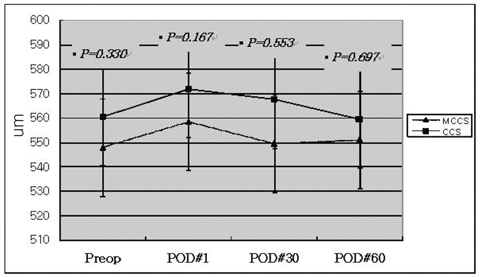
Figure 1.
Postoperative changes in corneal thickness (µm); The corneal thickness was increased at postoperative 1 day in both MCCS (microcoaxial cataract surgery) and CCS (conventional cataract surgery) group. After that, it was decreased gradually, but the differences between 2 groups were not statistically significant. The statistical analysis was performed using independent t-test. A P-value less than 0.05 is statistically significant.

Figure 2.
Postoperative changes in corneal endothelial cell count (cells/mm2); The corneal endothelial cell was gradually decreased postoperatively in both MCCS (microcoaxial cataract surgery) and CCS (conventional cataract surgery) group. But the differences between 2 groups were not statistically significant. The statistical analysis was performed using independent t-test. A P-value less than 0.05 was considered as statistically significant.

Figure 3.
Perioperative surgically induced astigmatism (Diopter); The surgically induced astigmatism (SIA) by vectorial analysis was significantly smaller in MCCS (microcoaxial cataract surgery) group than CCS (conventional cataract surgery) group at postoperative 1 day. And after that, smaller SIA continued in MCCS group, but the difference was not significantly different. The statistical analysis was performed using independent t-test. A P-value less than 0.05 was considered as statistically significant.

Table 1.
Patient characteristics (N=40)
| MCCS* | CCS† | P-value | |
|---|---|---|---|
| Number (eye) | 21 | 19 | |
| Gender (Male/Female) | 5/16 | 6/13 | |
| Age (year±SD) | 59.95±8.87 | 68.32±9.83 | 0.859 |
Table 2.
Surgical parameters used in the study with Infiniti Phacoemulsification PlatformⓇ
| MCCS* | CCS† | |
|---|---|---|
| Incision size (mm) | 2.2 mm | 3.0 mm |
| Capsulorrhexis diameter (mm) | 5.5 mm | 5.5 mm |
| Phacoemulsification tip | 30° | 30° |
| Prechopping | No | No |
| Settings | ||
| Power | 50% | 45% |
| Aspiration flow | 40 cm3/min | 40 cm3/min |
| Vacuum | 400 mmHg | 400 mmHg |
| Anterior chamber pressure | 97 cm3-H2 O | 92 cm3-H2 O |
| Mode | Hyperpulser (30 pps, 40% time on) | Hyperpulser (30 pps, 40% time on) |
| I/A of cortical remnants | 350 mmHg | 350 mmHg |
| I/A of viscoelastic | 150 mmHg | 150 mmHg |
Table 3.
| MCCS* | CCS† | P-value‡ | |
|---|---|---|---|
| Mean phaco time (sec) | 14.42±18.27 | 15.71±12.24 | 0.976 |
| Total phaco % | 4.74±2.39 | 5.42±2.28 | 0.708 |
| EPT (sec)§ | 1.00±2.12 | 1.01±1.00 | 0.696 |
| Total BSS∏ used (cc) | 51.2±4.68 | 58.9±6.73 | 0.453 |


 PDF
PDF ePub
ePub Citation
 Print
Print


 XML Download