

 PDF
PDF ePub
ePub Citation
 Print
Print


Abstract
Methods
We reviewed the ocular records of 29 consecutive patients with limbal dermoids who had undergone simple keratectomy in Seoul National University Children’s hospital from 1989 to 2006. The preoperative and postoperative visual acuity and astigmatism levels as well as the cosmetic outcomes were measured.
Results
The mean age at surgery was 28 months (range, 6~70 months), and the mean follow-up period was 56 months (range, 18~168 months). The mean visual acuity at the last follow-up was 20/30. The mean preoperative and postoperative cylinder was -2.64D and -2.12D, respectively, in the affected eye ( p=0.064); and -0.79D and -0.43D, respectively, in the fellow eye ( p=0.149). There was a significant correlation between preoperative astigmatism and the final visual acuity in the involved eye. No opaque lesions were visible at a distance of 1 meter in 16 of the 29 eyes (55.2%), and there were no significant complications related to the surgery.
References
1. Dailey EG, Lubowitx RM. Dermoids of the limbus and cornea. Am J Ophthalmol. 1962; 53:661–5.

2. Mansour AM, Barber JC, Reinecke RD, Wang FM. Ocular choristomas. Surv Ophthalmol. 1989; 33:339–58.
3. Mohan M, Mukherjee G, Panda A. Clinical evaluation and surgical intervention of limbal dermoid. Indian J Ophthalmol. 1981; 29:69–73.
4. Panton RW, Sugar J. Excision of limbal dermoids. Ophthalmic Surg. 1991; 22:85–9.
5. Sen DK, Mohan H, Gupta DK. The syndrome of Goldenhar. Acta Ophthalmol. 1969; 47:1044–8.
6. Baum JL, Feingold M. Ocular aspects of Goldenhar’s syndrome. Am J Ophthalmol. 1973; 75:250–6.
7. McKendrick AM, Brennan NA. Clinical evaluation of refractive techniques. J Am Optom Assoc. 1995; 66:758–65.
8. Harris WF, Malan DJ, Rubin A. Ellipsoidal confidence regions for mean refractive status. Optom Vis Sci. 1991; 68:950–3.
9. Harris WF. Dioptric power: its nature and its representation in three- and four-dimensional space. Optom Vis Sci. 1997; 74:349–66.
10. Harris WF. Representation of dioptric power in Euclidean 3-space. Ophthalmic Physiol Opt. 1991; 11:130–6.
11. Robb RM. Astigmatic refractive errors associated with limbal dermoids. J Pediatr Ophthalmic Strabismus. 1996; 33:241–3.
12. Scott JA, Tan DT. Therapeutic lamellar keratoplasty for limbal dermoids. Ophthalmology. 2001; 108:1858–67.
13. Cuttone JM, Durso F, Miller M, Evans LS. The relationship between soft tissue anomalies around the orbit and globe and astigmatic refractive errors: a preliminary report. J Pediatr Ophthalmol Strabismus. 1980; 17:29–36.
14. Panda A, Ghose S, Khokhar S, Das H. Surgical outcomes of epibulbar dermoids. J Pediatr Ophthalmol Strabismus. 2002; 39:20–5.
15. Shen YD, Chen WL, Wang IJ. . Full‐ thickness central corneal grafts in lamellar keratoscleroplasty to treat limbal dermoid. Ophthalmology. 2005; 112:1955.
16. Watts P, Michaeli-Cohen A, Abdolell M, Rootman D. Outcome of lamellar keratoplasty for limbal dermoids in children. J AAPOS. 2002; 6:209–15.
17. Yoon CK, Park SW, Song BY. The Role of Ultrasound Biomicroscopy in Operation for Limbal Dermoid. J Korean Ophthalmol Soc. 2004; 45:364–9.
Figure 1.
Corneal opacity grade after surgery. (A) Grade 1 opacity (invisible opacity at 1 meter), (B) Grade 2 opacity (visible opacity at 1 meter), (C) Grade 3 opacity (invasion into visual axis), (D) Grade 4 opacity (pseudopterygium or fibrovascular scar), (E) Photo of patient B taken before operation.

Figure 2.
Best corrected visual acuity change after surgery in affected eye Postoperative visual acuity increased in all of 6 patients. (All dots are located below the diagonal line.)
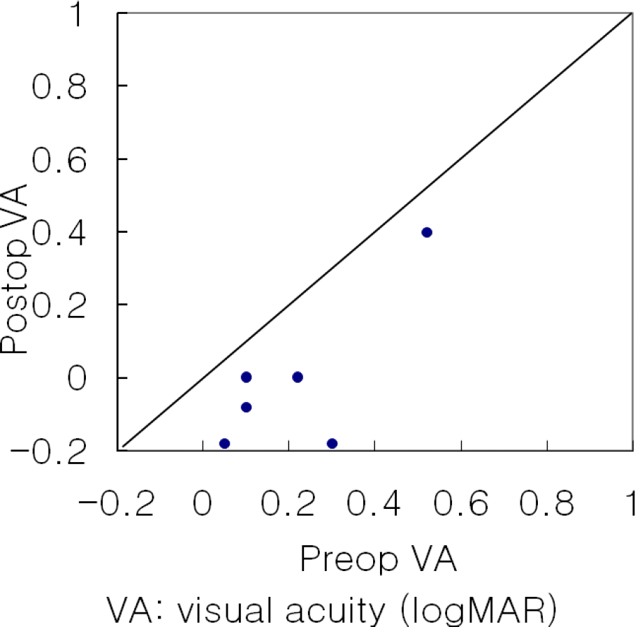
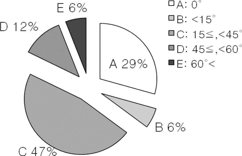
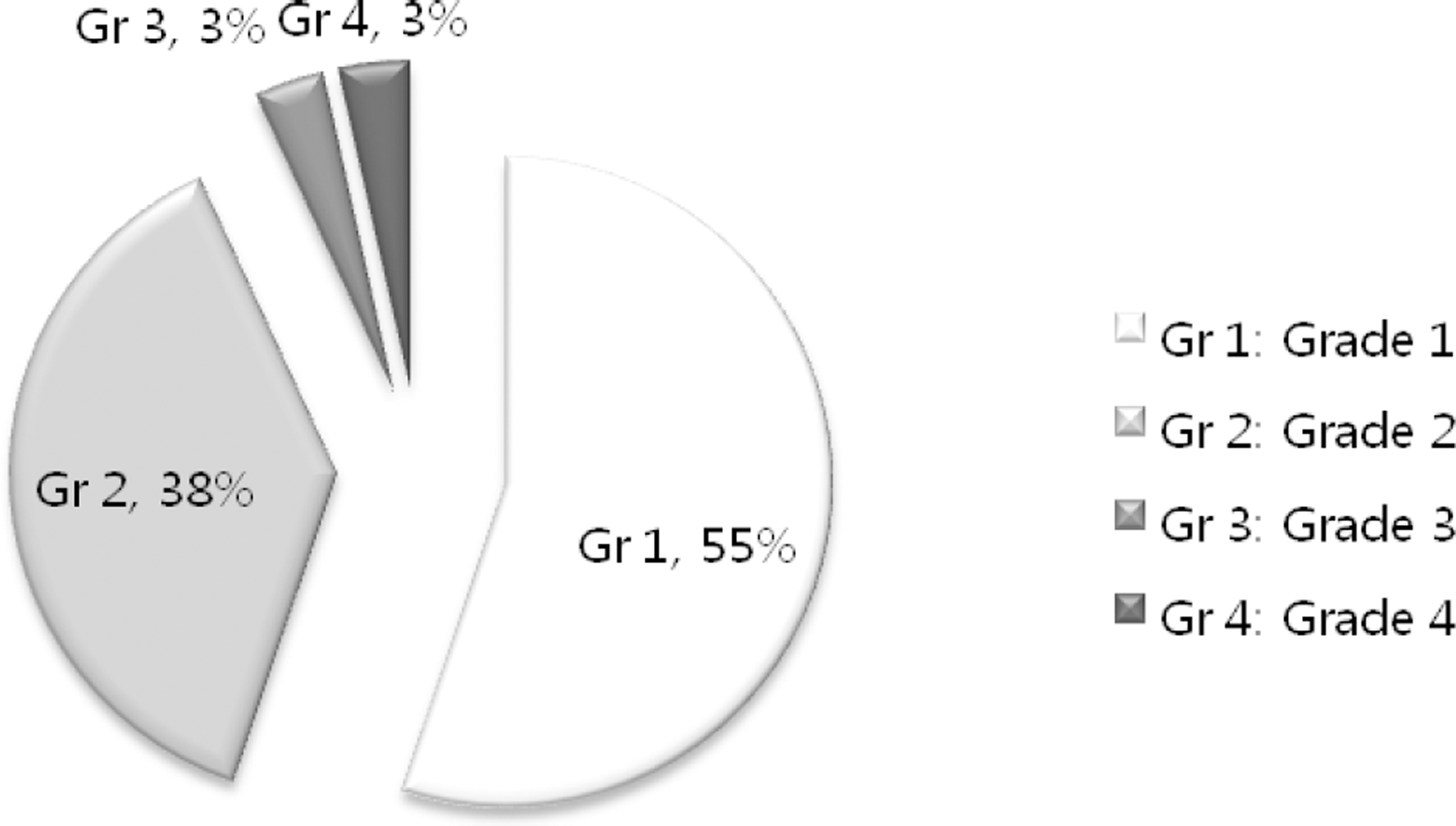
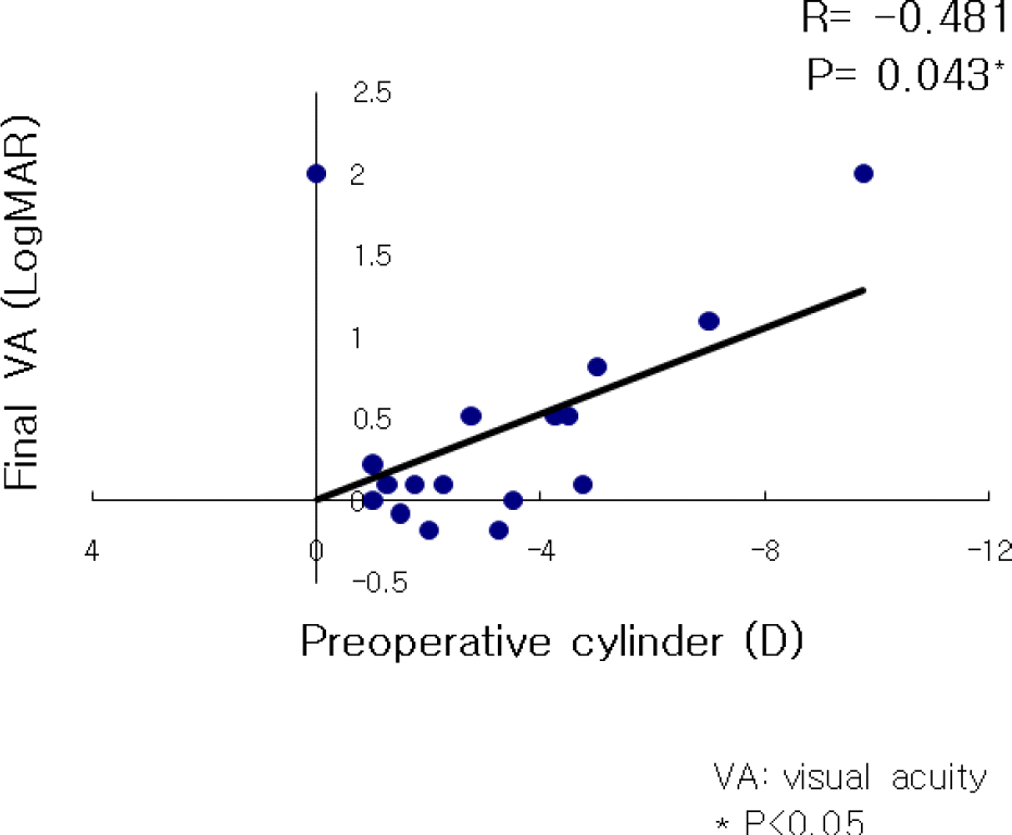
Table 1.
Visual acuity change after surgery (Postoperative data was obtained from all patients)
| Preop BCVA* | Postop BCVA | P‡ | |
|---|---|---|---|
| Affected eye | 0.21±0.17 | 0.42±0.56 | 0.623 |
| Fellow eye | 0.22±0.20 | 0.07±0.13 | 0.062 |
| P† | 0.844 | 0.000§ |
Table 2.
Visual acuity change after surgery (In 6 patients who had the preoperative visual acuity checked)
| Preop BCVA* | Postop BCVA | P† | |
|---|---|---|---|
| Affected eye | 0.21±0.17 | 0.00±0.20 | 0.027‡ |
| Fellow eye | 0.22±0.20 | -0.01±0.06 | 0.043‡ |
| P† | 0.752 | 1.000 |
Table 3.
Refractive error change after surgery (In 16 patients who had both preoperative and postoperative refraction)
| Preop (D) | Postop (D) | P* | |
|---|---|---|---|
| SE† in affected eye | 0.80±3.270 | -0.21±2.27 | 0.055 |
| SE† in fellow eye | 0.57±1.195 | -0.71±1.37 | 0.009‡ |
| P* | 0.365 | 0.127 | |
| Astigmatism in affected eye | -2.64±1.86 | -2.12±1.86 | 0.064 |
| Astigmatism in fellow eye | -0.79±0.99 | -0.43±0.65 | 0.149 |
| P* | 0.002‡ | 0.004‡ |
 XML Download