Abstract
Purpose
Paresis of the inferior oblique is the least likely to result in paralysis. We report a patient without a history of trauma successfully treated using contralateral IO recession and SR recession.
Case summary
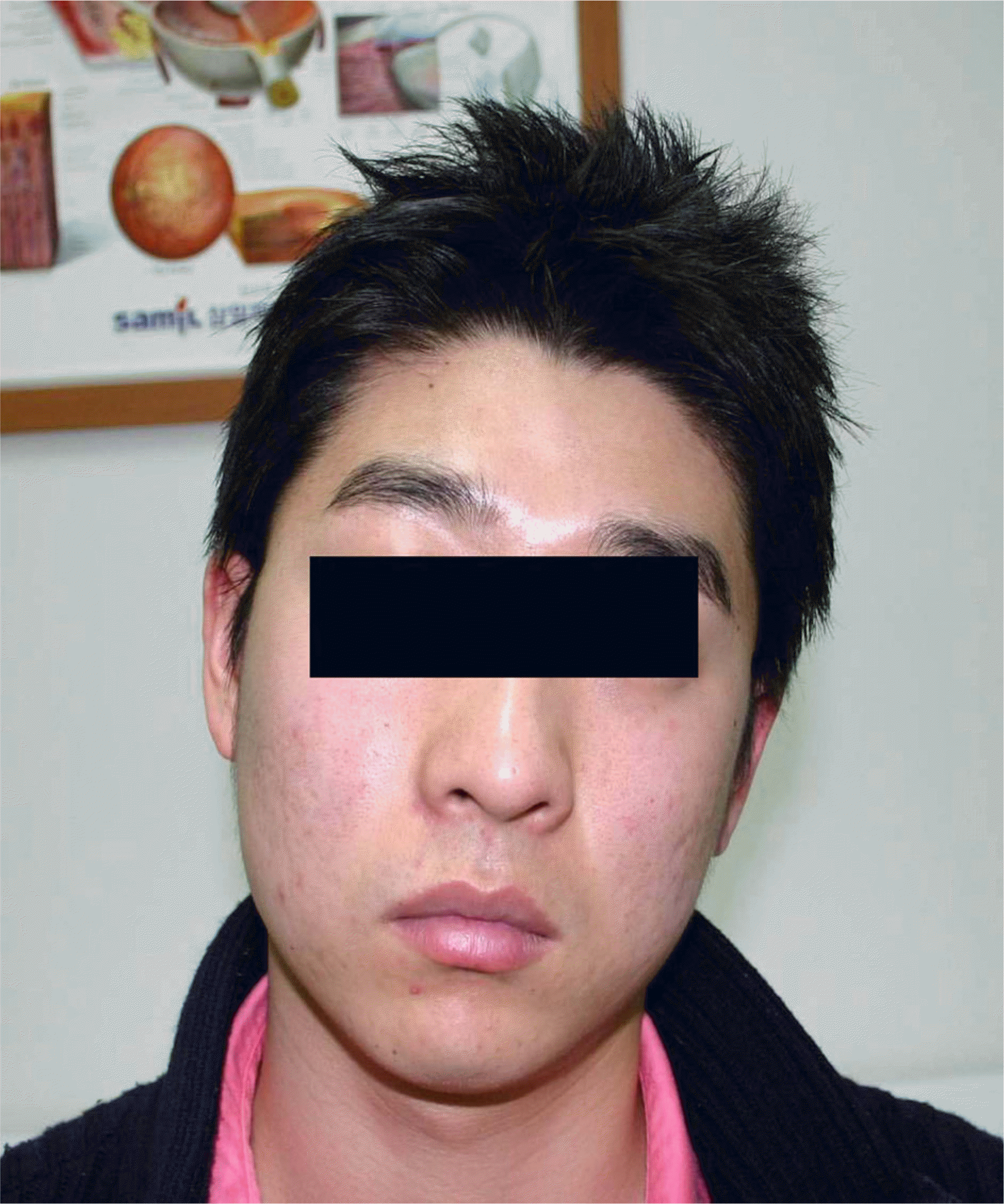
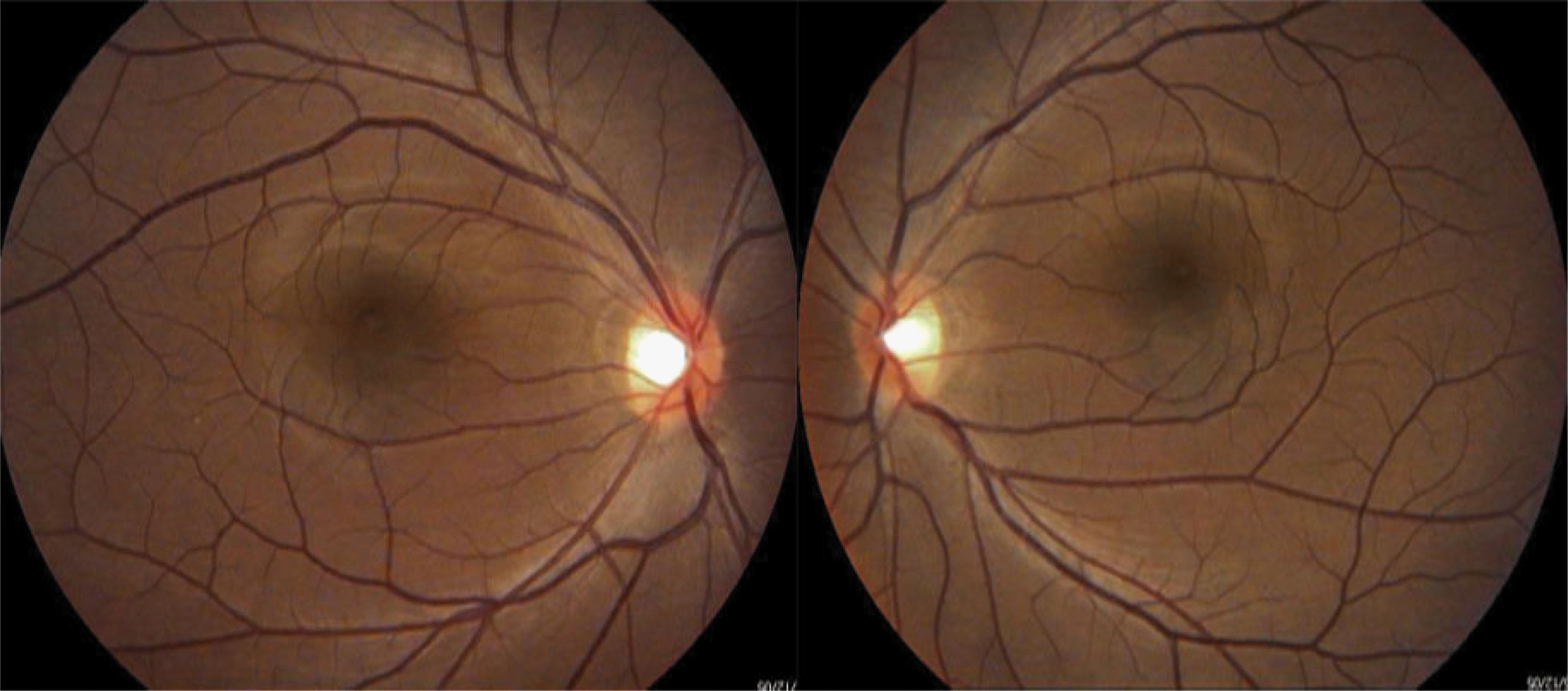
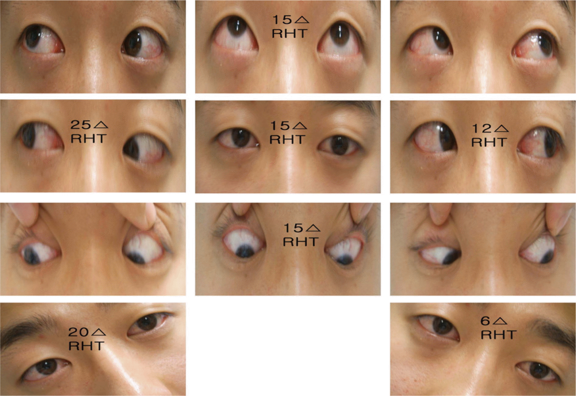
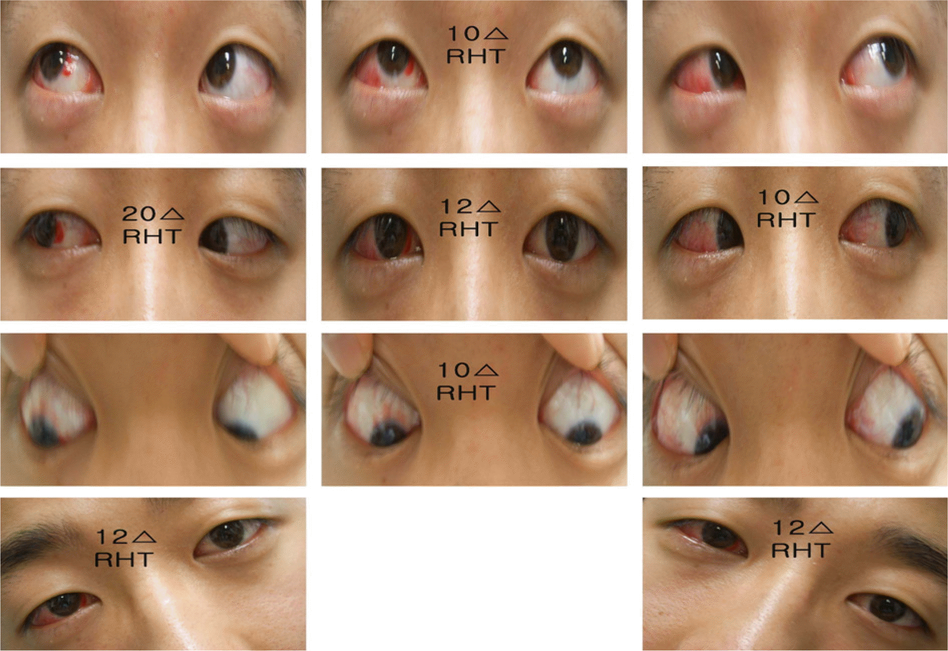
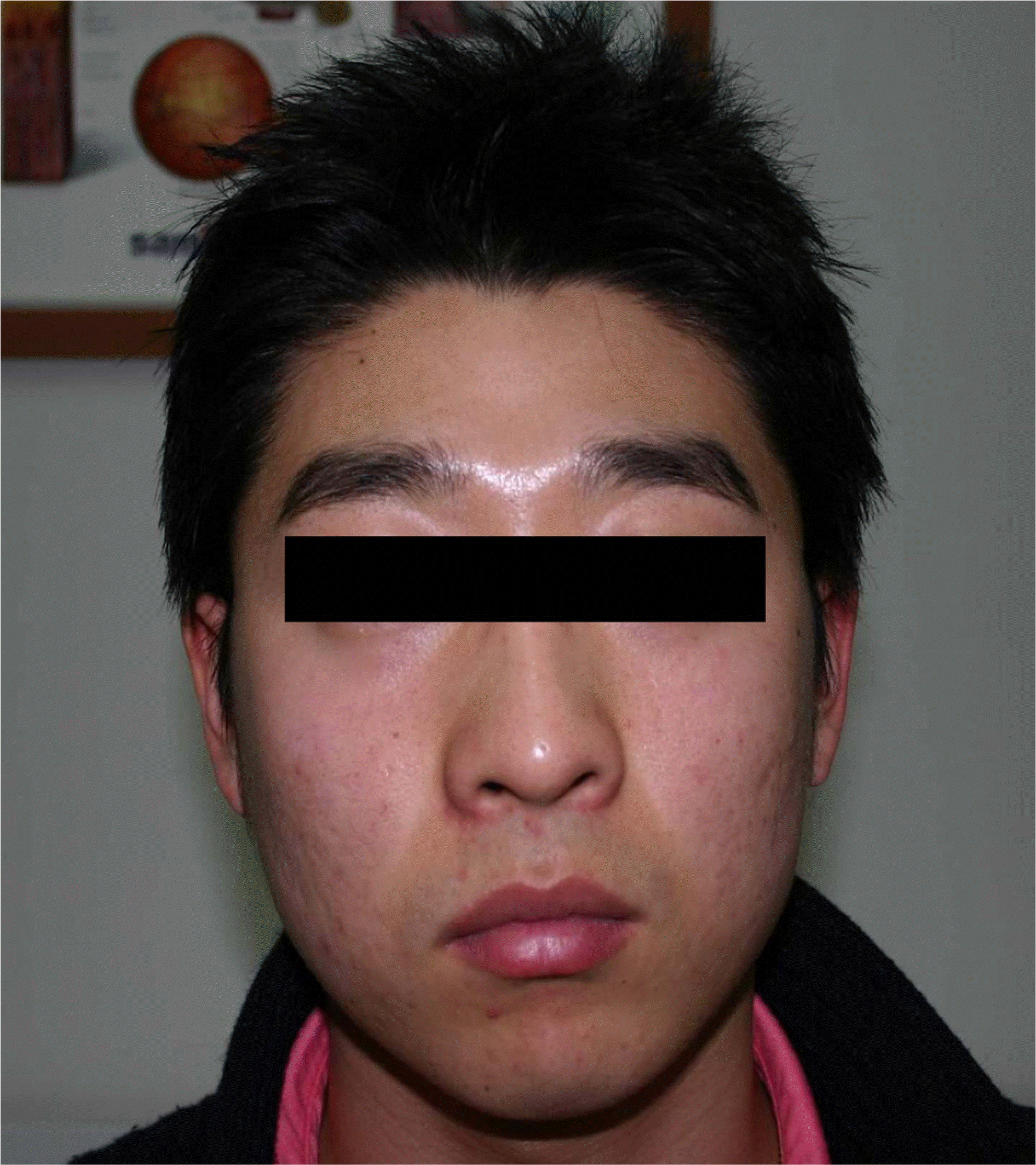
A 25-year-old male patient presented to us with an extended history of abnormal head posture, manifested by a marked habitual left head tilt with a face turn to the right. A cover test in the primary position demonstrated 15 prism diopter right hypertropia, which increased to 25 prism diopter right hypertropia in right gaze and 20 prism diopter right hypertropia in right head tilt. The patient was diagnosed with IO palsy, and a right IO recession was performed.
Results
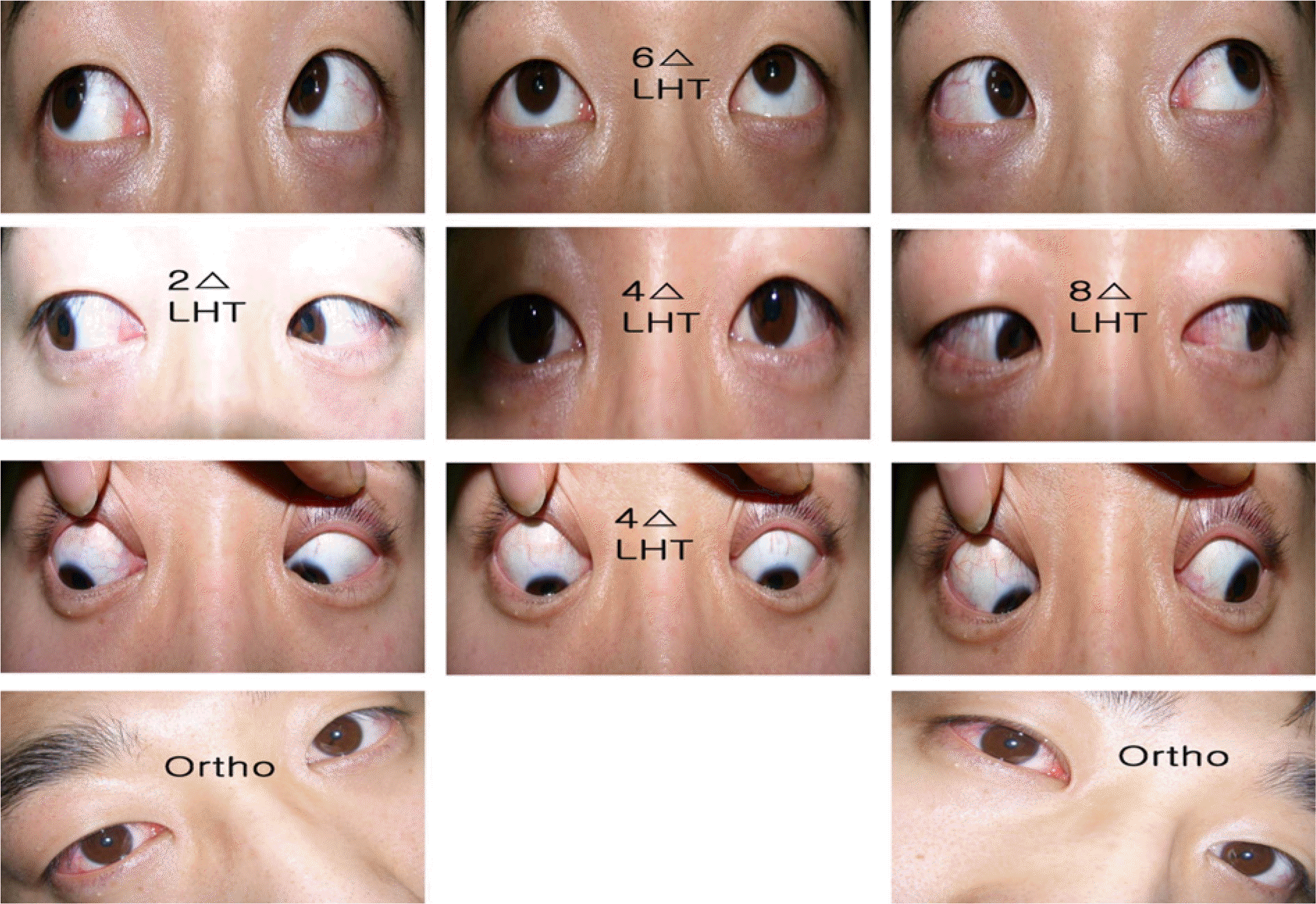
Following the IO recession, head tilt was completely resolved and face turn to the right was slightly resolved. Cover test in the primary position demonstrated 12 prism diopter right hypertropia, which increased to 20 prism diopter right hypertropia in right gaze. A head tilt test demonstrated a symmetrical 12 prism diopter right hypertropia. We performed a right SR recession to decrease face turn and hypertropia in the primary position.
References
2. Hunter DG, Lam GC, Guyton DL. Inferior oblique muscle injury from local anesthesia for cataract surgery. Ophthalmology. 1995; 102:501–9.

3. Harley RD, Nelson LM, Flanagan JC, Calhoun JH. Ocular motility disturbances following cosmetic blepharoplasty. Arch Ophthalmol. 1986; 104:542–4.

4. Lee JY, Lim HT, Ahn HS. 2 Cases of Traumatic Inferior Oblique Palsy. J Korean Ophthalmol Soc. 2000; 43:1349–54.
5. White JW, Brown HW. Occurrence of vertical anomalies associated with convergent and divergent anomalies. Arch Ophthalmol. 1939; 12:999.

7. Donahue SP, Lavin PJ, Mohney B, Hamed L. Skew deviation and inferior oblique palsy. Am J Ophthalmol. 2001; 132:751–6.

8. Olivier P, von Noorden GK. Results of superior oblique tenectomy in inferior oblique paresis. Arch Ophthalmol. 1982; 100:581–3.

9. Reese PD, Scott WE. Superior Oblique Tenotomy in the Treatment of Isolated Inferior Oblique Paresis. J Pediatr Ophthalmol Strabismus. 1987; 24:4–9.

11. Urist JM. Complications following bilateral superior oblique weakening surgical procedures for A pattern horizontal deviations. Am J Ophthalmol. 1970; 70:583–7.
12. McNeer KW. Untoward effects of superior oblique tenotomy. Ann Ophthalmol. 1972; 4:747–55.
13. Bedrossian E. Bilateral superior oblique tenotomy ofr the A pattern in strabismus. Arch Ophthalmol. 1967; 78:334–6.
14. Wright K. Superior oblique silicone expander for Brown syndrome and superior oblique overation. J Pediatr Ophthalmol Strabismus. 1991; 28:101–7.
15. Pollard ZF, Greenberg MF. Results and complications in 66 cases using a silicone tendon expander on overacting superior obliques with A pattern anisotropias. Binocul Vis Strabismus Q. 2000; 15:113–20.
16. Jung JI, Han SH. Anterior Transposition of the Inferior Oblique Muscle for Treatment of Hypertropia in Superior Oblique Muscle Palsy. J Korean Ophthalmol Soc. 1999; 40:242–7.


 PDF
PDF ePub
ePub Citation
 Print
Print


 XML Download