

 PDF
PDF ePub
ePub Citation
 Print
Print


Abstract
Case summary
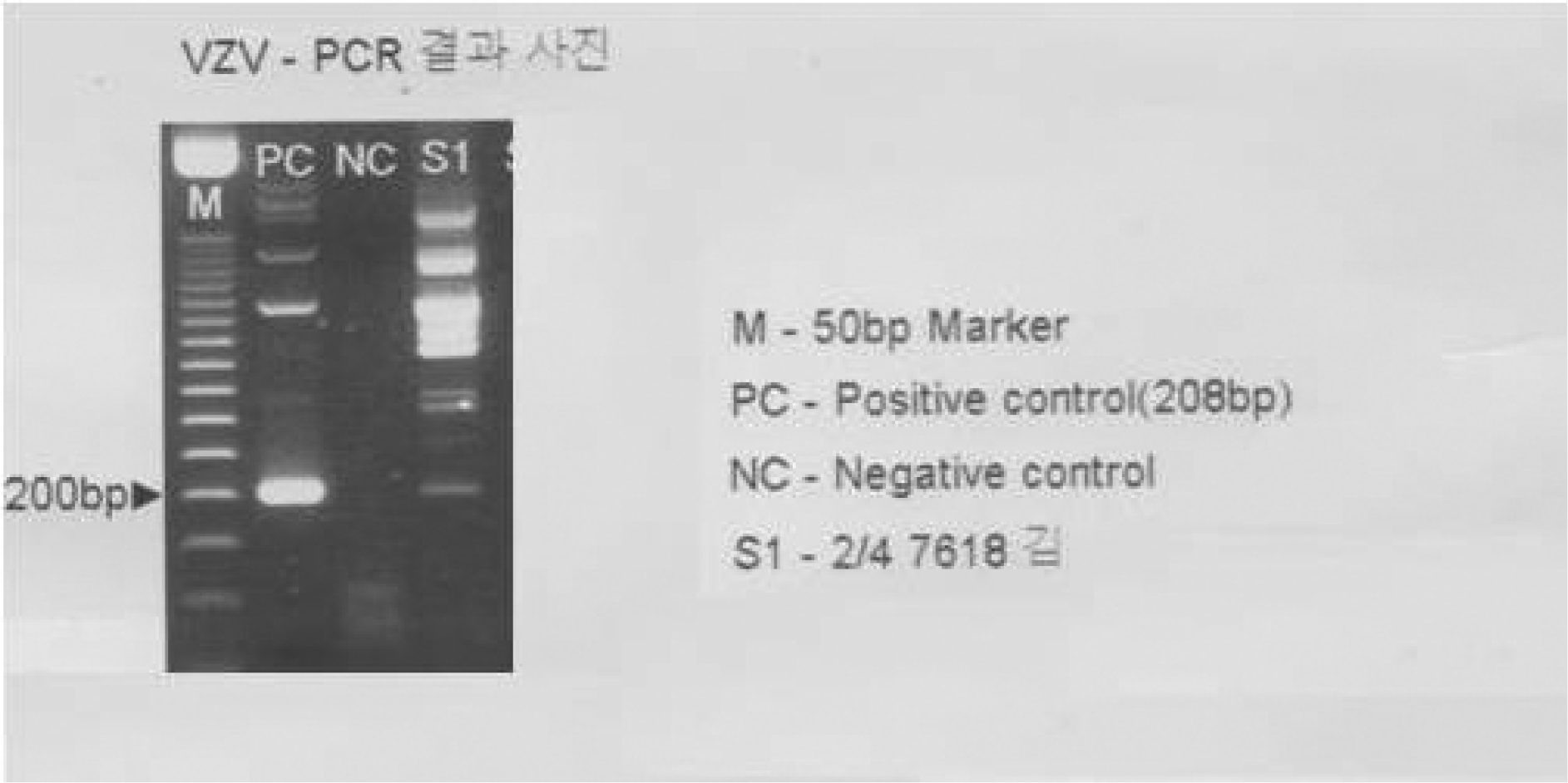
A 24-year-old female presenting with a history of centripetal eruption and erythema, followed by vesicle and eschar, was diagnosed with varicella and managed in a local medical clinic. Five days after the varicella eruption, she experienced decreased vision in her left eye. On initial exam visual acuity was light-sense positive in the left eye and 1.0 in the right eye; on fundus examination the patient was diagnosed with CRAO. We performed hematologic tests including thrombophilia studies, but there were no abnormal findings on routine hematologic tests, the carotid artery, or cardiovascular examinations. Antinuclear antibody, rheumatoid factor, and antiphospholipid antibody were negative. Skin biopsy and PCR results both corresponded with varicella, and the patient was diagnosed with CRAO associated with chickenpox.
References
1. Vyse AJ, Gay NJ, Hesketh LM, et al. Seroprevalence of antibody to varicella zoster virus in England and Wales in children and young adults. Epidemiol Infect. 2004; 132:1129–34.

2. Macleod J. Davidson's priciples and practice of medicine. 19th ed.1. Edinburgh: Churchill Livingstone;1984. p. 730.
3. Duke-Elder S. System of ophthalmology. 3rd ed.15. London: Kimpton;1976. p. 167.
4. Hall S, Maupin T, Seward J, et al. Second varicella infections: Are they more common than previously thought? Pediatrics. 2002; 19:1068–73.
5. Ostler HB, Thygeson P. The ocular manifestations of herpes zoster, varicella, infectious mononucleosis, and cytomegalovirus disease. Surv Ophthalmol. 1976; 21:148–59.
6. Liesegang TJ. The varicella-zoster virus: systemic and ocular features. J Am Acad Dermatol. 1984; 11:165–91.
7. Appel I, Frydman M, Savir H, et al. Uveitis and ophthalmoplegia complicating chickenpox. J Pediatr Ophthalmol. 1977; 14:346–8.
8. Chu W, Pavan-Langston D. Ocular surface manifestations of the major viruses. Int Ophthalmol Clin. 1979; 19:135–67.
9. Edwards T. Ophthalmic complications of varicella. J Pediatr Ophthalmol. 1965; 2:37–40.
10. Jordan DR, Noel LP, Clarke WN. Ocular involvement in varicella. Clin Pediatr. 1984; 23:434–6.
11. Matoba A. Ocular viral infections. Pediatr Infect Dis. 1984; 3:358–68.
12. Robb R. Cataracts acquired following varicella infection. Arch Ophthalmol. 1972; 87:352–4.
13. Yoser SL, Forster DJ, Rao NA. Systemic viral infections and their retinal and choroidal manifestations. Surv Ophthalmol. 1993; 37:313–52.
14. Garweg J, Bohnke M. Varicella zoster virus is strongly associated with atypical necrotizing herpetic retinopathies. Clin Infect Dis. 1997; 24:603–8.
15. Capone A Jr, Meredith TA. Central visual loss caused by chickenpox retinitis in a 2-year-old child. Am J Ophthalmol. 1992; 113:592–3.
16. Purvin V, Hrisomalos N, Dunn D. Varicella optic neuritis. Neurology. 1988; 38:501–3.
17. Hugkulstone CE, Watt LL. Branch retinal arteriolar occlusion with chickenpox. Br J Ophthalmol. 1988; 72:78–80.
18. Cho NC, Han HJ. Central retinal artery occlusion after varicella. Am J Ophthalmol. 1992; 114:235–6.
19. Friedberg MA, Micale AJ. Monocular blindness from central retinal artery occlusion associated with chickenpox. Am J Ophthalmol. 1994; 117:117–8.
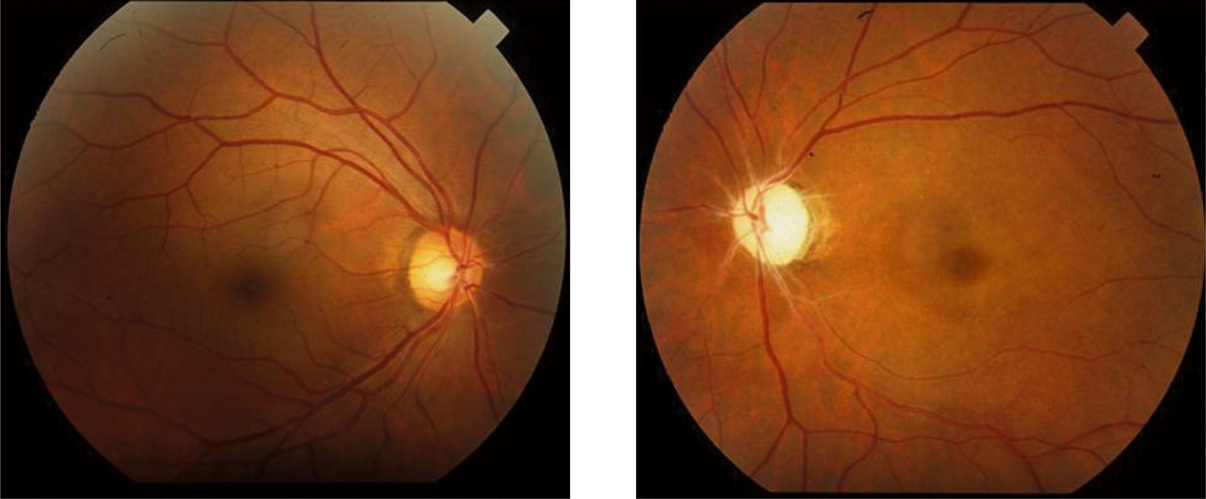
Figure 1.
Fundus photograph show whitening of the superficial retina and cherry-red spot in the left eye.
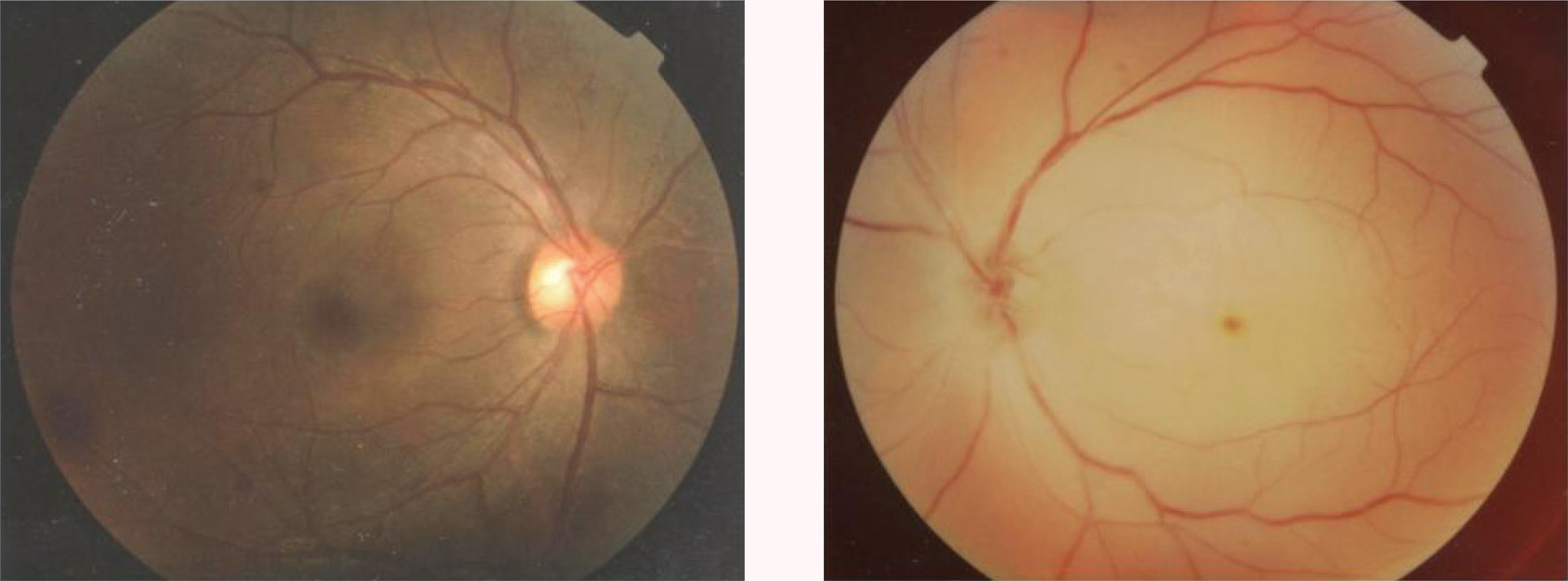
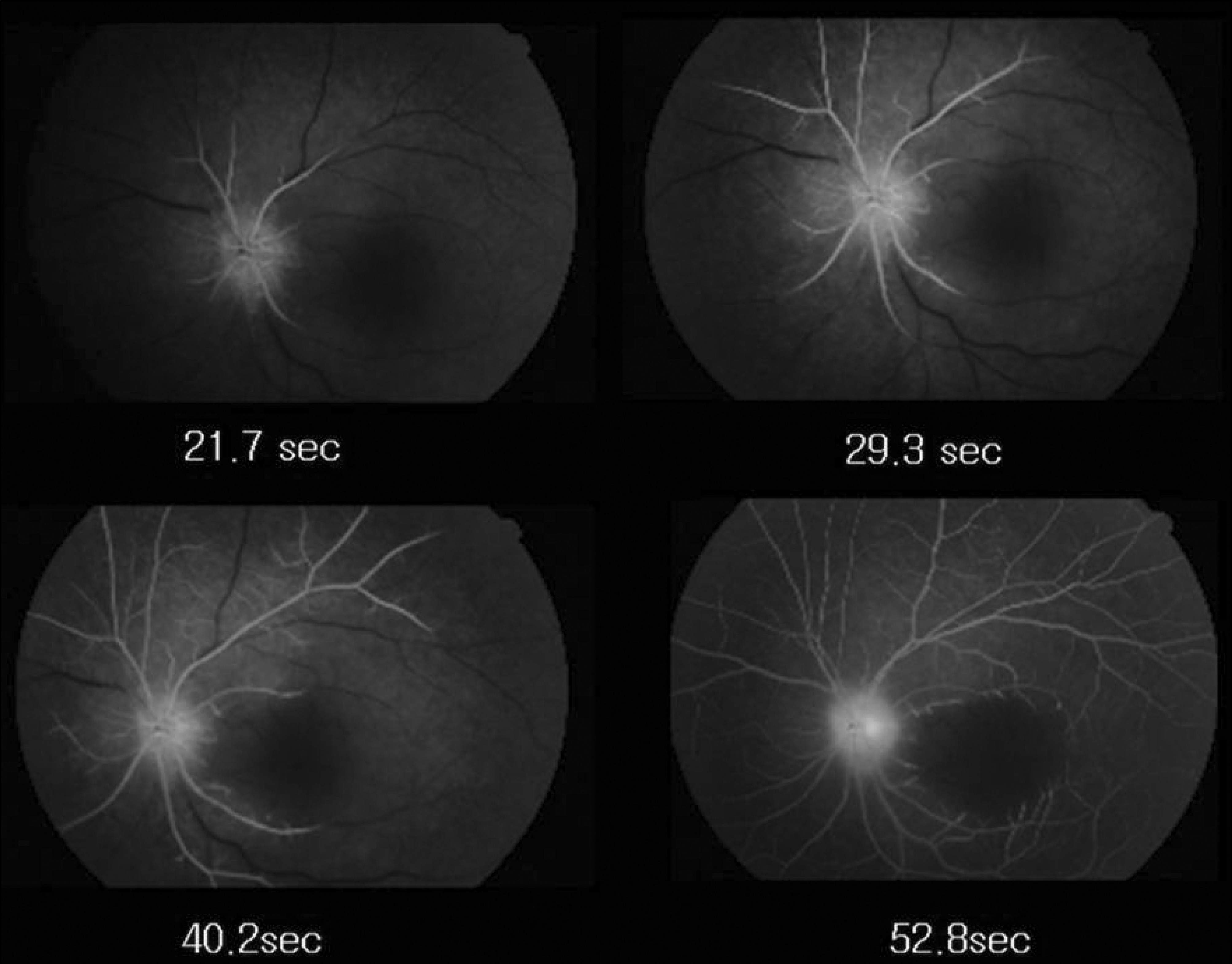
Figure 2.
Fluorescein angiograhs show delayed retinal arterial filling time and arteriovenous transit time in the left eye.
 XML Download