

 PDF
PDF ePub
ePub Citation
 Print
Print


Abstract
Case summary
A 44-year-old man visited our clinic complaining of decreased visual acuity in his left eye. His best corrected visual acuity was hand movement in his left eye, and rhegmatogenous retinal detachment involving the macula at the superior temporal site was found. We performed pars plana vitrectomy and attempted to reattach the retina using endolaser photocoagulation; however, the laser burn was not made, and we failed to reattach the retina. At that point, we carried out cryopexy around the retinal tear, and injected silicone oil into the vitreous cavity. Ten months after surgery, his best corrected visual acuity was 0.06, and there was no recurrent retinal detachment or proliferative vitreoretinopathy.
References
1. Dorey SE, Neveu MM, Burton LC, et al. The clinical features of albinism and their correlation with visual evoked potentials. Br J Ophthalmol. 2003; 87:767–72.

2. Susan MC, Raymond EB, Pamela JS, William VG. Albinism:modern molecular diagnosis. Br J Ophthalmol. 1998; 82:189–95.
3. King RA, Pietsch J, Fryer JP, et al. Tyrosinase gene mutations in oculocutaneous albinism 1 (OCA1): definition of the phenotype. Hum Genet. 2003; 113:502–13.
4. Guillery RW. Why do albinos and other hypopigmented mutants lack normal binocular vision, and what else is abnormal in their central visual pathway? Eye. 1996; 10:217–21.
5. Kim MH. Two cases of complete generalized albinism. J Korean Ophthalmol Soc. 1976; 17:150–63.
6. Park JC, Lee JH. Two cases of ocular albinism. J Korean Ophthalmol Soc. 1980; 21:271–3.
7. Lee KY, Ban MS, Song BR, Yoo JH. A case of oculocutaneous albinism. J Korean Ophthalmol Soc. 2000; 41:288–93.
8. Campochiaro PA, KAden IH, Vidaurri-Leal J, Glaser BM. Cryotherapy enhances intravitreal dispersion of variable pigment epithelial cells. Arch Ophthalmol. 1985; 103:434–6.
9. Wilkinson CP, Rice TA. Michels retinal detachment. 2nd ed.St Louis: Mosby;1997. p. 1067.
10. McDonald HR. Diagnostic and therapeutic challenges. Retina. 2001; 21:367–70.
Figure 1.
A 44-year-old man with oculocutaneous albinism. (A) The photogram of the right eye shows hypopigmented skin, eyelash, and iris. Note the visible intraocular IOL optic and haptics through the hypopigmented iris. (B) Fluorescein angiogram shows early faded-out dye in the retinal vessels and visible choroidal vessels through the window-defect. (C) B-scan sonogram of the left eye showed bullous retinal detachment. (D) Light microcopy from the skin at the trunk shows generalized absence of melanin pigment (HE stain, ×200).

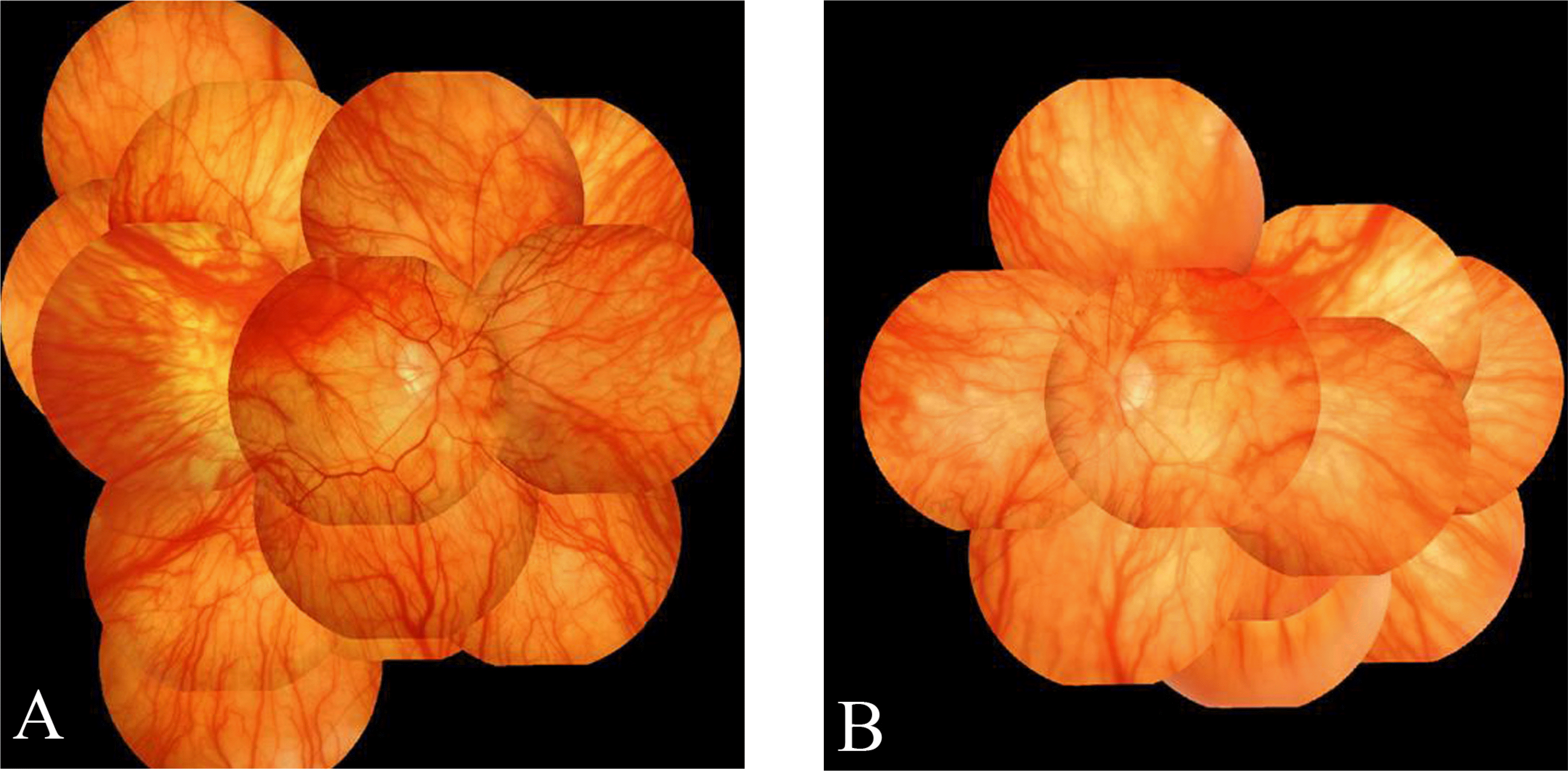
Figure 2.
The composite fundus photograms of a 44-year-old man with oculocutaneous albinism. (A) The fundus photograph of the right eye shows hypoplastic fovea and visible choroidal vasculature. (B) The fundus photograph of the left eye 6 months after retinal detachement operation shows well attached retina.
 XML Download