Abstract
Purpose
To report a case of an anterior fibrovascular membrane following cataract extraction and intraocular lens implantation in a patient with congenital aniridia.
Case summary
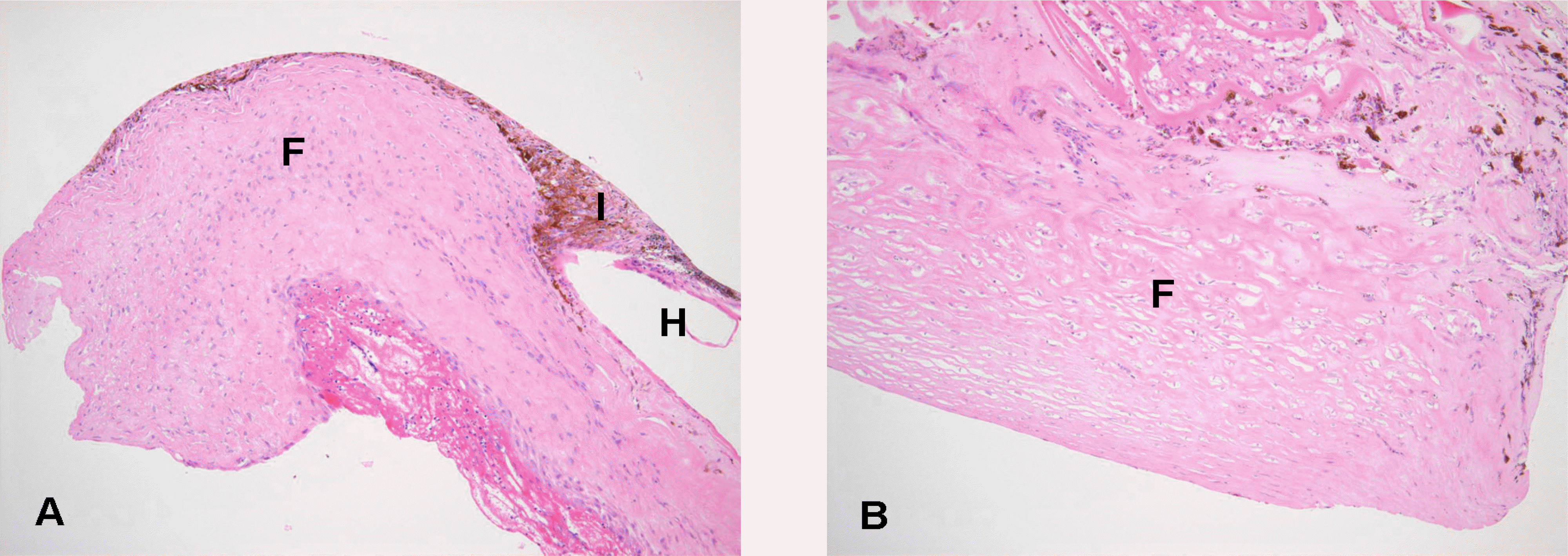
A 13-year-old girl presented with congenital aniridia and cataracts in both eyes. She underwent cataract extraction by phacoemulcification with intraocular lens implantation. Six months after cataract surgery, a progressive anterior chamber fibrovascular membrane was noted in both intraocular lens and rudimentary iris. Surgical excision of the fibrovascular membrane was performed, but there was recurrence after five weeks in both eyes. Subsequent surgical intervention on both eyes involved intraocular lens explantation combined with membranectomy to prevent recurrence and phthisis. Surgical findings indicated that the fibrovascular membrane involved the retrolenticular space, and histopathological evidence indicated that the extensive fibrotic tissue originated from the root of the rudimentary iris.
Conclusions
Patients with congenital aniridia should be monitored carefully for the development of intraocular fibrosis after intraocular lens implantation. If a fibrovascular membrane is noted, early surgical intervention is recommended, and the explantation of the intraocular lens should be considered during surgical intervention to prevent recurrence and complications.
Go to : 
References
1. Barratta G. Observazioni pratiche sulle principal: malattie degli orchid, Milano 1818 Tomo 2, 349 as cited by Jungke C: Veber den angeborren mangel der iris. J Chir Augen-Heilkunde. 1821; 2:677.
2. Shaw MM, Falla HF, Neel JV. Congenital aniridia. Am J Hum Genet. 1960; 12:389–415.
3. Axton R, Hanson I, Danes S, et al. The incidence of PAX6 mutation in patients with simple aniridia; an evaluation of mutation detection in 12 cases. J Med Genet. 1997; 34:279–86.

4. Collins ET. Congenital defects of the iris and glaucoma. Trans Ophthalmol Soc U K. 1883; 13:128–39.
5. Nwell FW. Opthalmology. 8th ed.St, Louis: CV Mosby;1996. p. 282–3.
6. Layman PR, Anderson DR, Flynn JT. Frequent occurrence of hypoplastic optic disks in patients with aniridia. Am J Ophthalmol. 1974; 77:513–6.

7. Elsas FJ, Maumenee IH, Kenyon KR, Yider F. Familial aniridia with preserved ocular function. Am J Ophthalmol. 1977; 83:718–24.

8. Lewallen WM Jr. Aniridia and related iris defects; a report of twelve cases with bilateral cataract extraction and resulting good vision in one. AMA Arch Ophthalmol. 1958; 59:831–9.
9. Chung SJ. A case of aniridia. J Korean Ophthalmol Soc. 1971; 12:50–3.
10. Ahn SK, Kang JS, Shyn KH. A Case of Congenital Aniridia. J Korean Ophthalmol Soc. 1989; 30:815–8.
12. Duke-Elder S. System of Ophthalmology. 3. St. Louis: CV Mosby;1971. p. 586–91.
13. Sundmacher R, Reinhard T, Althaus C. Black-diaphragm intraocular lens for correction of aniridia. Ophthalmic Surg. 1994; 25:180–5.

14. Park SJ, Kim HT, Kim SM, Chung SK. Implantation of Black Diaphragm Intraocular Lens in Cataract Surgery with Congenital Aniridia. J Korean Ophthalmol Soc. 1998; 39:1748–54.
15. Tsai JH, Freeman JM, Chan CC, et al. A progressive anterior fibrosis syndrome in patients with postsurgical congenital aniridia. Am J Ophthalmol. 2005; 140:1075–9.

16. Stefan C, Niculescu C. Congenital aniridia and cataract. Oftalmologia. 1994; 38:141–3.
17. Osher RH, Burk SE. Cataract surgery combined with implantation of an artificial iris. J Cataract Refract Surg. 1999; 25:1540–7.

18. Chan CC, Fujikawa LS, Rodrigues MM, et al. Immunohistochemistry and electron microscopy of cyclitic membrane: Report of a case. Arch Ophthalmol. 1986; 104:1040–5.
Go to : 
 | Figure 1.(A) Preoperative microscopic findings after intraocular lens implantation. (B) Progressive formation of fibrovascular membrane (F) with extension of the membrane anteriorly in the anterior chamber. C = cornea: I = black-diaphragm intraocular lens. (C) Intraocular lens (I) explantation combined with fibrovascular membrane removal. (D) Microscopic findings after fibrovascular membrane removal. |


 PDF
PDF ePub
ePub Citation
 Print
Print


 XML Download