

 PDF
PDF ePub
ePub Citation
 Print
Print


Abstract
Purpose
To report a case of a familial lecithin cholesterol acyltransferase (LCAT) deficiency patient with bilateral corneal opacities.
Case summary
A 26-year-old man with bilateral corneal opacities visited our hospital. We took slit lamp examination, corneal thickness measurement, corneal endothelial cell counts and fundus examination. Blood and urine tests were included. Kidney biopsy was done. The tissues were observed by a light microscopy and an electron microscopy. Hemolytic anemia, proteinuria, hematuria, hypertriglyceridemia, decreased HDL cholesterol level, and lecithin cholesterol acyltransferase (LCAT) deficiency were found. At kidney biopsy, electron-lucent vacuoles and lamellar inclusion body were found.
References
1. Gjone E, Norum KR. Familial serum cholesterol ester deficiency: Clinical study of a patient with a new syndrome. Acta Med Scand. 1968; 183:107–12.
2. Borysiewicz LK, Soutar AK, Evans DJ, et al. Renal failure in familial lecithin:cholesterol acyltransferase deficiency. Q J Med. 1982; 51:411–26.
3. Vergani C, Catapano AL, Roma P, Giudici G. A new case of familial LCAT deficiency. Acta Med Scand. 1983; 214:173–6.

4. Muthusethupathi MA, Padmanabhan R, Date A, et al. Familial lecithin:cholesterol acyltransferase deficiency with renal failure in two siblings: First case report from India. Nephron. 1999; 81:89–93.
5. Viestenz A, Schlötzer-Schrehardt U, Hofmann-Rummelt C, et al. Histopathology of Corneal Changes in Lecithin-Cholesterol Acyltransferase Deficiency. Cornea. 2002; 21:834–7.
6. Frohlich J, McLeod R, Hon K. Lecithin: Cholesterol Acyl Transferase (LCAT). Clin Biochem. 1982; 15:269–78.
7. Miller NE. Associations of high-density lipoprotein subclasses and apolipoproteins with ischemic heart disease and coronary atherosclerosis. Am Heart J. 1987; 113:589–97.
8. Silvia SF, Jeffrey MH, Gerd A, Bryan BJ. Lecithin Cholesterol Acyltransferase Deficiency and Fish Eye disease. Charles RS, Arthur LB, William SS, editors. The Metabolic & Molecular Bases of Inherited Disease. 8th ed.New York, London: McGraw-Hill;2001. 2:chap. 118.
9. Kenneth RK, Samuel EN, Christos H. Corneal Manifestations of Metabolic Diseases. Jay HK, Mark JM, Edward JH, editors. Cornea. 2nd ed.Elsevier: Mosby Year Book;2005. 1:chap. 64.
10. Kim JH, Myong YW. Histopathological Findings of Schnyder's Crystalline Corneal Dystrophy. J Korean Ophthalmol Soc. 1995; 36:1363–9.
11. Jung CS, Myong YW. A Case of Spontaneous Regression of Schnyder's Crystalline Corneal Dystrophy. J Korean Ophthalmol Soc. 2000; 41:1441–4.
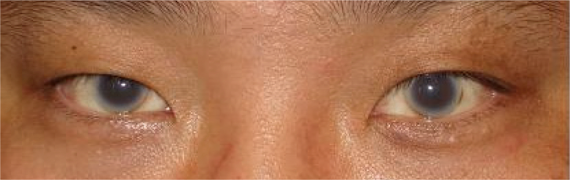
Figure 2.
Slit-lamp examination show diffuse, cloudy, drop-shaped opacities involving the entire cornea (A) Right cornea (B) Left cornea.
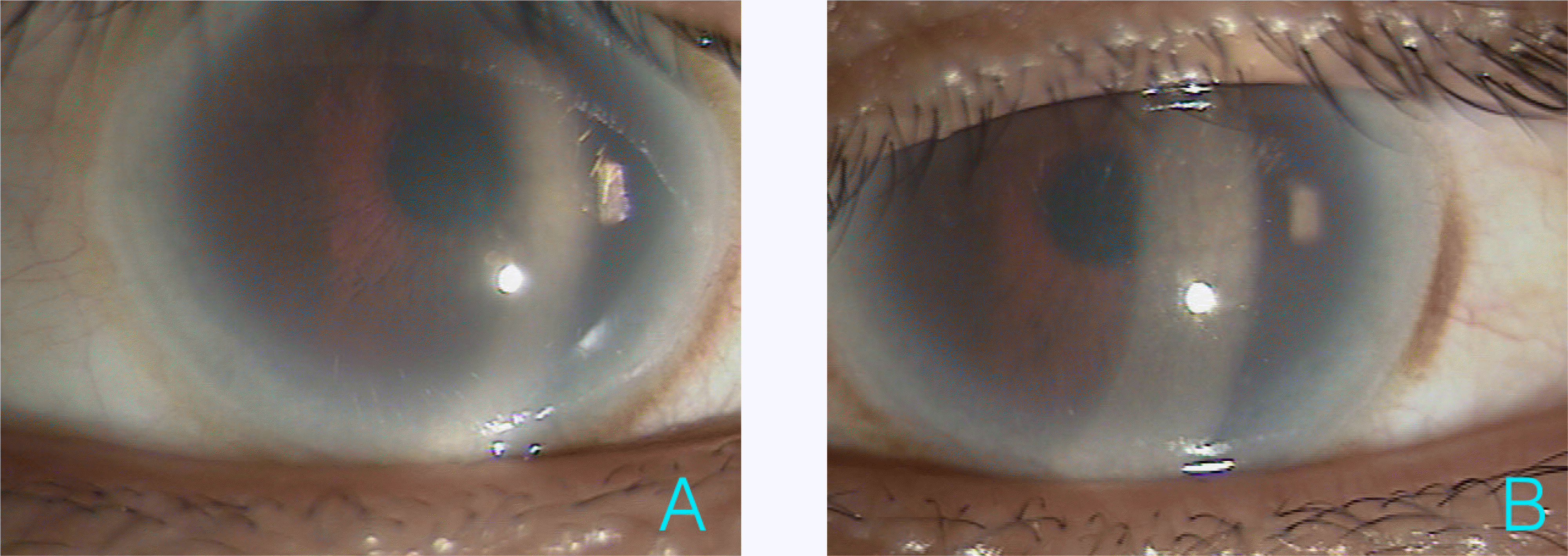
Figure 3.
Histopathologic findings of kidney were consistent with Familial lecithin cholesterol acyltransferase deficiency. (A) H&E stains showed capillary wall thickening and mild mesangial widening. (B) Characteristic EM finding was various sized electron-lucent vacuoles with lamellar inclusion body (arrow).

 XML Download