

 PDF
PDF ePub
ePub Citation
 Print
Print


Abstract
Methods
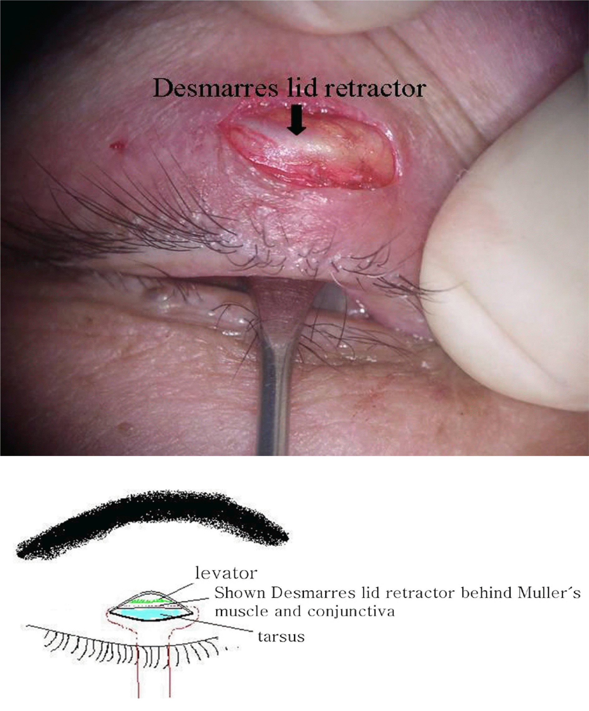
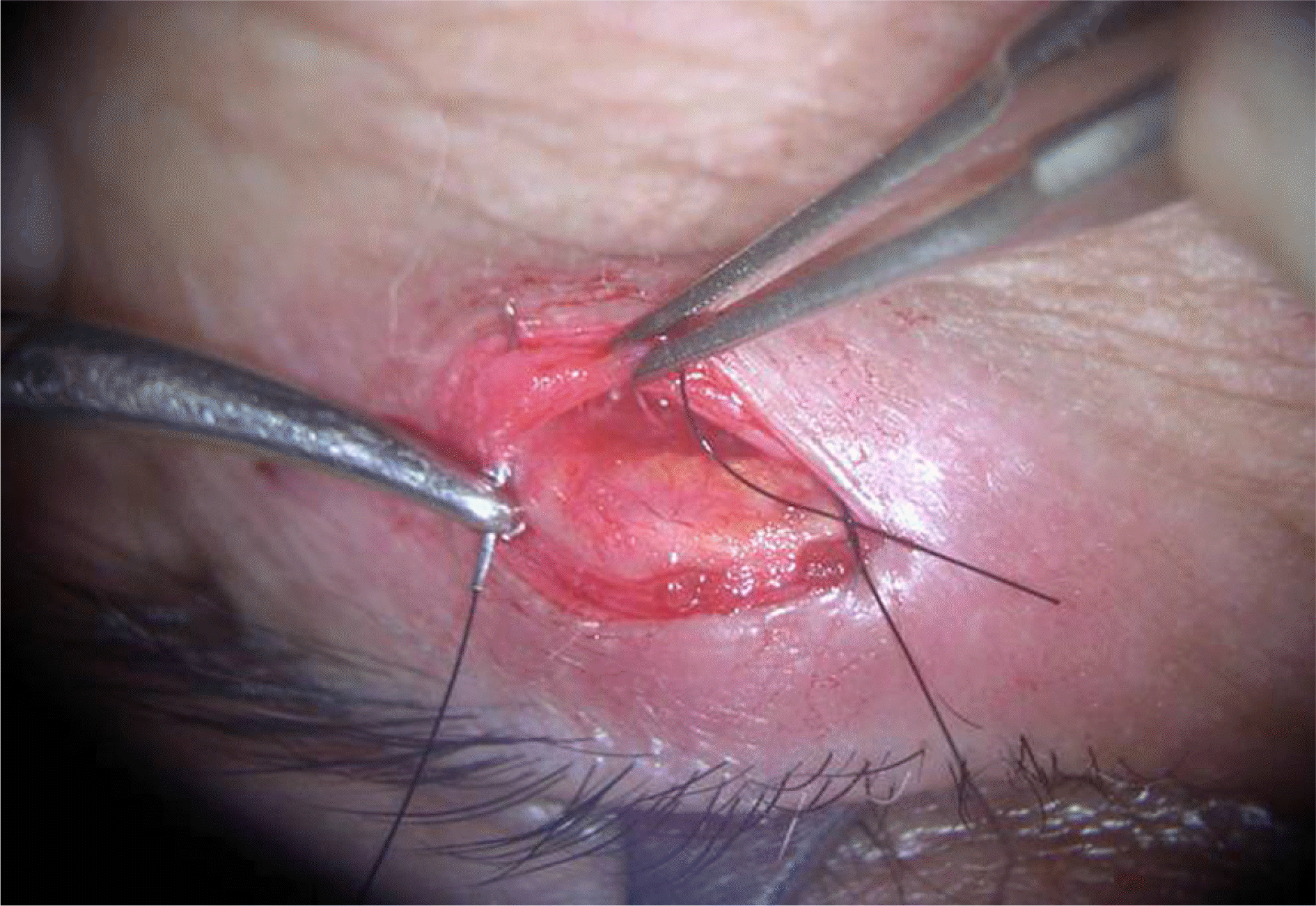
Twenty three ptotic eyelids of 17 patients were repaired through a small incision between March 2005 and December 2005. Levator function and pre- and postoperative upper eyelid marginal reflex distances (MRD1) were checked. The small incision was executed on the eyelid crease, with a length of approximately 10.0∼12.0 mm. Two 6-0 Ethilon stitches were placed to the tarsus after detecting levator aponeurosis with a Desmarres retractor.
Results
Of the 32eyes, 9 eyes had congenital ptosis and 23 eyes had acquired ptosis. The mean followup time was 12.1±3.4 months, with a levator function of 7.6±2.1 mm, preoperative MRD1 of 0.2±0.4 mm, and postoperative MRD1 of 2.6±0.3 mm. Two eyelids remained mildly undercorrected and required an additional operation.
Conclusions
The small incision technique for ptosis correction is safe and precise, allowing more rapid recovery from surgery. The Desmarres retractor allows the levator to be found easily without complications. In addition, the small incision technique for levator repair is useful for congenital ptosis with good levator function as well as acquired ptosis.
References
1. Frueh BR. The mechanistic classification of ptosis. Ophthalmology. 1980; 87:1019–21.

2. Beard C, Callahan MA. Beard's ptosis. 4th ed.1. Birmingham: Aesculapius Publishung Co.;1990. p. 52–87.
3. Youn DY, Sung MK, Lee KH. Clinical examination on the blepharoptosis and the resection of the levator muscle. J Korean Ophthalmol Soc. 1990; 31:125–33.
4. Lee UK, Chung HS. The effect of external levator resection in blepharoptosis with poor levator function. J Korean Ophthalmol Soc. 1998; 39:1062–8.
5. Fasanella RM, Servat J. Levator resection for minimal ptosis another simplified operation. Arch Ophthalmol. 1961; 65:493–6.
6. Anderson RL, Dixon RS. Aponeurotic ptosis surgery. Arch Ophthalmol. 1979; 97:1123–8.
7. Jeong S. Clinical study of simple levator resection in ptosis patients. J Korean Ophthalmol Soc. 2002; 43:551–5.
8. Liu D. ptosis repair by single suture aponeurotic tuck. Ophthalmology. 1993; 2:251–9.
9. Baek SH, Park MS, Park HJ. The effect of Simplified Single Suture Aponeurotic Tuck in Ptosis Patients. J Korean Ophthalmol Soc. 2003; 44:1011–6.
10. Older JJ. Levator aponeurosis tuck: a treatment for ptosis. Ophthalmic Surg. 1978; 97:1123–8.
11. Lucarelli MJ, Lemke BN. Small incision external levator repair: technique and early results. Am J Ophthalmol. 1999; 127:637–44.
12. Bernardini FP, de Conciliis C, Devoto MH. Mini invasive ptosis surgery. Orbit. 2006; 25:111–5.
13. Baroody M, Holds JB, Sakamoto DK, et al. Small incision transcutaneous levator aponeurotic repair for blepharoptosis. Ann Plast Surg. 2004; 52:558–61.
14. Frueh BR, Musch DC, McDonald HM. Efficacy and efficiency of a small incision, minimal dissection procedure versus a traditional approach for correcting aponeurotic ptosis. Ophthalmology. 2004; 111:2158–63.
15. Berris CE. Adjustable sutures for the correction of adult acquired ptosis. Ophthal Plast Reconstr Surg. 1988; 4:171–3.


 XML Download