Journal List > J Korean Ophthalmol Soc > v.49(1) > 1008255

Kang and Park: The Combined Effect of Subtenon Triamcinolone Injection and Panretinal Photocoagulation on Diabetic Retinopathy

Abstract

Purpose

To investigate the result of posterior sub-tenon triamcinolone acetonide injection combined with panretinal photocoagulation (PRP) in patients with diabetic retinopathy.

Methods

A prospective study was performed on patients with diabetic retinopathy who required PRP. The study group consisted of 12 patients (12 eyes) of diabetic retinopathy without clinically significant macular edema (CSME) and 13 patients (13 eyes) with CSME. All patients were injected posteriorly with sub-tenon triamcinolone acetonide (40 mg) one week before PRP. During a six-month follow-up, best-corrected visual acuity, the development of macular edema, changes in fluorescein angiography, and related complications were monitored.

Results

During a six-month follow-up, visual acuity was well preserved in patients with diabetic retinopathy without CSME (12 eyes). Only one patient in this group experienced temporary macular edema at 3 months after combined therapy. In the CSME group (13 eyes), the visual acuities of seven patients (53.8%) increased, those of five patients (38.5%) remained same, and one patient’s visual acuity (7.7%) decreased in a study period of six months. Fluorescein angiography showed that macular edema was resolved in most patients except in one patient in whom macular edema remained for up to 6 months. Complications from combined therapy occurred in two patients who showed slight and temporary increase of intraocular pressure.

Conclusions

Combined treatment with posterior sub-tenon triamcinolone acetonide injection and PRP may provide benefits for patients with diabetic retinopathy who require urgent PRP by preventing exacerbation of macular edema.

References

1. Early Treatment Diabetic Retinopathy Study Research Group. Early photocoagulation for diabetic retinopathy. Early Treatment Diabetic Retinopathy Study Report No 9. Ophthalmology. 1991; 98:766–85.
2. McDonald HR, Schatz H. Macular edema following panretinal photocoagulation. Retina. 1985; 5:5–10.
crossref
3. McDonald HR, Schatz H. Visual loss following panretinal photocoagulation for proliferative diabetic retinopathy. Ophthalmology. 1985; 92:388–93.
crossref
4. Higgins KE, Meyers SM, Jaffe MJ, et al. Temporary loss of foveal contrast sensitivity associated with panretinal photocoagulation. Arch Ophthalmol. 1986; 104:997–1003.
crossref
5. Henricsson M, Heiji A. The effect of panretinal photocoagulation on visual acuity, visual field and on subjective visual impairment in preproliferative and early proliferative diabetic retinopathy. Acta Ophthalmol. 1994; 72:570–5.
6. Jonas JB, Sofker A. Intraocular injection of crystalline cortisone as adjunctive treatment of diabetic macular edema. Am J Ophthalmol. 2001; 132:425–7.
crossref
7. Martidis A, Duker J, Greenberg PB, et al. Intravitreal triamcinolone for refractory diabetic macular edema. Ophthalmology. 2002; 109:920–7.
crossref
8. Jonas JB, Kreissig I, Sofker A, et al. Intravitreal injection of triamcinolone for diffuse diabetic macular edema. Arch Ophthalmol. 2003; 121:57–61.
crossref
9. Choi YJ, Oh IK, Oh JR, et al. Intravitreal versus posterior subtenon injection of triamcinolone acetonide for diabetic macular edema. Korean J Ophthalmol. 2006; 20:205–9.
crossref
10. Lee YH, Kim CG. Intravitreal triamcinolone acetonide injection for treatment of macular edema. J Korean Ophthalmol Soc. 2004; 45:2055–63.
11. Zacks DN, Johnson MW. Combined intravitreal injection of triamcinolone acetonide and panretinal photocoagulation for concomitant diabetic macular edema and proliferative diabetic retinopathy. Retina. 2005; 25:135–40.
crossref
12. Coles R, Krohn D, Breslin H, et al. Depo Medrol in treatment of inflammatory disease of the anterior segment of the eye. Am J Ophthalmol. 1962; 54:407–11.
13. Verma LK, Vivek MB, Kumar A, et al. A prospective controlled trial to evaluate the adjunctive role of posterior subtenon triamcinolone in the treatment of diffuse diabetic macular edema. J Ocul Pharmacol Ther. 2004; 20:277–84.
crossref
14. Freeman WR, Green RL, Smith RE. Echographic localization of corticosteroids after periocular injection. Am J Ophthalmol. 1987; 103:281–8.
crossref
15. Er H, Doganay S, Turkoz Y, et al. The levels of cytokine and nitric oxide in rabbit vitreous humor after retinal photocoagulation. Ophthalmic Surg Lasers. 2000; 31:479–83.
16. Shimura M, Yasuda K, Shiono T. Posterior sub-Tenon’s capsule injection of triamcinolone acetonide prevents panretinal photocoagulation-induced visual dysfunction in patients with severe diabetic retinopathy and good vision. Ophthalmology. 2006; 113:381–7.
crossref
17. Kim YG, Yu SY, Kwak HW. The effect of intravitreal triamcinolone acetonide injection according to the diabetic macular edema type. J Korean Ophthalmol Soc. 2005; 46:84–9.
18. Bakri SJ, Kaiser KP. Posterior subtenon triamcinolone acetonide for refractory diabetic macular edema. Am J Ophthalmol. 2005; 139:290–4.
crossref
19. Craig JH, Gray NH. The effects of posterior subtenon injection of triamcinolone acetonide in patients with intermediate uveitis. Am J Ophthalmol. 1995; 120:55–64.
20. Nozik RA. Periocular injection of steroids. Trans Am Acad Ophthalmol Otolaryngol. 1972; 74:178–88.
21. Kim YJ, Kang SW, Ahn BH, et al. The results of posterior subtenon steroid injection in uveitis patients. J Korean Ophthalmol Soc. 2003; 44:66–72.
22. Verma LK, Vivek MB, Kumar A, et al. A prospective controlled trial to evaluate the adjunctive role of posterior subtenon triamcinolone in the treatment of diffuse diabetic macular edema. J Ocul Pharmacol Ther. 2004; 20:277–84.
crossref
23. Jonas JB, Degenring RF, Kreissig I, et al. Intraocular pressure elevation after intravitreal triamcinolone acetonide injection. Ophthalmology. 2005; 112:593–8.
crossref
24. Helm CJ, Holland GN. The effect of posterior subtenon injection of triamcinolone acetonide in patients with intermediate uveitis. Am J Ophthalmol. 1995; 120:55–64.
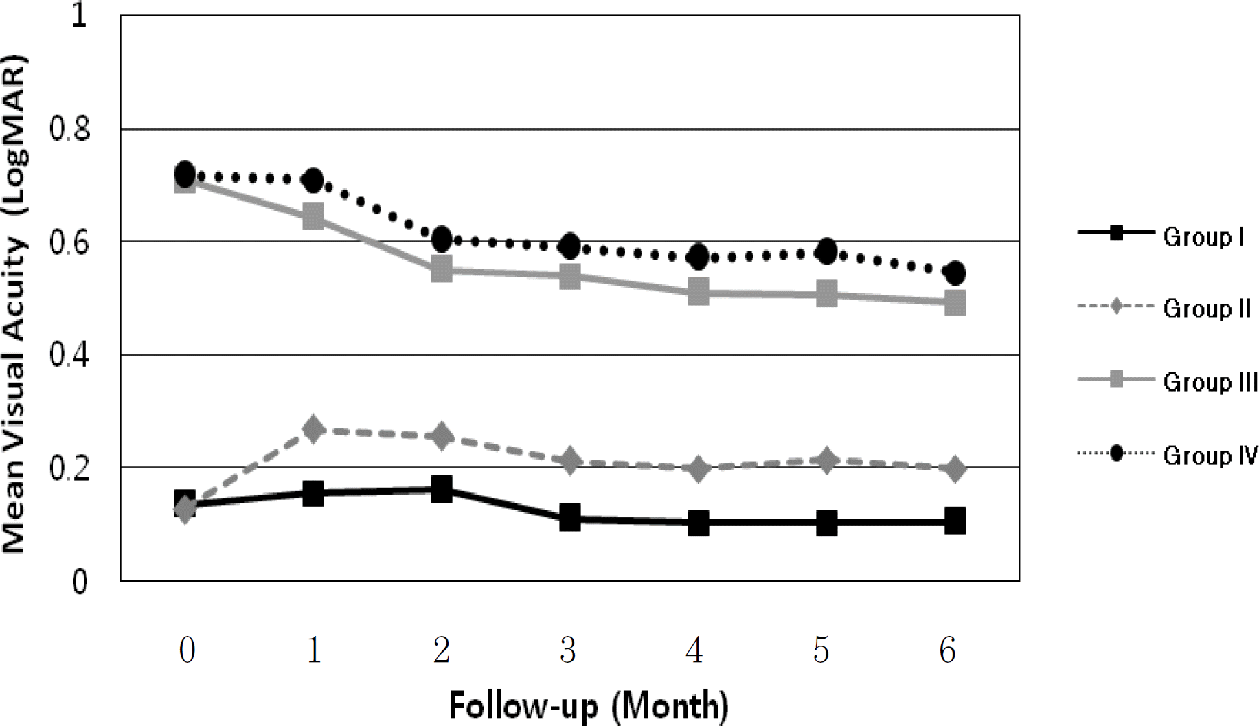
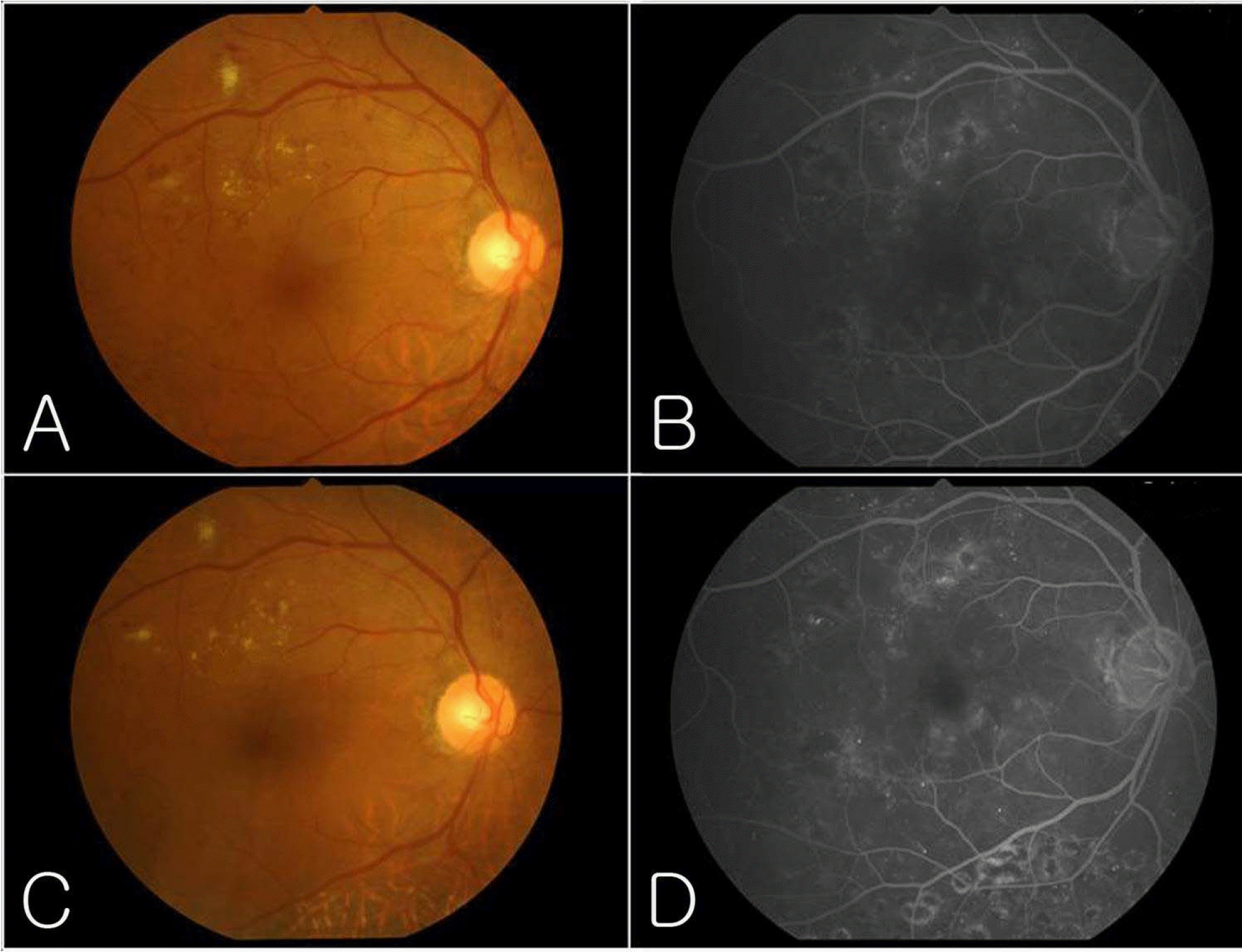
Figure 1.
Graph illustrating the change of mean best- corrected visual acuity (logMAR) in four groups. Group I: patients received combined therapy of triamcinolone sub-tenon injection and panretinal photocoagulation. Group II: patients received only panretinal photocoagulation. Group III: patients who had CSME received combined therapy of triamcinolone sub-tenon injection and panretinal photocoagulation. Group IV: patients who had CSME received focal or grid retinal photocoagulation before panretinal photocoagulation. In Group I and II, there was no difference of visual acuity between baseline and at 6 month. In Group III, compared with its baseline, values of visual acuity (logMAR) were significantly lower at 4, 5 and 6 months (p=0.019, p=0.044, p=0.042, respectively). In Group IV, compared with its baseline, values of visual acuity (logMAR) were significantly lower only at 6 months (p=0.028).
jkos-49-71f1.tif
Figure 2.
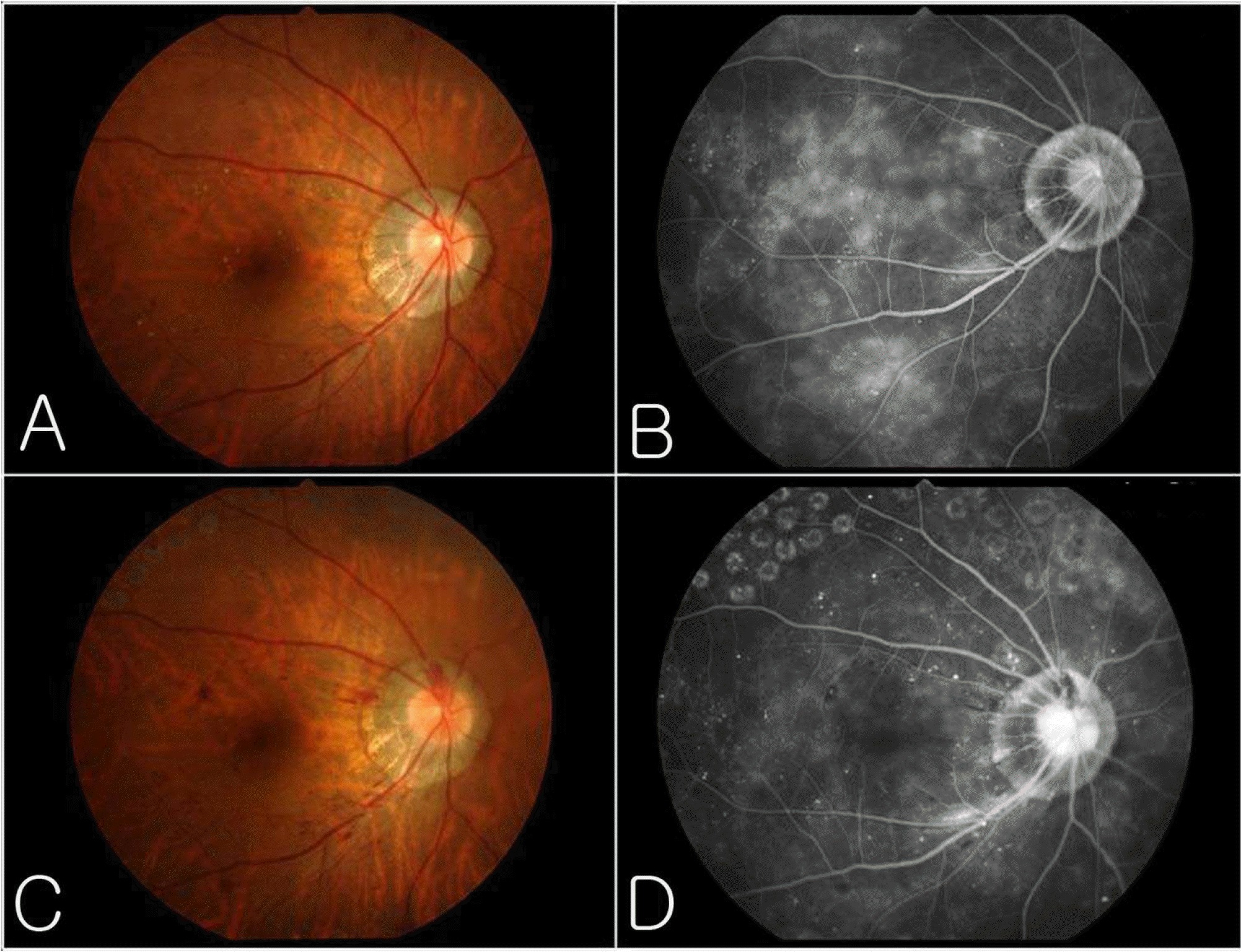
Color fundus photographs and late-phase fluorescein angiograms of a male patient aged 58 without clinically significan macular edema. Fundus photograph (A) and fluorescein angiogram (B) before posterior sub-tenon injection of triamcinolon acetonide. (C, D) Same patients 6 months after combined sub-tenon injection of triamcinolone acetonide and panretina photocoagulation. Note that there was no macular edema before and after combined therapy.
jkos-49-71f2.tif
Figure 3.
Color fundus photographs and late-phase fluorescein angiograms of a male patient aged 54 with clinically significant macular edema. Fundus photograph (A) and fluorescein angiogram (B) before posterior sub-tenon injection of triamcinolone acetonide showing macular edema and fluorescein leakage at macula (C, D) Same patient 6 months after combined sub-tenon injection of triamcinolone acetonide and panretinal photocoagulation. Note the decreased amount of late leakage in the macula as seen on the fluorescein angiogram. The macular edema improved.
jkos-49-71f3.tif
Table 1.
Demographic data and diabetic retinopathy status of each treatment group without CSME
Variables Group I (n=12) Group II (n=12) P-value*
Age (years)
 Range 47-69 39-68
 Mean±S.D. 57.5±7.89 56.8±8.48 0.932
Sex
 Male 3 3
 Female 9 9
Duration of DM (years)
 Range 4-21 5-20
 Mean±S.D. 8.56±5.58 9.23±6.12 0.486
DM status (No. of eyes)
 PDR 2 3
 Severe NPDR 10 9

Group I: patients received combined therapy of triamcinolone sub-tenon injection and panretinal photocoagulation. Group II: patients received only panretinal photocoagulation. CSME=clinically significant macular edema; PDR=proliferative diabetic retinopathy; NPDR=non-proliferative diabetic retinopathy; S.D.=standard deviation;

P-value*=the significance of the difference was assessed by the Mann-Whitney U test.

Table 2.
Demographic data and diabetic retinopathy status of each treatment group with CSME
Variables Group III (n=13) Group IV (n=13) P-value*
Age (years)
 Range 36-73 43-63
 Mean±S.D. 55.1±9.71 56.5±6.80 0.794
Sex
 Male 8 8
 Female 5 5
Duration of DM (years)
 Range 3-28 4-25
 Mean±S.D. 9.89±6.24 9.01±7.36 0.756
DM status (No. of eyes)
 PDR 6 7
 Severe NPDR 7 6

Group III: patients who have CSME received combined therapy of triamcinolone sub-tenon injection and panretinal photocoagulation. Group IV: patients who have CSME received focal retinal photocoagulation before panretinal photocoagulation. CSME=clinically significant macular edema; PDR=proliferative diabetic retinopathy; NPDR=non-proliferative diabetic retinopathy; S.D.=standard deviation;

P-value*=the significance of the difference was assessed by the Mann-Whitney U test.

Table 3.
Mean visual acuity and IOP during in both groups without CSME
Study Peroid Group I (n=12)
Group II (n=12)
VA (logMAR±S.D.) IOP (mmHg±S.D.) VA (logMAR±S.D.) IOP (mmHg±S.D.)
Baseline 0.135±0.10 16.67±3.73 0.127±0.09 15.85±4.21
1 month 0.155±0.14 15.50±4.35 0.268±0.18* 16.02±4.02
2 months 0.161±0.14 14.91±3.02 0.256±0.13* 15.41±3.62
3 months 0.111±0.12 15.27±3.07 0.213±0.11 15.73±3.97
4 months 0.103±0.12 16.17±3.16 0.199±0.11 15.89±3.41
5 months 0.103±0.12 15.92±3.48 0.214±0.10 15.76±3.01
6 months 0.104±0.11 15.83±4.24 0.199±0.12 16.01±3.84

Group I: patients received combined therapy of triamcinolone sub-tenon injection and panretinal photocoagulation. Group I patients received only panretinal photocoagulation. CSME=clinically significant macular edema; VA=visual acuity IOP=intraocular pressure; S.D.=standard deviation;

* =the statistical difference between baseline VA and VA at each follow-up was assessed by Wilcoxon signed rank test (p<0.05).

Table 4.
Mean visual acuity and IOP during study peroid in both groups with CSME
Study Peroid Group III (n=13)
Group IV (n=13)
VA (logMAR±S.D.) IOP (mmHg±S.D.) VA (logMAR±S.D.) IOP (mmHg±S.D.)
Baseline 0.708±0.46 16.23±4.00 0.717±0.43 15.33±3.53
1 month 0.643±0.48 14.58±3.38 0.709±0.45 15.58±4.01
2 months 0.549±0.32 15.15±3.28 0.603±0.53 16.15±3.87
3 months 0.539±0.33 15.53±2.90 0.590±0.40 15.94±3.31
4 months 0.509±0.34* 14.53±3.04 0.571±0.36 15.37±4.03
5 months 0.506±0.37* 14.92±3.45 0.580±0.39 15.27±3.59
6 months 0.492±0.38* 14.61±3.42 0.545±0.38* 15.21±3.58

Group III: patients who have CSME received combined therapy of triamcinolone sub-tenon injection and panretinal photocoagulation. Group IV: patients who have CSME received focal retinal photocoagulation before panretinal photocoagulation. CSME=clinically significant macular edema; VA=visual acuity; IOP=intraocular pressure; S.D.=standard deviation;

* =the statistical difference between baseline VA and VA at each follow-up was assessed by Wilcoxon signed rank test (p<0.05).

Table 5.
Summary of patients who received re-treatment after combined therapy
Group No. of eyes Re-treatment Interval from baseline Comments
without CSME (n=12) 1 Subtenon injection 4 months ME was developed at 3 months and resolved at 5 months after subtenon re-injection.
with CSME (n=13) 4 Subtenon injection 1 month Because initial CSME was just slightly resolved after combined therapy, subtenon re-injection was done again at 1 month. However, CSME has not decreased until 6 months. Thin ERM was developed.
Grid laser 7 weeks Initial CSME was nearly subsided after combined therapy. CSME re-appeared at 6 weeks and was subsided at 4 months with grid laser treatment. After re-treatment, retina was flat up to 6 months.
Subtenon injection 2 months CSME was decreased much but small amout of CSME was still remained up to 2 months. Subtenon re-injection was performed at 2 months, which resulted in disappearing remained CSME.
Subtenon injection 3 months CSME was remained slightly up to 3 months. It was completely subsided 2 weeks after subtenon re-injection.

CSME=clinically significant macular edema; ME=macular edema; ERM=epiretinal membrane.

TOOLS
Similar articles