Journal List > J Korean Ophthalmol Soc > v.49(4) > 1008244

Lee, Ryu, Moon, Kim, Woo, Cho, and Koh: The Efficacy of Intravitreal Gatifloxacin in Experimental S. epidermidis Endophthalmitis

Abstract

Purpose

To compare the efficacy of intravitreal gatifloxacin with intravitreal vancomycin in the treatment of Staphylococcus epidermidis endophthalmitis in a rabbit model.

Methods

Albino rabbits (n=30), infected with an intravitreal inoculum of S. epidermidis (105 colony forming unit/0.1 mL), were divided into 6 groups (n=5). Groups I and IV received 200 µg/0.1 mL of intravitreal gatifloxacin, and groups II and V were injected 1000 µg/0.1 mL of vancomycin intravitreally. Intravitreal balanced salt solutions (untreated control) were given to Groups III and VI. Intravitreal antibiotic therapy commenced 24 hours after bacterial inoculation. The bactericidal efficacy was determined by electroretinography (ERG), clinical grading, bacterial culture of vitreous aspirates and histopathologic grading. ERGs and clinical gradings were performed only for groups I, II, and III and bacterial cultures were done only for groups IV, V, and VI.

Results

Eyes in the gatifloxacin groups showed similar appearance to those in the vancomycin treated groups clinically, histologically, and functionally as proved with ERG. All aspirates from the gatifloxacin and vancomycin groups were culture‐ negative at 5 days after bacterial inoculation, whereas all eyes in the untreated control group were culture‐ positive.

Conclusions

This study demonstrated that intravitreal injection of 200 µg /0.1mL gatifloxacin appeared to be equally effective compared to intravitreal 1000 µg /0.1 mL vancomycin in the treatment of S. epidermidis endophthalmitis. If proven safe and efficacious after further study in humans, intravitreal injection of gatifloxacin could be considered an effective alternative to vancomycin for the treatment of S. epidermidis endophthalmitis.

References

1. O'Day DM, Jones DB, Patrinely J, Elliott JH. Staphylococcus epidermidis endophthalmitis. Visual outcome following noninvasive therapy. Ophthalmology. 1982; 89:354–60.
2. Results of the Endophthalmitis Vitrectomy Study. A randomized trial of immediate vitrectomy and of intravenous antibiotics for the treatment of postoperative bacterial endophthalmitis. Endophthalmitis Vitrectomy Study Group. Arch Ophthalmol. 1995; 113:1479–96.
3. Aydin E, Kazi AA, Peyman GA, Esfahani MR. Intravitreal toxicity of moxifloxacin. Retina. 2006; 26:187–90.
crossref
4. Ermis SS, Cetinkaya Z, Kiyici H, Ozturk F. Treatment of Staphylococcus epidermidis endophthalmitis with intravitreal moxifloxacin in a rabbit model. Tohoku J Exp Med. 2005; 205:223–9.
crossref
5. Roth DB, Flynn HW Jr. Antibiotic selection in the treatment of endophthalmitis: the significance of drug combinations and synergy. Surv Ophthalmol. 1997; 41:395–401.
crossref
6. Gao H, Pennesi ME, Qiao X, et al. Intravitreal moxifloxacin: retinal safety study with electroretinography and histopathology in animal models. Invest Ophthalmol Vis Sci. 2006; 47:1606–11.
crossref
7. Bower KS, Kowalski RP, Gordon YJ. Fluoroquinolones in the treatment of bacterial keratitis. Am J Ophthalmol. 1996; 121:712–5.
crossref
8. Donnenfeld ED, Schrier A, Perry HD, et al. Penetration of topically applied ciprofloxacin, norfloxacin, and ofloxacin into the aqueous humor. Ophthalmology. 1994; 101:902–5.
crossref
9. Rowen S. Preoperative and postoperative medications used for cataract surgery. Curr Opin Ophthalmol. 1999; 10:29–35.
crossref
10. Goldstein MH, Kowalski RP, Gordon YJ. Emerging fluoroquinolone resistance in bacterial keratitis: a 5‐ year review. Ophthalmology. 1999; 106:1313–8.
11. Mather R, Karenchak LM, Romanowski EG, Kowalski RP. Fourth generation fluoroquinolones: new weapons in the arsenal of ophthalmic antibiotics. Am J Ophthalmol. 2002; 133:463–6.
12. Ednie LM, Jacobs MR, Appelbaum PC. Activities of gatifloxacin compared to those of seven other agents against anaerobic organisms. Antimicrob Agents Chemother. 1998; 42:2459–62.
crossref
13. Hariprasad SM, Mieler WF, Holz ER. Vitreous penetration of orally administered gatifloxacin in humans. Trans Am Ophthalmol Soc. 2002; 100:153–9.
14. Hariprasad SM, Mieler WF, Holz ER. Vitreous and aqueous penetration of orally administered gatifloxacin in humans. Arch Ophthalmol. 2003; 121:345–50.
crossref
15. Solomon R, Donnenfeld ED, Perry HD, et al. Penetration of topically applied gatifloxacin 0.3%, moxifloxacin 0.5%, and ciprofloxacin 0.3% into the aqueous humor. Ophthalmology. 2005; 112:466–9.
crossref
16. Kazi AA, Jermak CM, Peyman GA, et al. Intravitreal toxicity of levofloxacin and gatifloxacin. Ophthalmic Surg Lasers Imaging. 2006; 37:224–9.
crossref
17. Loewenstein A, Zemel E, Lazar M, Perlman I. Drug‐ induced retinal toxicity in albino rabbits: the effects of imipenem and aztreonam. Invest Ophthalmol Vis Sci. 1993; 34:3466–76.
18. Mino De Kaspar H, Hoepfner AS, Engelbert M, et al. Antibiotic resistance pattern and visual outcome in experimentally-induced Staphylococcus epidermidis endophthalmitis in a rabbit model. Ophthalmology. 2001; 108:470–8.
19. Engelbert M, Mino de Kaspar H, Thiel M, et al. Intravitreal vancomycin and amikacin versus intravenous imipenem in the treatment of experimental Staphylococcus aureus endophthalmitis. Graefes Arch Clin Exp Ophthalmol. 2004; 242:313–20.
crossref
20. Domagala JM. Structure‐ activity and structure‐ side‐ effect relationships for the quinolone antibacterials. J Antimicrob Chemother. 1994; 33:685–706.
21. Hwang DG. Fluoroquinolone resistance in ophthalmology and the potential role for newer ophthalmic fluoroquinolones. Surv Ophthalmol. 2004; 49:79–83.
crossref
22. Ozkiris A, Evereklioglu C, Esel D, et al. The efficacy of intravitreal piperacillin/tazobactam in rabbits with experimental Staphylococcus epidermidis endophthalmitis: a comparison with vancomycin. Ophthalmic Res. 2005; 37:168–74.
23. Maxwell DP Jr, Brent BD, Orillac R, et al. A natural history study of experimental Staphylococcus epidermidis endophthalmitis. Curr Eye Res. 1993; 12:907–12.
24. Meredith TA, Aguilar HE, Miller MJ, et al. Comparative treatment of experimental Staphylococcus epidermidis endophthalmitis. Arch Ophthalmol. 1990; 108:857–60.
crossref
25. Meredith TA, Trabelsi A, Miller MJ, et al. Spontaneous sterilization in experimental Staphylococcus epidermidis endophthalmitis. Invest Ophthalmol Vis Sci. 1990; 31:181–6.
26. Chung KH, Shin JP, Kim IT. Spontaneous sterilization in experimental Staphylococcus and Pseudomonas endophthalmitis. J Korean Ophthalmol Soc. 1995; 36:1895–902.
27. Kwok AK, Hui M, Pang CP, et al. An in vitro study of ceftazidime and vancomycin concentrations in various fluid media: implications for use in treating endophthalmitis. Invest Ophthalmol Vis Sci. 2002; 43:1182–8.
28. Hui M, Kwok AK, Pang CP, et al. An in vitro study on the compatibility and precipitation of ciprofloxacin and vancomycin in human vitreous. Br J Ophthalmol. 2004; 88:218–22.
29. Choi KS, Kim JS, Nam KR. Effect of intravitreal ciprofloxacin in the treatment of experimental Bacillus endophthalmitis. J Korean Ophthalmol Soc. 2002; 43:890–7.
30. Recchia FM, Busbee BG, Pearlman RB, et al. Changing trends in the microbiologic aspects of postcataract endophthalmitis. Arch Ophthalmol. 2005; 123:341–6.
crossref
31. Esmaeli B, Holz ER, Ahmadi MA, et al. Endogenous endophthalmitis secondary to vancomycin-resistant enterococci infection. Retina. 2003; 23:118–9.
crossref
32. Saenz Gonzalez MC, Valero Juan LF. [Enterococcus sp. resistance, a growing problem. Epidemiologic study (1987–1993)]. Med Clin (Barc). 1994; 103:485–9.
33. Schwalbe RS, Stapleton JT, Gilligan PH. Emergence of vancomycin resistance in coagulase‐ negative staphylococci. N Engl J Med. 1987; 316:927–31.
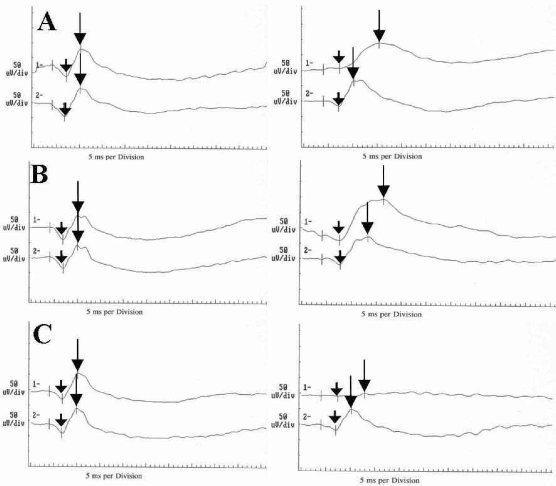
Figure 1.
ERG responses obtained from eyes of groups I (A), II (B), and III (C). In each graph, the upper curves (wave 1) correspond to the ERG of experimental eyes and lower curves (wave 2) represent ERG from contralateral normal eyes. Baseline ERG before inoculation of S. epidermidis is on the left and the ERG of the same eyes 7 days after bacterial inoculation (6th day of antibiotics injection) is on the right. In gatifloxacin (A) and vancomycin (B) administered groups, the b‐ wave amplitude ratios (experimental/contralateral) are not changed, but b-wave implicit time ratios (experimental/contralateral) increase 7 days after bacterial inoculation. In infected control group (C), the wave of experimental eye is nearly extinguished. Short arrows, trough of a‐ waves; long arrows, peak of b-waves.
jkos-49-651f1.tif
Figure 2.
Histopathologic features of the retina, ciliary body, and vitreous in three eyes from each group (H & E). Gatifloxacin (A, group I) and vancomycin (B, group II) treated eyes shows relatively clear vitreous and well preserved retinal layers. In infected control group (C, group III), the retina, ciliary body, and vitreous are infiltrated with many inflammatory cells. Left column shows the retina, choroid, and sclera. ×200 (A and B), ×100 (C). Right column represents the ciliary body and vitreous base. ×40(A, B, and C). V, vitreous cavity; R, retina; C, choroids; S, sclera; CB, ciliary body.
jkos-49-651f2.tif
Figure 3.
Histopathologic features of the retina, ciliary body, and vitreous in groups 4 (A), 5 (B), and 6 (C) (H & E). Gatifloxacin (A, group IV) and vancomycin (B, group V) treated eyes show relatively clear vitreous and well preserved retinal layers. Some inflammatory cells are observed in the vitreous cavity. In untreated infected control group (C, group VI), the retina, ciliary body, and vitreous are infiltrated with dense inflammatory cells and no retinal layer is intact. Left column shows the retina, choroids, and sclera. ×200 (A and B), ×100 (C). Right column represents the ciliary body and vitreous base. ×40 (A, B, and C). V, vitreous cavity; R, retina; C, choroids; S, sclera; CB, ciliary body.
jkos-49-651f3.tif
Table 1.
Grading scheme for clinical observations*
Score Cornea Iris and anterior chamber Vitreous and retina
0 Normal appearance Normal appearance Normal appearance
1 Mild stromal haze Mild hyperemia, cells in anterior chamber White condensation in vitreous, petechial hemorrhages along nerve-vessel-streaks
2 Moderate stromal haze Moderate hyperemia, exudate in anterior chamber Vitreous haze, optic disc and large vessels still visible
3 Cornea opaque Iris vessels engorged and massive exudate in anterior chamber, or iris not visualizable because of anterior chamber inflammation No red reflex

* Cornea, iris and anterior chamber, and vitreous and retina were assigned points according to the described features. The score for anterior chamber and iris findings was multiplied by a factor of 2, and the score for vitreous findings by a factor of 3, before being added up. This score was then divided by 6 to give a range from 0 (normal appearance) to 3 (severe inflammatory changes).

Table 2.
Grading scheme for histopathologic evaluation*
Score Cornea and limbus Ciliary body Vitreous base Retina
1 Normal Normal Normal Normal
2 0–1 Inflammatory cells in cornea, mild limbal infiltrate 0–50 Inflammatory cells per 40× field 0–20 Inflammatory cells per 40× field Cystoid changes, thickness increased up to twice normal, few infiltrates
3 1–3 Corneal infiltrates, marked limbal infiltrates 50–100 Inflammatory cells per 40× field 20–50 Inflammatory cells per 40× field Thickness increased to more than twice normal, mild infiltrate, photoreceptors recognizable
4 3–5 Infiltrates, marked limbal infiltrates 100–200 Inflammatory cells per 40× field 50–150 Inflammatory cells per 40× field Retinal layers discernible, marked leukocytic infiltrates, no photoreceptors recognizable
5 3–5 Infiltrates, severe limbal infiltrates and swelling >200 Inflammatory cells per 40× field >150 Inflammatory cells per 40× field No discernible retinal layers, massive inflammatory infiltrate

* Cornea, ciliary body, vitreous base, and retina were assigned points according to the described features. To emphasize the importance of the findings in the posterior segment, vitreous points were multiplied by 2 and retinal points by 3. Points were added up, and the mean total score calculated. The total score was then divided by 7 to give a range from 1 (no pathologic changes) to 5 (severe inflammatory changes).

Table 3.
Statistical summary of the clinical scores and histopathologic results
Group Clinical scores§ at
Histopathologic scores
day 2 day 3 day 4 day 7
I, Gatifloxacin (200 µg/0.1 mL) 0.95±0.23* 1.53±0.44 1.92±0.18 0.81±0.22 1.78±0.40
II, Vancomycin (1000 µg/0.1 mL) 1.13±0.27* 1.37±0.42 1.80±0.22 0.87±0.40 1.90±0.28
III, Infected control (BSS 0.1 mL) 1.25±0.29 2.04±0.09 2.46±0.09 2.67±0.13 3.04±0.52

* p>0.05, compare with infected control group (Kruskal-Wallis Test)

p<0.05, compare with infected control group (Kruskal-Wallis Test)

p>0.05, compare between gatifloxacin and vancomycin treated groups (Mann-Whitney U Test)

§ Mean clinical scores are calculated on day 2, 3, 4, and 7 which corresponds to 1, 2, 3, and 6 days after bacterial inoculation, respectively.

Table 4.
Average ERG ratio of each study group
Group Preinoculation 7 days after inoculation p§
I, Gatifloxacin (200 µg/0.1 mL) 1.09±0.17* 0.98±0.13 0.116
II, Vancomycin (1000 µg/0.1 mL) 0.99±0.13* 0.90±0.36 0.498
III, Infected control (BSS 0.1 mL) 1.07±0.11 0.29±0.15 0.068

* p>0.05, compare with infected control group (Kruskal‐ Wallis test)

p<0.05, compare with infected control group (Kruskal-Wallis test); Wilcoxon signed ranks test

Π b-wave amplitude ratio of eyes inoculated with S. epidermidis compared with contralateral control

p>0.05, compare between gatifloxacin and vancomycin treated groups (Mann-Whitney U Test)

§ Wilcoxon signed ranks test

b-wave amplitude ratio of eyes inoculated with S. epidermidis compared with contralateral control eyes.

Table 5.
Average ERG ratio§ of each study group
Group Preinoculation 7 days after inoculation p
I, Gatifloxacin (200 µg/0.1 mL) 1.01±0.11* 1.45±0.15* 0.028
II, Vancomycin (1000 µg/0.1 mL) 1.08±0.04* 1.34±0.10* 0.043
III, Infected control (BSS 0.1 mL) 1.07±0.12 1.34±0.30 0.114

* p>0.05, compare with infected congrol group (Kruskal-Wallis test)

p>0.05, compare between gatifloxacin and vancomycin treated groups (Mann‐ Whitney U Test)

Wilcoxon signed ranks test

§ b-wave implicit time ratio of eyes inoculated with S. epidermidis compared with contralateral control eyes.

Table 6.
Culture results
Group CFU/mL
day 3 day 4 day 6 day 8
IV, Gatifloxacin (200 µg/0.1 mL) 6586±10521.11* 228±487.77* 0* 0*
V, Vancomycin (1000 µg/0.1 mL) 65±92.33* 2.0±4.47* 0* 0*
VI, Infected control (BSS 0.1 mL) 1.25×10⁷±1.04×10⁷ 7.75×10⁷±1.02×10⁷ 2.75×10⁵±5.74×10⁵ 2.96×10⁶±5.70×10⁶

Day 3, 4, 6, and 8 corresponds to 36 h, 3, 5, and 7 days after inoculation, respectively

* p<0.05, compare with infected control group (Kruskal‐Wallis Test)

p>0.05, compare between gatifloxacin and vancomycin treated groups (Mann-Whitney U Test)

After 36 h, 3, 5, and 7 days of inoculation, vitreous aspirates were obtained and inoculated on the blood agar plate and incubated for 48 h at 37℃. After the incubation period, surface colonies were counted and converted into CFU/mL.

Table 7.
Culture results expressed as culture positivity# and histopathologic scores
Group No. of eyes that shows culture positive after 48 h of incubation Histopathologic
day 3 day 4 day 6 day 8 Scores
IV, Gatifloxacin (200 µg/0.1 mL), N=5 4 (80%)* 2 (40%)* 0 (0%) 0 (0%) 2.02±0.39§
V, Vancomycin (1000 µg/0.1 mL), N=5 3 (60%)* 1 (20%)* 0 (0%) 0 (0%) 2.04±0.39§
VI, Infected control (BSS 0.1 mL), N=5 5 (100%) 5 (100%) 5 (100%) 4 (80%) 4.14±0.57

Day 3, 4, 6, and 8 corresponds to 36 h, 3, 5, and 7 days after inoculation, respectively

* p>0.05, compare with infected control group (Chi-Square Test)

p<0.05, compare with infected control group (Chi-Square Test)

p>0.05, compare between gatifloxacin and vancomycin treated groups (Chi-Square Test)

§ p<0.05, compare with infected control group (Kruskal-Wallis Test)

p>0.05, compare between gatifloxacin and vancomycin treated groups (Mann-Whitney U test)

# After 36 h, 3, 5, and 7 days of inoculation, vitreous aspirates were obtained and inoculated on the blood agar plate and incubated for 48 h at 37℃. After the incubation period, number of eyes that shows culture positive are counted.

TOOLS
Similar articles