Abstract
Case summary
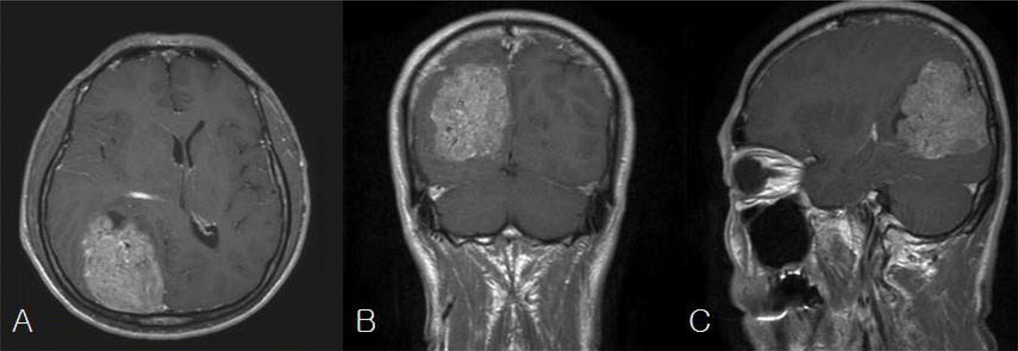
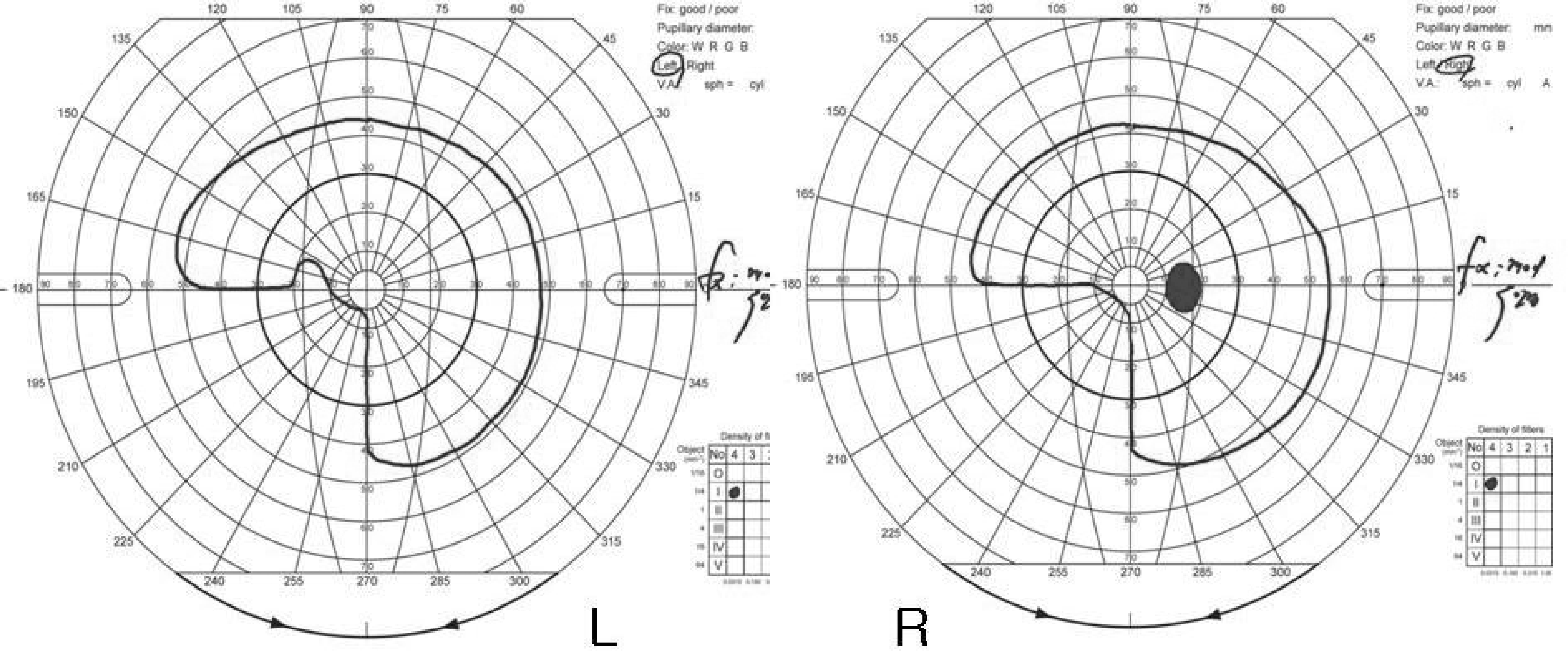
The authors reviewed the medical record, brain magnetic resonance image, and Goldmann visual field test of a 56-year-old male patient who was diagnosed with a meningioma in the right parietal and occipital lobe and underwent resection of the tumor. The preoperative Goldmann visual field test showed homonymous left inferior quadrantanopsia. Subtotal resection of the mass in the right parietal and occipital lobe was performed, and postoperative histopathologic examination confirmed the diagnosis of a meningioma. Postoperatively, the patient complained of visual hallucination in an area of the eye with visual field defects. However, his consciousness and orientations were intact, and other cognitive functions were also normal.
References
1. Menon GJ, Rahman I, Menon SJ, Dutton GN. Complex visual hallucinations in the visually impaired: the Charles Bonnet syndrome. Surv Ophthalmol. 2003; 48:58–72.
2. Rovner BW. The Charles Bonnet syndrome: a review of recent research. Curr Opin Ophthalmol. 2006; 17:275–7.

3. Dodd J, Heffeman A, Blake J. Visual hallucinations associated with Charles Bonnet Syndrome-an ever increasing diagnosis. Ir Med J. 1999; 92:344–5.
4. Choi EJ, Lee JK, Kang JK, Lee SA. Complex visual hallucinations after occipital cortical resection in a patient with epilepsy due to cortical dysplasia. Arch Neurol. 2005; 62:481–4.

5. Lee JS, Kim SY. A Case of Charles Bonnet Syndrome Following Cerebral Infarction in the Right Occipital Lobe. J Korean Neurol Assoc. 2006; 24:577–80.
6. Damas-Mora J, Skelton-Robinson M, Jenner FA. The Charles Bonnet syndrome in perspective. Psychol Med. 1982; 12:251–61.

7. Gold K, Rabins PV. Isolated visual hallucinations and the Charles Bonnet syndrome: a review of the literature and presentation of six cases. Compr Psychiatry. 1989; 30:90–8.

8. Beniczky S, Keri S, Voros E, et al. Complex hallucinations following occipital lobe damage. Eur J Neurol. 2002; 9:175–6.

9. Chen CS, Lin SF, Chong MY. Charles Bonnet syndrome and multiple sclerosis. Am J Psychiatry. 2001; 158:1158–9.

10. Mewasingh LD, Kornreich C, Christiaens F, et al. Pediatric phantom vision (Charles Bonnet) syndrome. Pediatr Neurol. 2002; 26:143–5.

11. Siatkowski RM, Zimmer B, Rosenberg PR. The Charles Bonnet syndrome: visual perceptive dysfunction in sensory deprivation. J Clin Neuroophthalmol. 1990; 10:215–8.
12. Manford M, Andermann F. Complex visual hallucinations. Clinical and neurobiological insights. Brain. 1998; 121:1819–40.

13. Takata K, Inoue Y, Hazama H, Fukuma E. Night– time hypnopompic visual hallucinations related to REM sleep disorder. Psychiatry Clin Neurosci. 1998; 52:207–9.
14. Poggel DA, Muller– Oehring EM, Gothe J, et al. Visual hallucinations during spontaneous and training– induced visual field recovery. Neuropsychologia. 2007; 45:2598–607.
15. Teunisse RJ, Cruysberg JR, Hoefnagels WH, et al. Visual hallucinations in psychologically normal people: Charles Bonnet syndrome. Lancet. 1996; 347:794–7.
16. Tan CS, Lim VS, Ho DY, et al. Charles Bonnet syndrome in Asian patients in a tertiary ophthalmic center. Br J Ophthalmol. 2004; 88:1325–9.


 PDF
PDF ePub
ePub Citation
 Print
Print


 XML Download