

 PDF
PDF ePub
ePub Citation
 Print
Print


Abstract
Purpose
Subcutaneous fat necrosis is rare. To our knowledge, there is no report of subcutaneous fat necrosis on the lower eyelid. We report a case of subcutaneous fat necrosis of the lower eyelid related to anemia.
Case summary
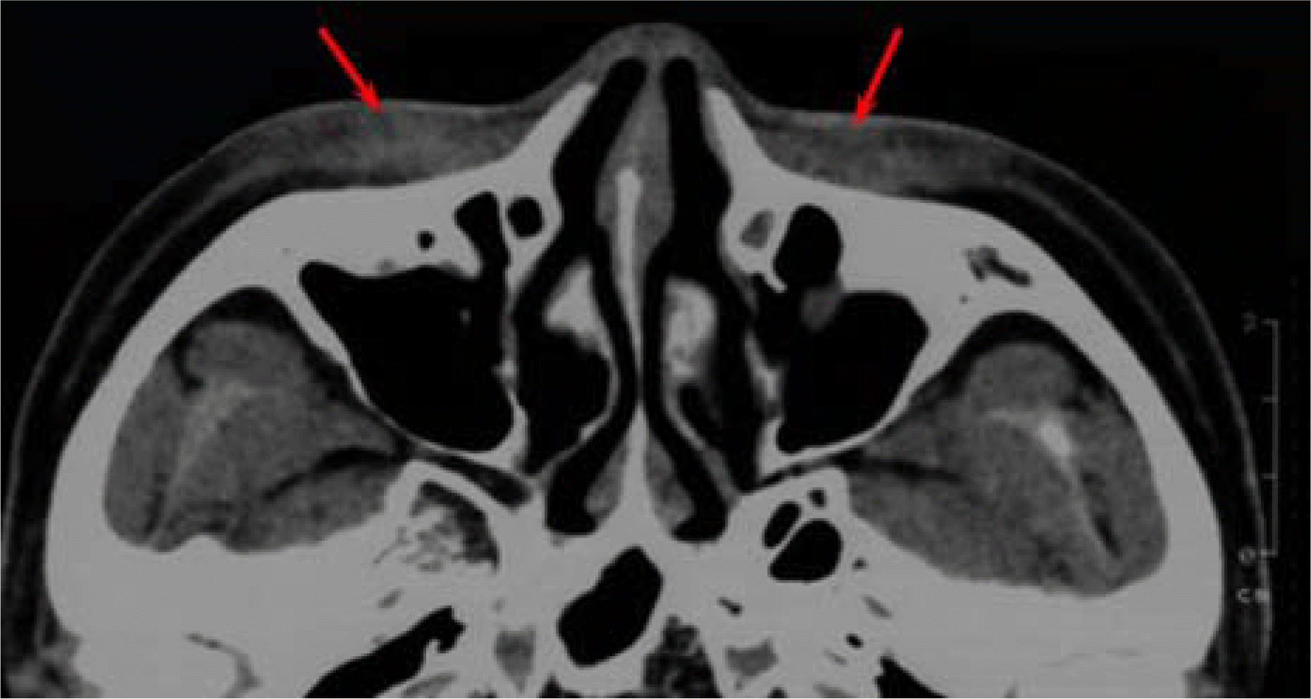
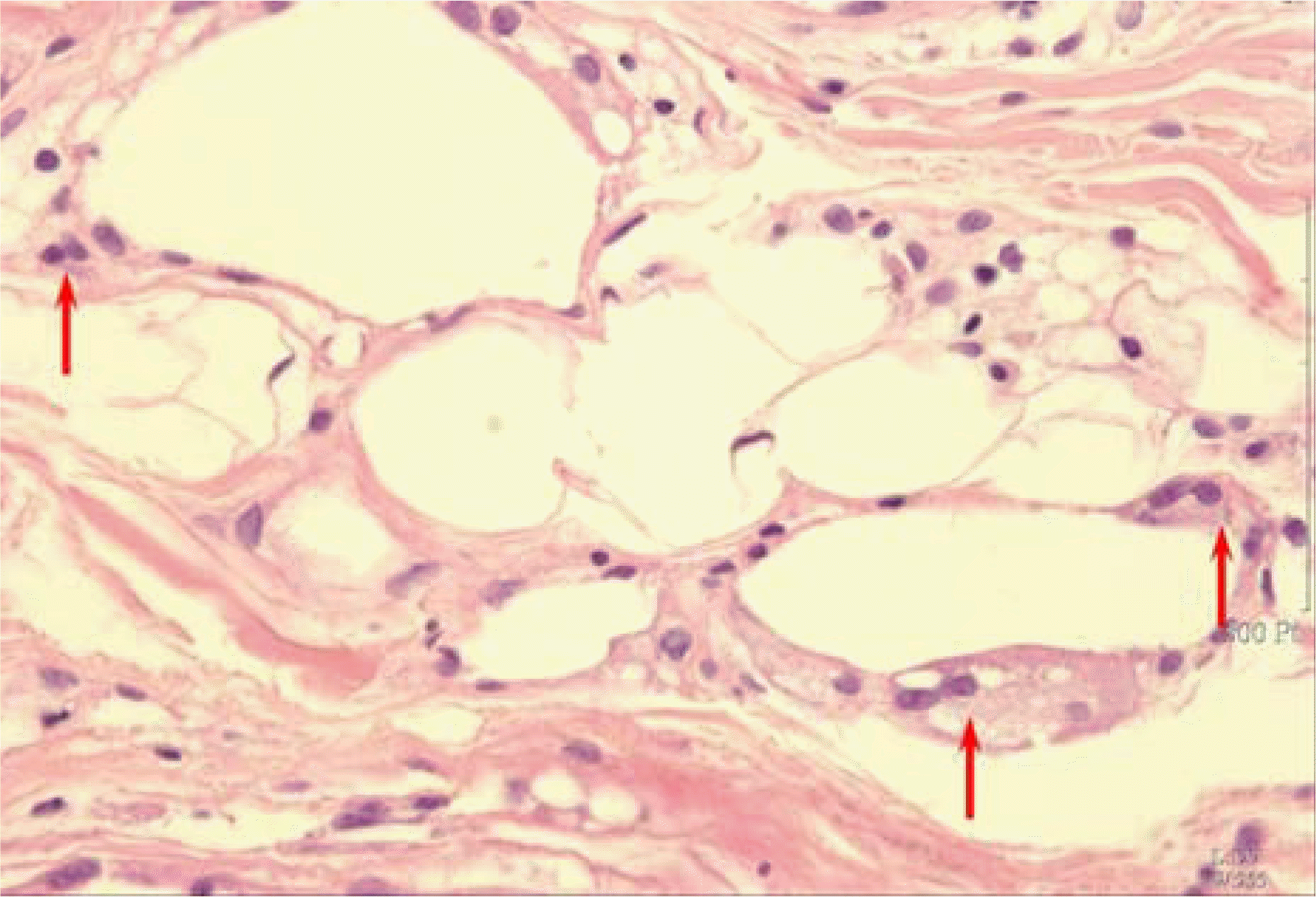
A 52-year-old female patient presented with palpable masses in both lower eyelids that had persisted for the past year. The thin, shallow, plaque-like mass with a well-demarcated border was palpated on the subcutaneous tissue of both lower eyelids. There was no tenderness or signs of inflammation. histopathologically, there were variously sized fat vacuoles with mild cellular infiltration. The mass was determined to be caused by subcutaneous fat necrosis. The patient had no unusual past history except a total gastrectomy 2 years previously. We performed a blood test and detected chronic anemia.
Go to : 
References
1. Burden AD, Krafchik BR. Subcutaneous fat necrosis of the newborn: a review of 11 cases. Pediatr Dermatol. 1999; 16:384–7.
2. Tan PH, Lai LM, Carrington EV, et al. Fat necrosis of the breast: review. Breast. 2006; 15:313–8.
3. Marsh Rde W, Hagler KT, Carag HR, Flowers FP. Pancreatic panniculitis. Eur J Surg Oncol. 2005; 31:1213–5.
4. Hicks MJ, Levy ML, Alexander J, Flaitz CM. Subcutaneous fat necrosis of the newborn and hypercalcemia: case report and review of the literature. Pediatr Dermatol. 1993; 10:271–6.

5. Vonk J, Janssens PM, Demacker PN, Folkers E. Subcutaneous fat necrosis in a neonate in association with aberrant plasma lipid and lipoprotein values. J Pediatr. 1993; 123:462–4.
6. Varan B, Gurakan B, Ozbek N, Emir S. Subcutaneous fat necrosis of the newborn associated with anemia. Pediatr Dermatol. 1999; 16:381–3.
7. Cook JS, Stone MS, Hansen JR. Hypercalcemia in association with subtutaneous fat necrosis of the newborn: studies of calcium regulating hormones. Pediatrics. 1992; 90:93–6.
8. Sharata H, Postellon DC, Hashimoto K. Subcutaneous fat necrosis, hypercalcemia, and prostaglandin E. Pediatr Dematol. 1995; 12:43–7.
9. Hanif Z, Ahmad M. Subcutaneous fat necrosis presenting as a large mass. Eur J Emerg Med. 2006; 13:106–7.
10. Bilgen IG, Ustun EE, Memis A. Fat necrosis of the breast: clinical, mammographic and sonographic features. Eur J Radiol. 2001; 39:92–9.
11. Avram MM, Avram AS, James WD. Subcutaneous fat in normal and diseased states: 1. introduction. J Am Acad Dermatol. 2005; 53:663–70.
12. Scales JW, Krowchuk DP, Schwartz RP, Jorizzo JL. An infant with firm, fixed plaques. Arch Dermatol. 1998; 134:425–6.
13. Tran JT, Sheth AP. Complications of subcutaneous fat necrosis of the newborn: a case report and review of the literature. Pediatr Dermatol. 2003; 20:257–61.
14. White WL, Wieselthier JS, Hitchcock MG. Panniculitis: recent developments and observations. Semin Cutan Med Surg. 1996; 15:278–99.
15. Requena L. Normal subcutaneous fat, necrosis of adipocytes and classification of the panniculitides. Semin Cutan Med Surg. 2007; 26:66–70.
Go to :
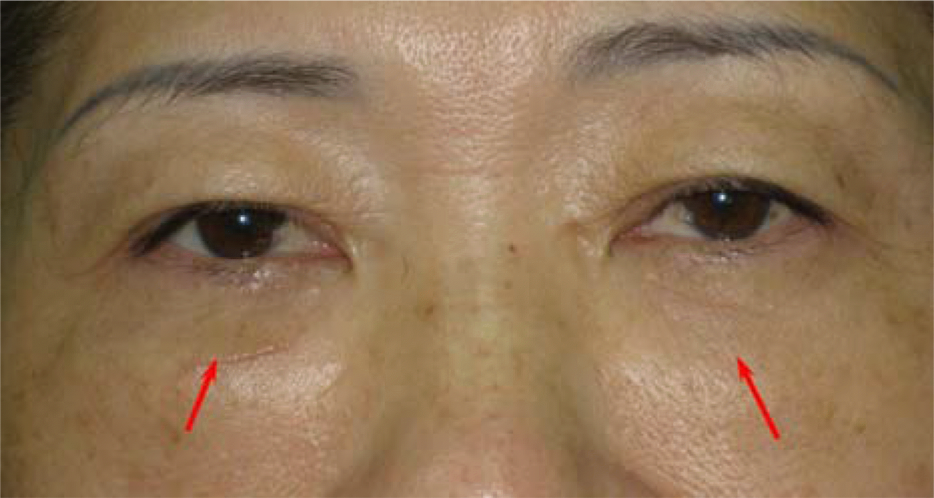 | Figure 1.Clinical photograph of the 52-year-old woman with a shallow plaque-shaped palpable mass on each lower eyelid (arrows). |
 XML Download