Abstract
Purpose
To evaluate the incidence of retinal choroidal collateral circulation after radial optic neurotomy (RON) with central retinal vein occlusion (CRVO) patients and to correlate these collaterals with changes in visual acuity.
Methods
We conducted a retrospective study of 17 eyes of 17 consecutive patients diagnosed with CRVO who underwent RON after a standard three port-vitrectomy. Fundus examination and, FAG were performed to evaluate the incidence of retinal choroidal collateral circulation according to preoperative best corrected visual acuity. We evaluated changes in best corrected visual acuity according to chorioretinal circulation formation.
Results
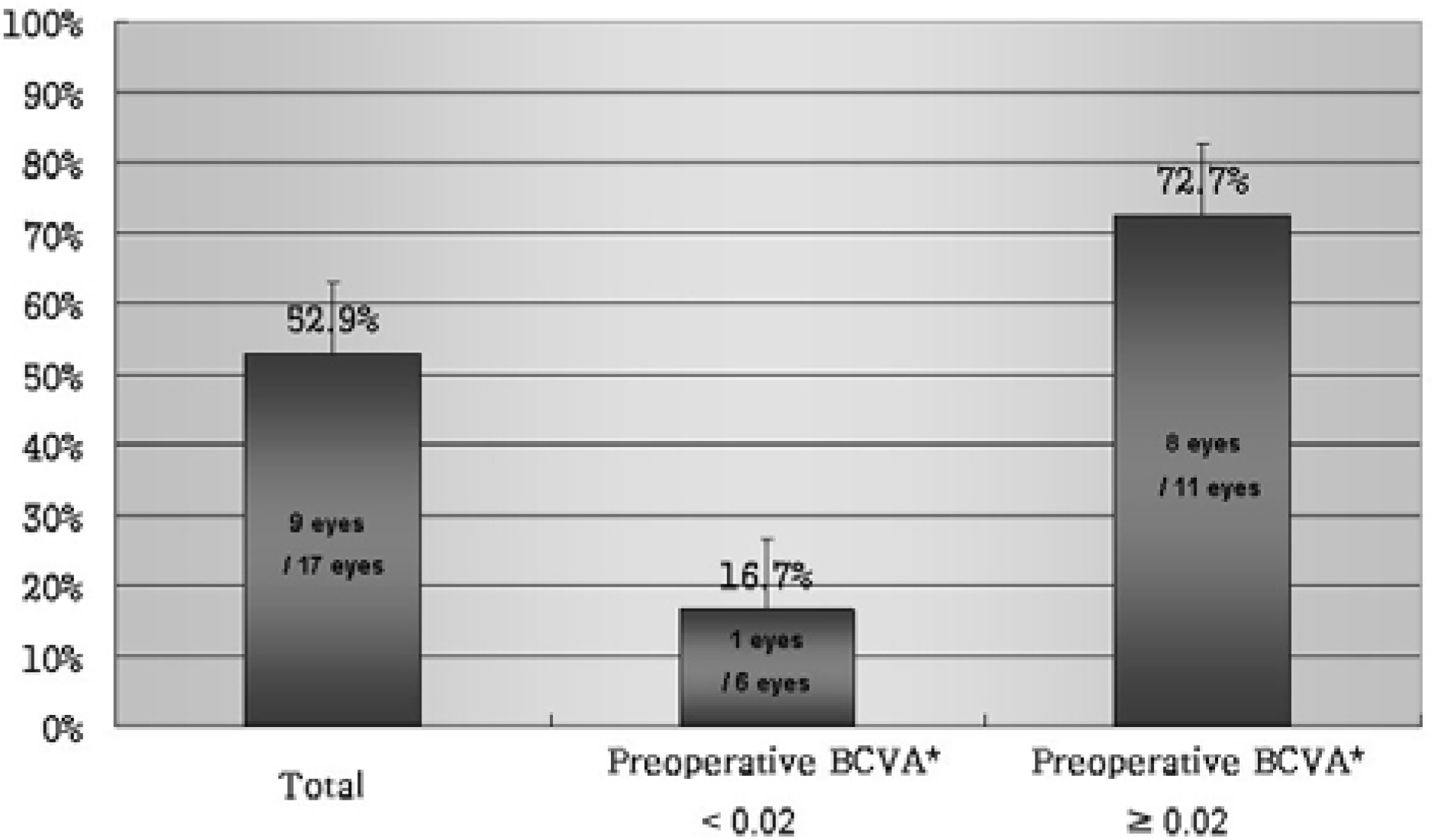
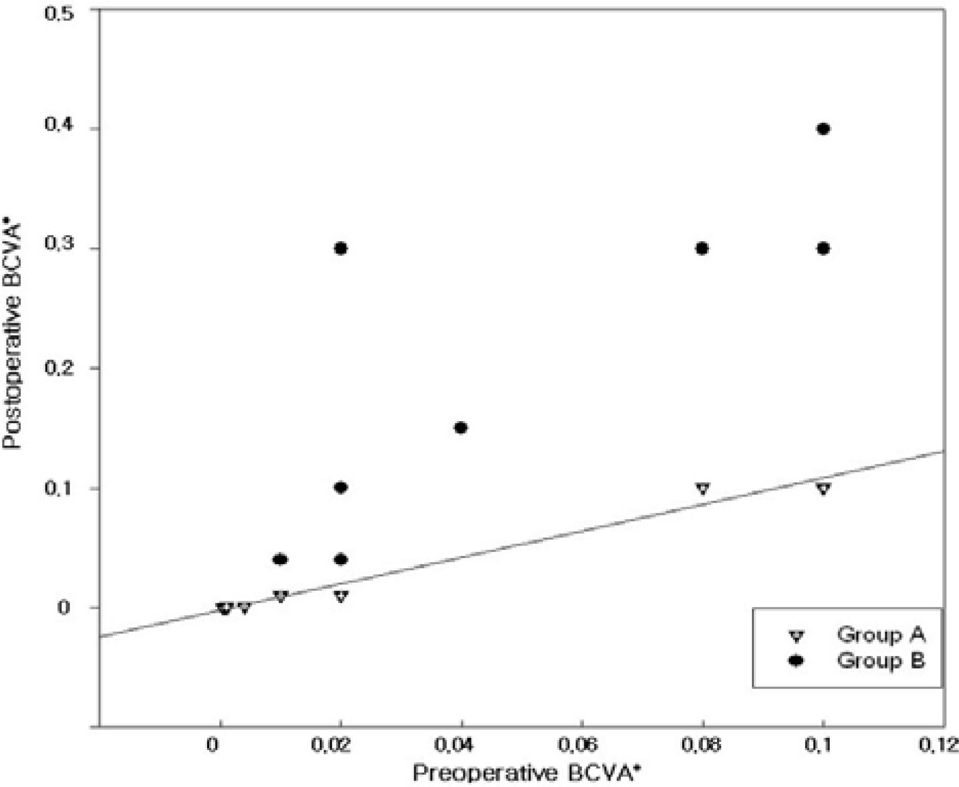
Retinochoroidal shunts developed in 9 eyes (52.9%) at the site of radial optic neurotomy. The group whose initial visual acuity was better than 0.02 (72.7%) developed more shunts than the group whose initial visual acuity was under 0.02 (16.7%) (P=0.043). Changes in visual acuity were highly correlated with the development of collaterals from the retinal to choroidal circulation (P=0.008).
Conclusions
Patients whose that initial visual acuity is better than 0.02 have more retinal choroidal collaterals. Surgical induction of retinochoroidal venous anastomosis may result in visual acuity improvement. Randomized studies are needed to compare the current study modality with the natural course of central retinal vein occlusion.
Go to : 
References
1. Byun DN, Yoon HM, Ji NC. A Clinical study of Central retinal vein occlusion. J Korean Ophthalmol Soc. 1991; 32:770–5.
2. Gutman FA. Evaluation a patient with central retinal vein occlusion. Ophthalmology. 1983; 90:481–3.
3. Operemcak EM, Bruce RA, Lomeo MD, et al. Radial optic neurotomy for central vein occlusion: A retrospective pilot study of 11 consecutive cases. Retina. 2001; 21:408–15.
4. Garcia Arumi J, Boixadera A, Martinez Castillo V, et al. Chorioretinal anastomosis after radial optic neurotomy for central retinal vein occlusion. Arch Ophthalmol. 2003; 121:1385–91.

5. Mirshahi A, Roohipoor R, Lashay A, et al. Surgical induction of chorioretinal venous anastomosis in ischaemic central retinal vein occlusion: a non-randomised controlled clinical trial. Br J Ophthalmol. 2005; 89:64–9.

6. Zegarra H, Gutman FA, Conforto J. The natural course of central retinal vein occlusion. Ophthalmology. 1979; 86:1931–8.

7. The Central Vein Occlusion Study Group. Natural history and clinical management of central retinal vein occlusion. Arch Ophthalmol. 1997; 115:486–91.
9. McAllister IL, Douglas JP, Constable IJ, Yu DY. Laser induced chorioretinal venous anastomosis for nonischemic central vein occlusion: evaluation of the complications ans their risk factors. Am J Ophthalmol. 1998; 126:219–29.
10. Fekrat S, Goldberg MF, Finkelstein D. Laser induced chorioretinal venous anastomosis for nonischemic central or branch retinal vein occlusion. Arch Ophthalmol. 1998; 116:43–52.
11. Browning DJ, Antoszyka N, McAllister IL. Laser Chorioretinal venous anastomosis for nonischemic central retinal vein occlusion. Ophthalmology. 1998; 105:670–9.
12. Hattenbach LO, Wellermann G, Steinkamp GW, et al. Visual outcome after treatment with low dose recombinant tissue palsmonogen activator or hemodilution in ischemic central retinal vein occlusion. Ophthalmologica. 1999; 213:360–6.
13. Weiss JN, Bynoe LA. Injection of tissue plasminogen activator into a branch retinal vein in eyes with central retinal vein occlusion. Ophthalmology. 2001; 108:2249–57.

14. Vannas S, Raitta C. Anticoagulation treatment of retinal venous occlusion. Am J Ophthalmol. 1966; 62:874–81.
15. Iturralde D, Spaide RF, Meyerle CG, et al. Intravitreal bevacizumab(Avastin) treatment of macular edema in central retinal vein occlusion: a short-term study. Retina. 2006; 26:279–84.
16. Vasco-Posada J. Modification of the circulation in the posterior pole of the eye. Ann Ophthalmol. 1972; 1:48–59.
17. Arciniegas A. Treatment of the occlusion of the central retinal vein occlusion by section of the posterior ring. Ann Ophthalmol. 1984; 16:1081–6.
18. Spaide RF, Klancnik JM Jr, Gross NE. Retinal choroidal collateral circulation after radial optic neurotomy correlated with the lessening of macular edema. Retina. 2004; 24:356–9.

19. Traynor MP, Conway BP. Collateral vessel formation after radial optic neurotomy. Retina. 2004; 24:616–7.

20. Dev S, Buckley EG. Optic nerve sheath decompression for progressive central retinal vein occlusion. Ophthalmic Surg Laser. 1999; 30:181–4.

21. Williamson TH, Poon W, Whitefield L, et al. A pilot study of pars plana vitrectomy, intraocular gas, and radial neurotomy in ischemic central retinal vein occlusion. Br J Ophthalmol. 2003; 87:1126–9.
22. Opremcak EM, Rehmar AJ, Ridenour CD, et al. Radial optic neurotomy for central retinal vein occlusion: 117 consecutive case. Retina. 2006; 26:297–305.
23. Garcia Arumi J, Boixadera A, Martinez Castillo V, et al. Radial optic neurotomy in central retinal vein occlusion: comparison of outcome in younger vs older patients. Am J Ophthalmol. 2007; 143:134–40.
24. Musser GL, Rosen S. Localization of carbonic anhydrase activity in the vertebrate retina. Exp Eye Res. 1973; 15:105–19.

25. Le Rouick JF, Becquet F, Zanlonghi X, et al. Radial optic neurotomy for severe central retinal vein occlusion: Preliminary results. J Fr Ophthalmol. 2003; 26:577–85.
26. Friedman SM. Optociliary venous anastomosis after radial optic neurotomy for central retinal vein occlusion. Ophthalmic Surg Lasers Imaging. 2003; 34:315–7.

27. Kaderli B, Avci R, Gelisken O. Radial optic neurotomy in central retinal vein occlusion:preliminary results. Int Ophthalmol. 2004; 25:215–23.
Go to : 
 | Figure 1.Chorioretinal collateral circulation formation correlated with the preoperative Best corrected visual acuity. P-value=0.043; * BCVA=best corrected visual acuity. |
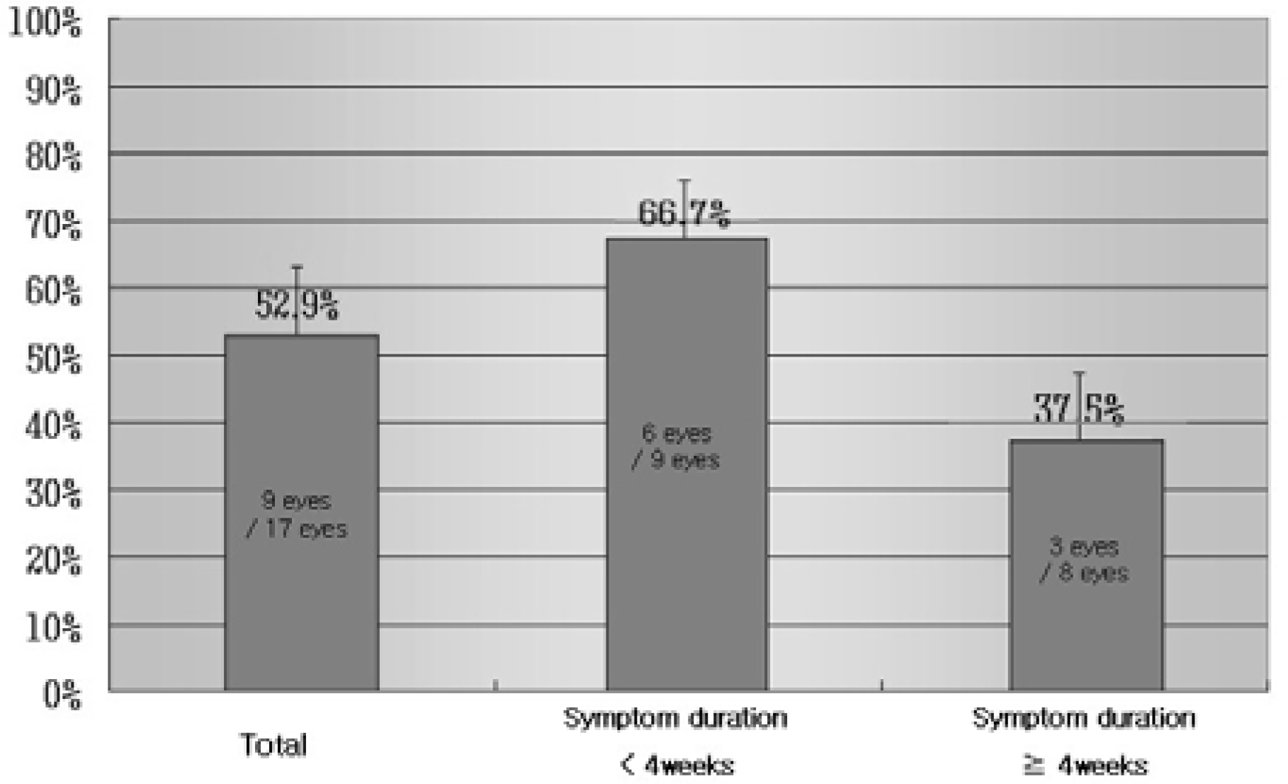
 | Figure 2.Chorioretinal collateral circulation formation correlated with symptom duration. P-value=0.347. |
 | Figure 3.Case No 16. (A) Preoperative fundus view of the right eye of a 69-year-old man with central retinal vein occlusion. His preoperative BCVA* was 0.1. (B) In fluorescein angiography, flame-shaped retinal hemorrhage and macular edema was seen. (C) Fundus view 3 months later, Remained retinal hemorrhage and decreased macula edema was seen, also chorioretinal collateral circulation was formed (white arrow), His Postoperative BCVA* improved to 0.3. (D) Fluorescein angiography 3 months later, shows drainage of the retinal veins through the chorioretinal anastomosis. E) Fundus view. Thirty months later, Some dot like retinal hemorrhages were detected and chorioretinal collateral circulation formation was also seen (white arrow). His postoperative BCVA* was 0.3. (F) Fluorescein angiography 30 months later, Some chorioretinal scar was seen at macular area and chorioretinal collateral circulation formation was also seen; * BCVA=best corrected visual acuity. |
 | Figure 4.Scatter plot of preoperative and postoperative BCVA* with Central Retinal Vein Occlusion according to chorioretinal circulation formation. The Group A is the patients who deveoped chorioretinal collateral circulation and The Group B is the patients who did not deveoped chorioretinal collateral circulation. Hand movement corresponds to 0.001 and light perception to 0.0005, No light perception to 0. P-value=0.008; * BCVA=best corrected visual acuity. |
Table 1.
Demorgaphic data for 17 patients with radial optic neurotomy for central retinal vein occlusion
| Case | Sex/Affected eye |
Symptom Duration (weeks) |
Retinal Ischemia |
BCVA* |
Collateral Circulation Formation |
Follow-up (months) |
IOP† Preop. (3 months) |
|
|---|---|---|---|---|---|---|---|---|
| Postop. |
Postop. (3 months) |
|||||||
| 1 | M/Left | 8 | No | FC50‡ | FC50 | No | 40 | 37 |
| 2 | F/Right | 1 | Yes | LP§ | NLP∏ | No | 15 | 15 |
| 3 | M/Right | 12 | No | HM# | NLP | No | 20 | 11 |
| 4 | F/Right | 2 | Yes | FC20 | HM | No | 4 | 29 |
| 5 | M/Right | 4 | No | 0.08 | 0.1 | No | 14 | 7 |
| 6 | M/Left | 8 | No | HM | LP | No | 8 | 38 |
| 7 | F/Right | 8 | Yes | 0.1 | 0.1 | No | 4 | 11 |
| 8 | F/Right | 3 | Yes | 0.02 | FC50 | No | 3 | 10 |
| 9 | M/Left | 8 | Yes | 0.02 | 0.3 | Yes | 40 | 11 |
| 10 | F/Left | 1 | Yes | FC50 | 0.04 | Yes | 3 | 13 |
| 11 | M/Right | 16 | No | 0.04 | 0.15 | Yes | 11 | 17 |
| 12 | F/Left | 2 | No | 0.1 | 0.3 | Yes | 3 | 11 |
| 13 | M/Right | 2 | Yes | 0.02 | 0.1 | Yes | 6 | 17 |
| 14 | M/Left | 8 | Yes | 0.02 | 0.04 | Yes | 3 | 7 |
| 15 | M/Left | 2 | No | 0.08 | 0.3 | Yes | 4 | 17 |
| 16 | F/Right | 2 | No | 0.1 | 0.3 | Yes | 32 | 20 |
| 17 | M/Left | 1 | No | 0.1 | 0.4 | Yes | 7 | 9 |


 PDF
PDF ePub
ePub Citation
 Print
Print


 XML Download