Abstract
Methods
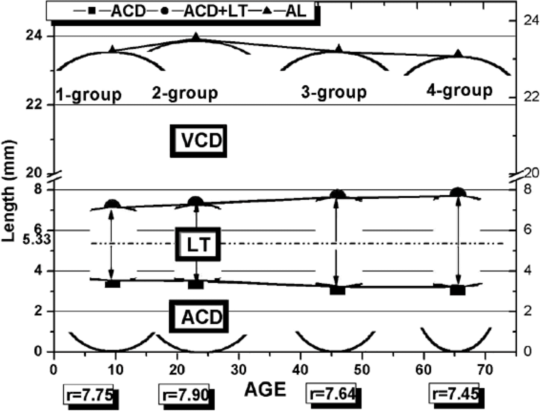
We examined the refraction, corneal curvature, and biometry in 150 subjects from 5 to 75 years old with spherical equivalent refractions under ±0.75 diopter (D). Ocular dimensions were measured by A-scan ultrasonography and keratometry. We analysed the distribution and change of ocular dimensions according to age (1: 0∼19-year-old group, 2: 20∼39-year-old group, 3: 40∼59-year-old group, 4: 60∼79-year-old group).
Results
The values for corneal radius (CR), vitreous chamber depth (VCD) and axial length (AL) were highest in group 2. Lens thickness (LT) increased with increasing age, whereas anterior chamber depth (ACD) decreased with increasing age (P<0.05). CR, VCD, AL (P<0.05) and ACD (P=0.10) seem to have higher values in males, while LT seems to have a higher value in females (P=0.06).
Conclusions
Axial length increases with increasing age in subjects aged 0 to 39 years in emmetropia. In subjects aged 40 years or older, axial length becomes smaller with age. In each age group compensational changes to achieve emmetropia according to AL change are shown in ocular dimensions like CR, VCD, ACD, LT.
References
1. William JB. Borish's clinical refraction. 1st ed.Philadelphia: WB Saunders;1998. p. 2–17.
2. Troilo D. Neonatal eye growth and emmetropization. Eye. 1992; 6:154–60.
3. David AA, George S. Optics of the human eye. 1st ed.Oxford: Butterworth-Heinemann;2000. p. 39–47.
4. Kim CS, Kim MY, Kim HS, Lee YC. Change of corneal astigmatism with aging in Korean with normal visual acuity. J Korean Ophthalmol Soc. 2002; 43:1956–62.
5. Sung PJ. Optometry. 2nd ed.Seoul: Daehakseolim;2002. p. 104–6.
6. Vaughan D, Asbury T, Tabbara KT. General Ophthalmology. 12th ed.USA: Prentice-Hall International Inc;1989. p. 364–6.
7. Naess RO. Optics for Technology Students. 1st ed.New Jersey: Prentice-Hall Inc;2001. p. 364–6.
8. Duke-Elder S. System of Ophthalmology. 1st ed.Vol. 2. St. Louis: CV Mosby;1961. p. 80–1.
9. Duane T. Clinical Ophthalmology. 3rd ed.Vol 1. Philadelphia: Harper & Row Publishers;1978. p. 1–3.
10. Kim SD, Lee DS, Kim JD. Study of the corneal refractive power and axial length of the adult korean eyeball. J Korean Ophthalmol Soc. 1990; 31:1365–9.
11. Lim KJ, Choi WS, Youn DH. Aging and Ocular Dimension. J Korean Ophthalmol Soc. 1992; 33:653–61.
12. Clemmesen V, Olurin O. Lens thickness in western nigeria a comparative ultrasonic study in negroes and danes. Acta Ophthalmol. 1985; 63:274–6.
13. Larsen JS. The sagittal growth of the eye. III. Ultrasonic measurement of the posterior segment (axial length of the vitreous) from birth to puberty. Acta Opthalmol. 1971; 49:873–86.
14. Sorsby A. Biology of the eye as an optical system. Duane TD, editor. Clinical Ophthalmology. 7th ed.Philadelphia: Harper & Row Publishers;1983. 1:chap. 34.
15. Grosvenor T. Reduction in axial length with age: An emme-tropizing mechanism for the adult eye? Am J Optom Physiol Opt. 1987; 64:657–63.
16. Koretz JF, Cook A, Kaufman PL. Accommodation and pres-byopia in the human eye: change in anterior segment and crystalline lens with focus. Invest Ophthalmol Vis Sci. 1997; 38:569–78.
17. Kim JB, Kim JM. Relationships between corneal curvature and refractive errors in korea. J Korean Ophthalmol Soc. 1977; 18:39–44.
18. Kiely PM, Smith G, Carney LG. Meridional variations of corneal shape. Am J Optom Physiol Optic. 1984; 61:619–25.

19. Hayahi K, Hayahi H, Hayahi F. Topographic analysis of the changes in corneal shape due to aging. Cornea. 1995; 14:527–32.

20. Choi HH. A consideration for corneal curvature, Its thickness and anterior chamber depth. J Korean Ophthalmol Soc. 1978; 19:417–22.
21. Wong TY, Foster PJ, Ng TP, et al. Variations in Ocular biometry in an adult chinese population in singapore: The tanjong pagar survey. Invest Ophthalmol Vis Sci. 2001; 42:73–80.
22. Wickremasinghe S, Foster PJ, Uranchimeg D, et al. Ocular biometry and refraction in mongolian adults. Invest Ophthalmol Vis Sci. 2004; 45:776–83.

23. Rengstorff RH, Arner RS. Refractive changes in the cornea: Mathematical considerations. Am J Optom Arch Am Acad Optom. 1971; 48:913–8.
24. Erickson P. Mathematical model for predicting dioptric effects of optical parameter changes in the eye. Am J Optom Physiol Opt. 1977; 54:226–33.

25. Smith G, Atchison DA. Pierscionek BK. Modeling the power of the aging human eye. J Opt Soc Am A. 1992; 9:2111–7.
26. Kim YK, Kim JS, Kim JD. Study on lens thickness and anterior chamber depth during accommodation and weak cycloplegic eyes. J Korean Ophthalmol Soc. 1991; 32:160–6.
27. Sorsby A, Leary GA, Richards MJ. The optical components in anisometropia. Vision Res. 1962; 2:43–51.

28. Brown N. The human lens in relation to cataract. Ciba Foundation Symposium 15. 1st ed.1. Amsterdam: Elsevier;1973. p. 65–78.
29. Friedman NE, Mutti DO, Zadnik K. Corneal changes in schoolchildren. Optom Vis Sci. 1996; 73:552–7.

30. Mcbrien NA, Adams DW. A longitudinal investigation of adult-onset and adult-progression of myopia in an occupational group: refractive and biometric findings. Invest Ophthalmol Vis Sci. 1997; 38:321–33.
31. Lin LL, Shih YF, Lee YC, et al. Changes in ocular refraction and its components among medical students: A 5 year longitudinal study. Optom Vis Sci. 1996; 73:495–8.
32. Grosvenor T, Scott R. Three-year changes in refraction and its components in youth-onset myopia. Optom Vis Sci. 1993; 70:677–83.
33. Attebo K, Rebecca Q, Lvers , Mitchell P. Refractive errors in an older population: The mountains eye study. Ophthalmology. 1999; 106:1066–72.
Table 1.
Distribution of ocular dimensions in each age group
| G* |
Age M†(Range) |
N |
CR |
ACD |
LT |
ACD+LT |
VCD |
AL |
CP |
LP |
TP |
|---|---|---|---|---|---|---|---|---|---|---|---|
| Mean±SD (mm) |
A-constant: 118.00 Mean±SD (D‡) |
||||||||||
| All | |||||||||||
| G 1 | 9.5 (5–19) | 30 | 7.82±0.22 | 3.55±0.29 | 3.56±0.20 | 7.11±0.25 | 16.29±0.79 | 23.40±0.86 | 43.20±1.24 | 20.63±1.5 | 60.18±1.92 |
| G 2 | 23.0 (20–36) | 36 | 7.98±0.17 | 3.47±0.24 | 3.81±0.24 | 7.28±0.25 | 16.52±0.38 | 23.81±0.55 | 42.32±1.27 | 20.40±1.0 | 59.18±1.12 |
| G 3 | 46.0 (40–59) | 48 | 7.68±0.18 | 3.20±0.45 | 4.41±0.52 | 7.62±0.37 | 15.76±0.67 | 23.38±0.64 | 43.92±1.12 | 20.03±0.9 | 60.36±1.48 |
| G 4 | 65.5 (60–75) | 36 | 7.58±0.19 | 3.17±0.30 | 4.55±0.27 | 7.72±0.22 | 15.58±0.52 | 23.30±0.45 | 44.56±1.19 | 19.64±0.8 | 60.61±1.08 |
| total | 43.0 (5–75) | 150 | 7.75 | 3.34 | 4.09 | 7.43 | 16.00 | 23.42 | 43.50 | 20.15 | 60.08 |
| ANOVA test | p=0.00 | p=0.00 | p=0.00 | p=0.00 | p=0.00 | p=0.07 | p=0.00 | p=0.11 | p=0.02 | ||
| Male | |||||||||||
| G 1 | 10.0 (5–18) | 15 | 7.89±0.20 | 3.57±0.28 | 3.50±0.26 | 7.07±0.09 | 16.65±0.70 | 23.72±0.76 | 42.72±1.12 | 20.20±1.46 | 59.45±1.66 |
| G 2 | 23.0 (20–34) | 22 | 8.02±0.19 | 3.53±0.23 | 3.74±0.20 | 7.27±0.24 | 16.62±0.38 | 23.89±0.55 | 42.11±1.55 | 21.27±1.08 | 58.98±1.18 |
| G 3 | 46.0 (40–59) | 32 | 7.71±0.12 | 3.24±0.54 | 4.41±0.62 | 7.65±0.43 | 15.87±0.67 | 23.47±0.54 | 43.80±0.94 | 19.90±0.91 | 60.13±1.16 |
| G 4 | 65.0 (60–70) | 22 | 7.66±0.20 | 3.18±0.36 | 4.47±0.31 | 7.65±0.18 | 15.76±0.52 | 23.41±0.50 | 44.09±1.10 | 19.79±1.01 | 60.31±1.25 |
| total | 43.0 (5–70) | 91 | 7.82 | 3.38 | 4.03 | 7.39 | 16.21 | 23.62 | 43.19 | 20.31 | 59.72 |
| Female | |||||||||||
| G 1 | 9.0 (8–19) | 15 | 7.75±0.22 | 3.54±0.33 | 3.61±0.13 | 7.15±0.36 | 15.39±0.73 | 23.03±0.86 | 43.60±0.86 | 21.06±1.63 | 60.91±2.00 |
| G 2 | 25.5 (20–36) | 14 | 7.90±0.12 | 3.36±0.24 | 3.93±0.27 | 7.29±0.31 | 16.35±0.40 | 23.64±0.60 | 42.70±0.96 | 20.44±1.15 | 59.59±1.39 |
| G 3 | 50.0 (42–56) | 16 | 7.64±0.30 | 3.12±0.19 | 4.42±0.20 | 7.55±0.17 | 15.59±0.80 | 23.13±0.84 | 44.20±1.70 | 20.35±0.71 | 60.90±1.80 |
| G 4 | 70.0 (65–75) | 14 | 7.45±0.09 | 3.16±0.21 | 4.68±0.19 | 7.84±0.12 | 15.30±0.22 | 23.14±0.28 | 45.30±0.40 | 19.40±0.39 | 61.09±0.69 |
| total | 42.0 (8–75) | 55 | 7.69 | 3.30 | 4.16 | 7.45 | 15.79 | 23.23 | 43.95 | 20.31 | 60.62 |
|
|
|||||||||||
| T-test | Male & Female | p=0.02 | p=0.10 | p=0.06 | p=0.40 | p=0.03 | p=0.03 | p=0.03 | p=0.99 | p=0.02 | |
Table 2.
Difference in ocular dimensions by gender in each age group
| Group (age) | N | Age | CR | ACD | LT | ACD+LT | VCD | AL | CP | LP | TP |
|---|---|---|---|---|---|---|---|---|---|---|---|
| * Difference (mm) | Difference (D†) | ||||||||||
| 1-Group | 30 | 0–19 | 0.15 | 0.03 | −0.11 | −0.08 | 0.72 | 0.64 | −0.82 | −0.86 | −1.46 |
| 2-Group | 36 | 20–39 | 0.12 | 0.17 | −0.19 | −0.02 | 0.27 | 0.25 | −0.62 | −0.07 | −0.62 |
| 3-Group | 48 | 40–59 | 0.07 | 0.12 | −0.02 | 0.10 | 0.24 | 0.34 | −0.43 | −0.45 | −0.77 |
| 4-Group | 36 | 60–79 | 0.21 | 0.02 | −0.21 | −0.19 | 0.46 | 0.27 | −1.19 | 0.39 | −0.78 |
| Mean | 0.14 | 0.09 | −0.13 | −0.05 | 0.42 | 0.38 | −0.77 | −0.25 | −0.91 | ||
| M&F T-test | p=0.02 | p=0.10 | p=0.06 | p=0.40 | p=0.03 | p=0.03 | p=0.03 | p=0.99 | p=0.02 | ||


 PDF
PDF ePub
ePub Citation
 Print
Print


 XML Download