

 PDF
PDF ePub
ePub Citation
 Print
Print


Abstract
Purpose
To report the first cases of Descemet’s stripping endothelial keratoplasty in patients with pseudophakic bullous keratopathy.
Methods
Four eyes of 4 patients who had corneal edema from pseudophakic bullous keratopathy were enrolled. We performed femtosecond laser-assisted Descemet’s membrane stripping endothelial keratoplasty (DSEK). We then evaluated preoperative and postoperative best corrected visual acuities (BCVA), manifest refractions, keratometries, topographic astigmatisms, corneal pachymetries, and perioperative complications.
Results
The mean follow-up period was 6 months. Three of the four eyes had successful transplantations. Since the operation, these patients have shown improvement in visual acuity and have recovered from corneal edema. However, in one patient, the transplanted corneal disc was dislocated, and he needed an additional procedure for reattachment.
Conclusions
The lack of corneal incisions or sutures in DSEK was helpful for maintaining corneal curvature and globe integrity after surgery. The surgery resulted in rapid visual rehabilitation and prevented suture-induced complications. With a femtosecond laser, we could prepare donor cornea in an automated and standardized fashion. In conclusion, femtosecond laser-assisted DSEK has significant advantages over standard penetrating keratoplasty for patients with pseudophakic bullous keratopathy.
References
1. Pineros O, Cohen EJ, Rapuano CJ, Laibson PR. Long term results after penetrating keratoplasty for Fuchs' endothelial dystrophy. Arch Ophthalmol. 1996; 114:15–8.
2. Melles GR, Lander F, Beekhuis WH, et al. Posterior lamellar keratoplasty for a case of pseudophakic bullous keratopathy. Am J Ophthalmol. 1999; 127:340–1.
3. Terry MA, Ousley PJ. Replacing the endothelium without corneal surface incisions or sutures. The first United States clinical series using the deep lamellar endothelial keratoplasty procedure. Ophthalmology. 2003; 110:755–64.
4. Melles GR, Wijdh RH, Nieuwendaal CP. A technique to excise the descemet membrane from a recipient cornea (descemetorhexis). Cornea. 2004; 23:2286–8.

5. Price FW Jr, Price MO. Descemet's stripping with endothelial keratoplasty in 50 eyes; a refractive neutral cornea transplant. J Refract Surg. 2005; 21:339–45.
6. Price FW Jr, Price MO. Descemet's stripping with endothelial keratoplasty: Comparative outcomes with microkeratome dissected and manually dissected donor tissue. Ophthalmology. 2006; 113:1936–42.
7. Durrie DS, Kezirian GM. Femtosecond laser versus mechanical keratome flap in wavefront guided laser in situ keratomileusis: prospective contralateral eye study. J Catarct Refract Surg. 2005; 31:120–6.
8. Cheng YY, Pels E, Nuijts RM. Femtosecond laser assisted Descemet's stripping endothelial keratoplasty. J Cataract Refract Surg. 2007; 33:152–5.
9. Melles GRJ, Lander F, van Dooren BT, et al. Preliminary clinical results of posterior lamellar keratoplasty through a sclerocorneal pocket incision. Ophthalmology. 2000; 107:1850–6.
10. Terry MA, Ousley PJ. Deep lamellar endothelial keratoplasty in the first United States patients; early clinical results. Cornea. 2001; 20:239–43.
11. Ousley PJ, Terry MA. Stability of vision, topography, and endothelial cell density from 1 year to 2 year after deep lamellar endothelial keratoplasty surgery. Ophthalmology. 2005; 112:50–7.
12. Koenig SB, Covert DJ. Early result of small incision Descemet's stripping and automated endothelial keratoplasty. Ophthalmology. 2007; 114:221–6.
13. Seitz B, Langenbucher A, Hofmann-Rummelt C, et al. Nonmechanical posterior lamellar keratoplasty using the Femtosecond laser (femto-PLAK) for corneal endothelial decompensation. Am J Ophthalmol. 2003; 136:769–72.
14. Sarayba MA, Juhasz T, Chuck RS, et al. Femtosecond laser posterior lamellar keratoplasty; a laboratory model. Cornea. 2005; 24:328–33.
15. Price FW, Price MO. Descemet's stripping with endothelial keratoplasty (DSEK) in 200 eyes: early challenges and techniques to promote donor adherence. J cataract Refract Surg. 2006; 32:411–8.
16. Koenig SB, Dupps WJ, Covert DJ, et al. Simple technique to unfold the donor corneal lenticule during Descemet's stripping and automated endothelial keratoplasty. J Cataract Refract Surg. 2007; 33:189–90.
17. Langenbucher A, Seitz B, Nguyen NX, Naumann GO. Corneal endothelial cell loss after nonmechanical penetrating keratoplasty depends on diagnosis: a regression analysis. Graefes Arch Clin Exp Ophthalmol. 2002; 240:387–92.
18. Reinhard T, Bohringer D, Enczmann J, et al. HLA class I/II matching and chronic endothelial cell loss in penetrating normal risk keratoplasty. Acta Ophthalmol Scand. 2004; 82:13–8.
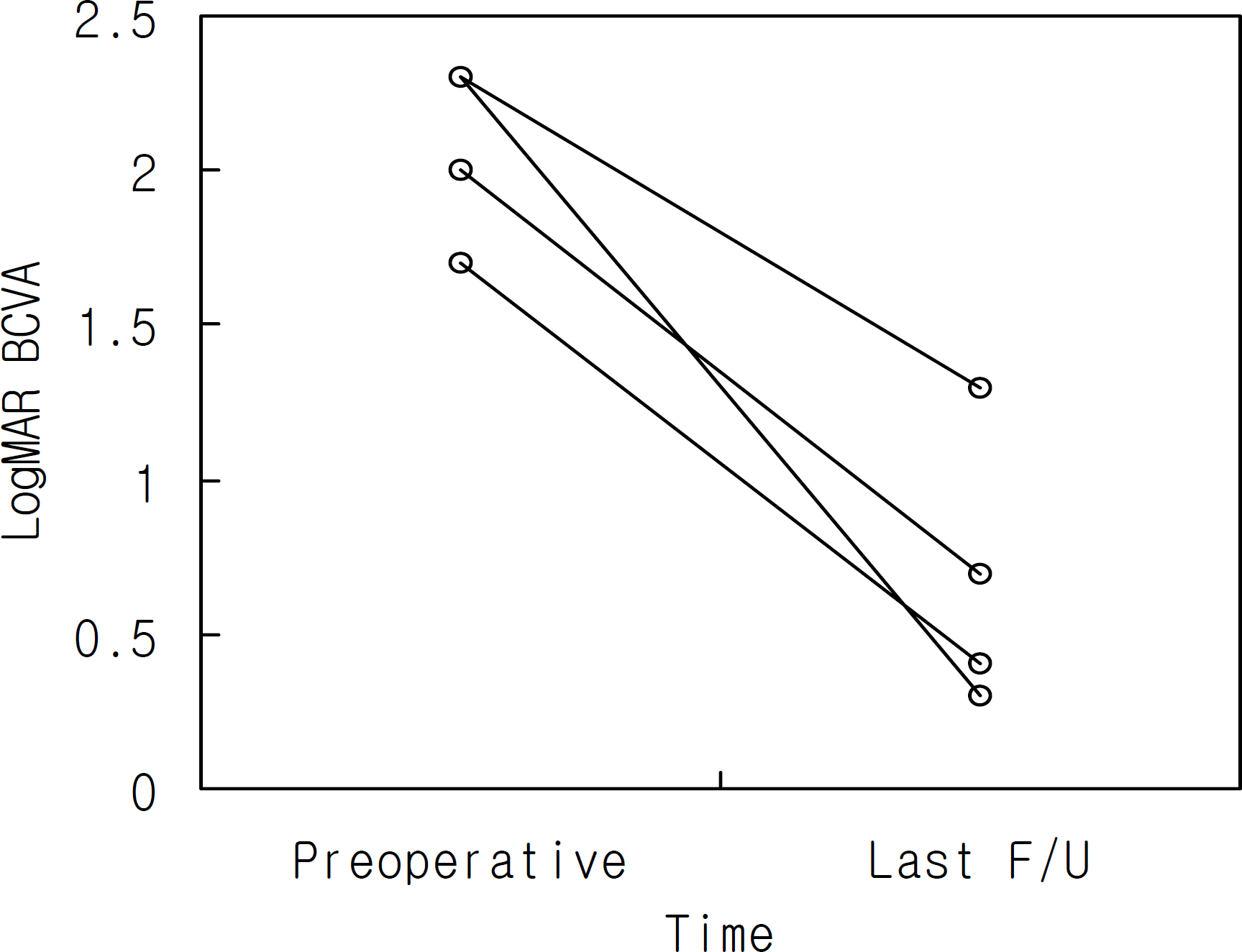
Figure 2.
Preoperative photo of patient 1 showing corneal edema and haze from pseudophakic bullous keratopathy. Intraocular lens haptic (arrow) is protruded through the previous iridectomy site.
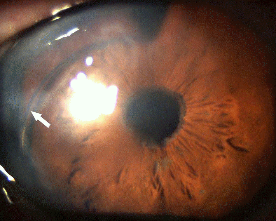
Figure 3.
Slitlamp photograph of DSEK graft 2 weeks after surgery. Note the clear central cornea with clear interface and smooth surface. The edge of the donor tissue is illuminated (arrow).
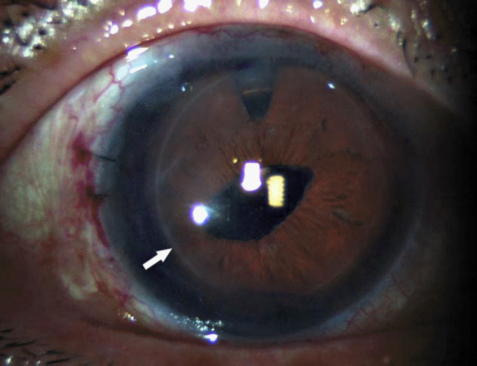
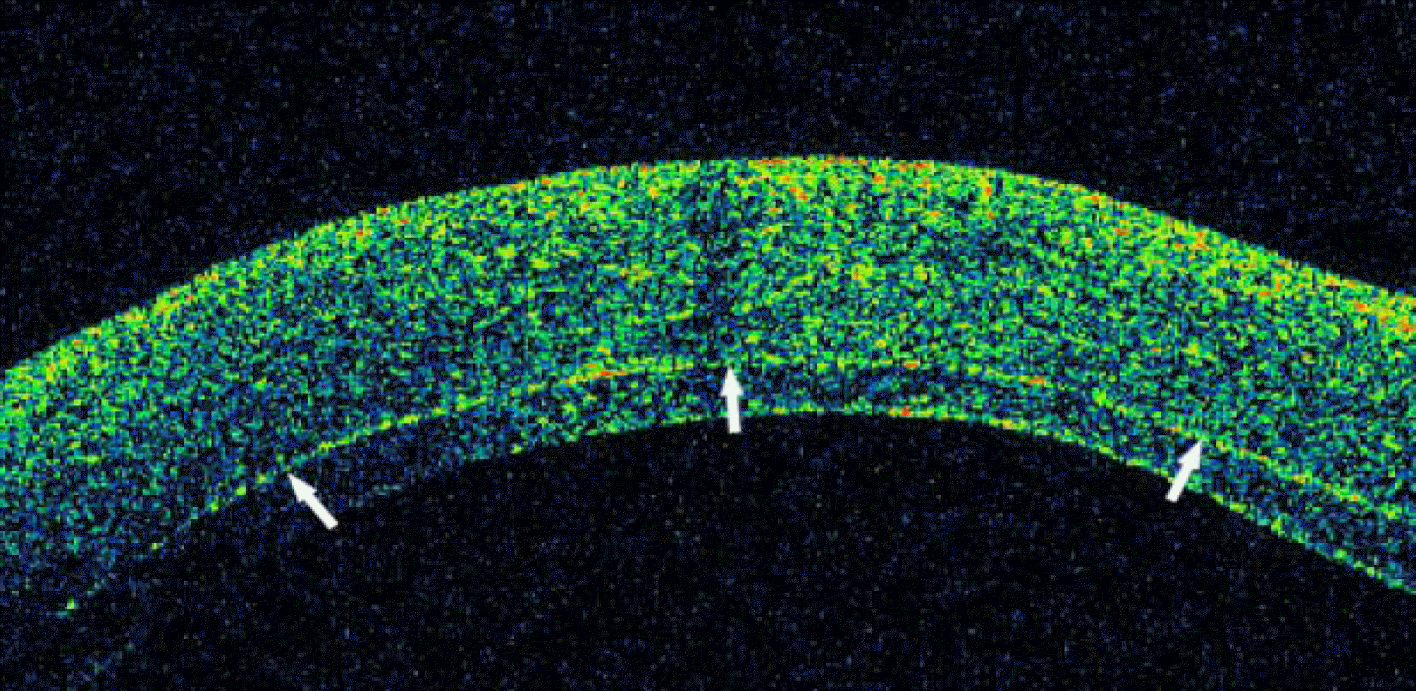
Figure 5.
Dislocated corneal disc is sutured with 10-0 nylon at 1,3 and 10 o’clock (arrow). Corneal edema is persisted at detached corneal disc (asterisk).
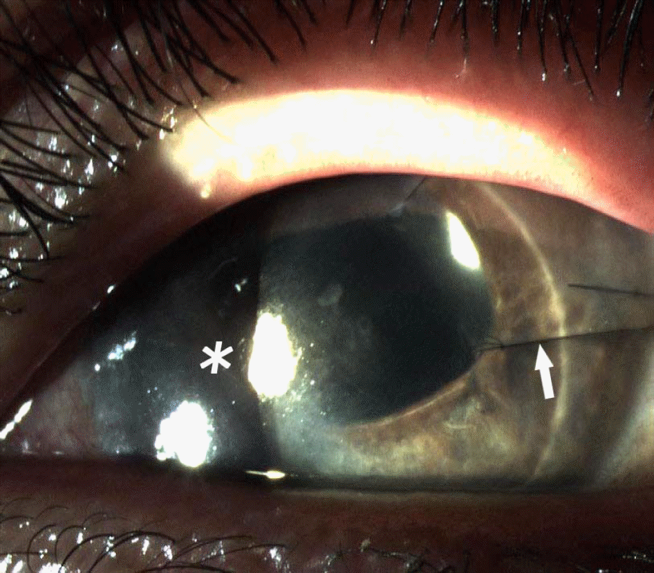
Table 1.
Preoperative and postoperative clinical details
| case | Dx* | Preop BCVA†(logMAR) | (logMAR) Postop BCVA | Preop specular (cells/mm2) | Postop specular (cells/mm2) | (µm) Preop pachymetry | (µm) Postop pachymetry | F/U (month) |
|---|---|---|---|---|---|---|---|---|
| 1 | PBK‡ | 2.3 | 0.3 | 177 | 553 | 684 | 729 | 8 |
| 2 | PBK | 1.7 | 0.4 | NA§ | 1206 | 750 | 756 | 7 |
| 3 | PBK | 2.3 | 1.3 | NA | NA | 821 | NA | 6 |
| 4 | PBK | 2.0 | 0.7 | NA | 1503 | NA | 730 | 3 |
 XML Download