

 PDF
PDF ePub
ePub Citation
 Print
Print


Abstract
Purpose
We report a case of a 38-year-old man who suffered a blowout fracture of the orbital wall with an intact eyeball entrapped within the maxillary sinus after trauma.
Case summary
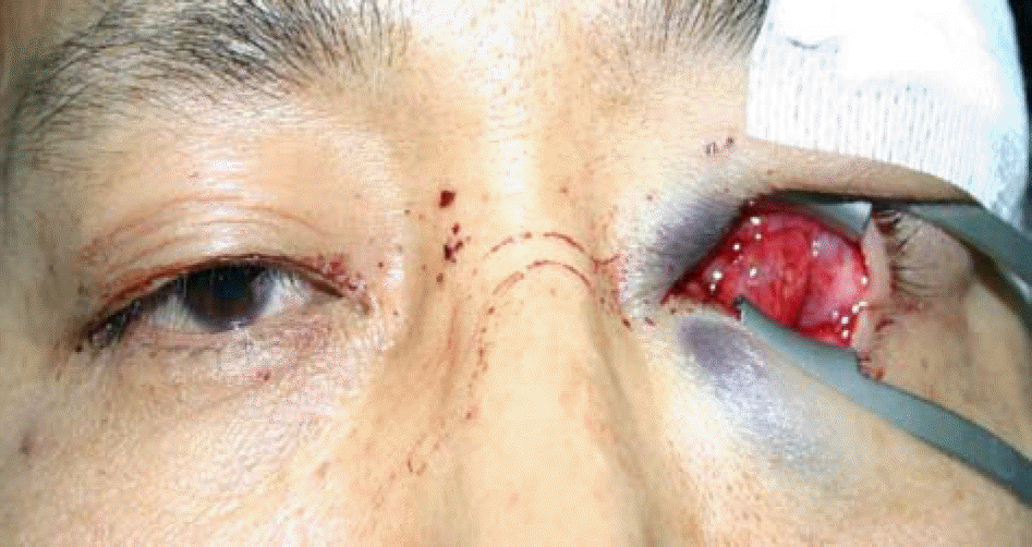
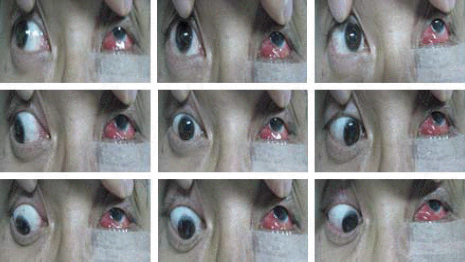
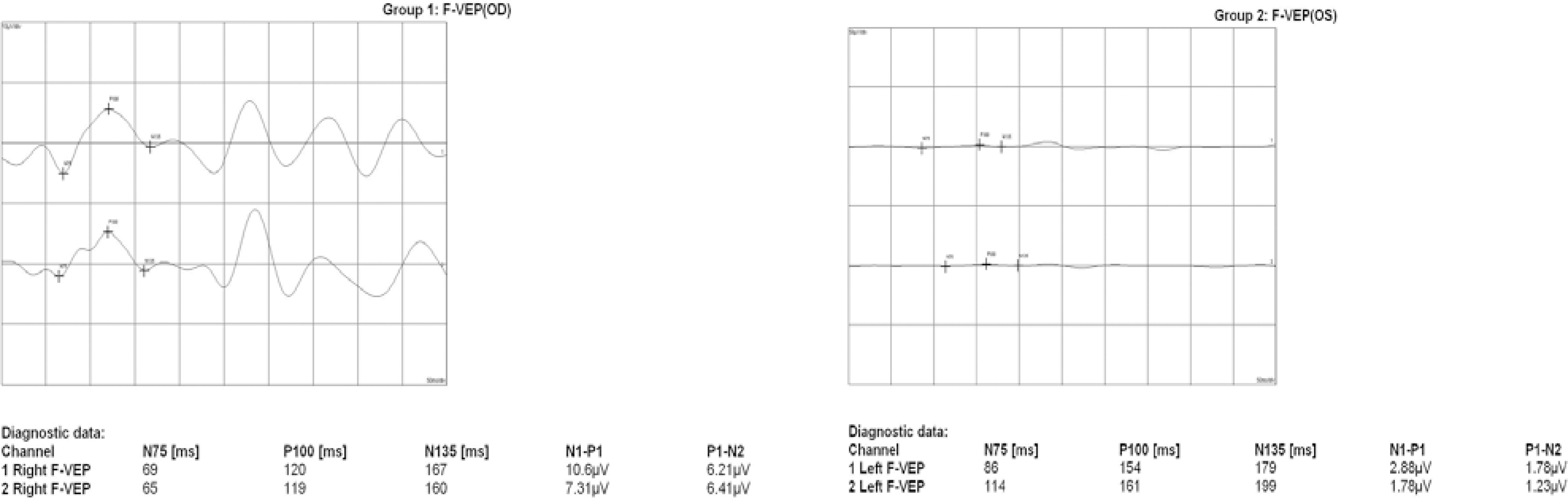
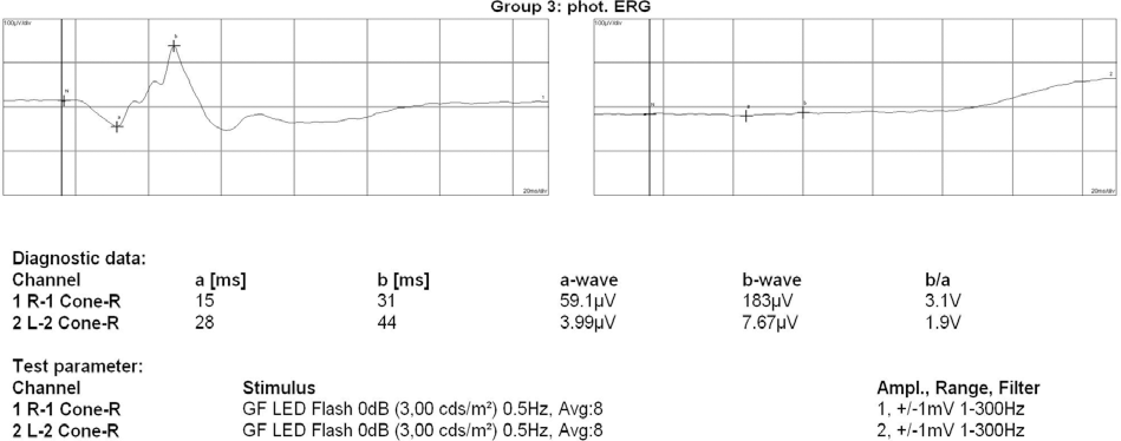
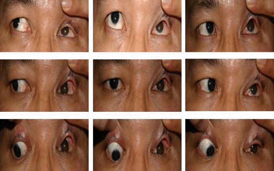
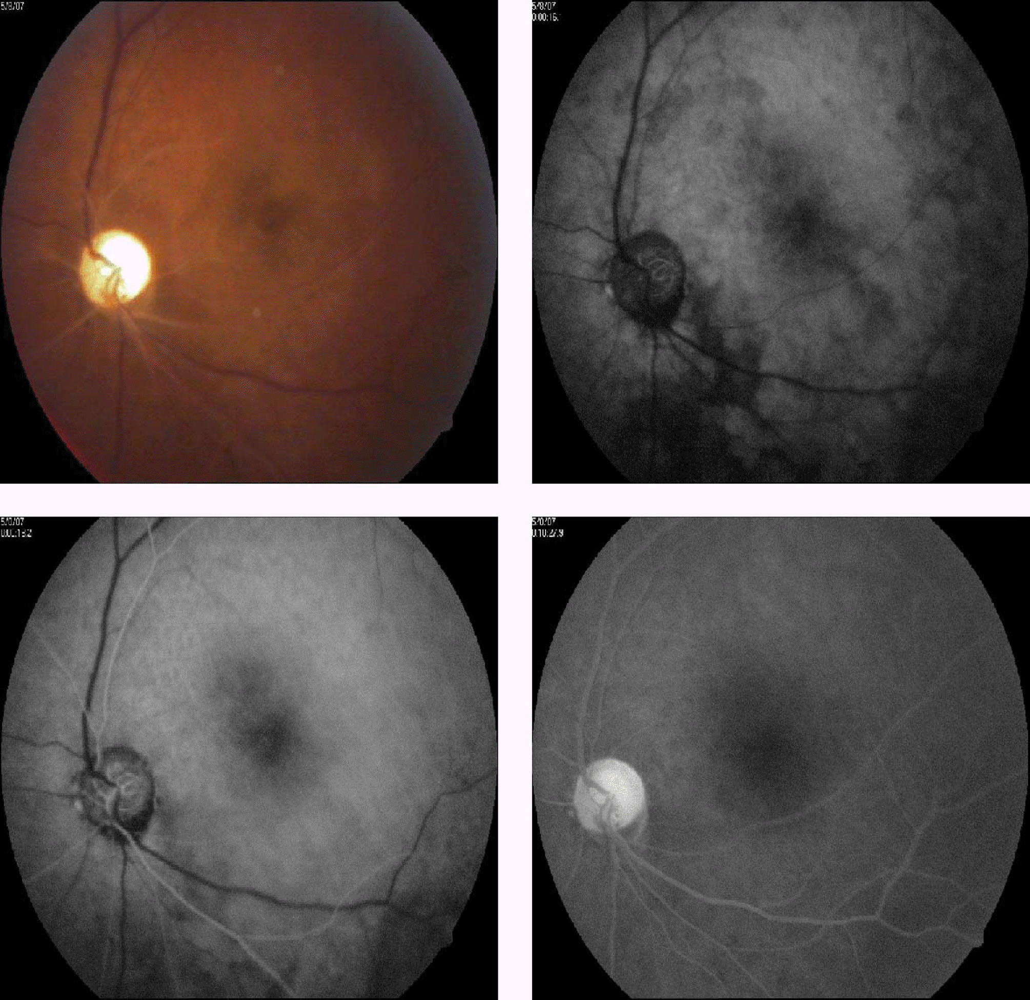
The 38-year-old man was admitted to the emergency room after sustaining a work-related trauma. His chief complaints were loss of vision and bleeding from the left periorbital area. He had no light perception and no eyeball was found in the orbit. Facial CT revealed that the intact eyeball was entrapped within the maxillary sinus. The condition of the optic nerve was difficult to ascertain. Ten hours after post-trauma, reduction surgery was done with a graft from the iliac bone. Ruptured extraocular muscles were not primarily sutured. After four months, vitrectomy was performed on the left eye. The eyeball was repositioned in its place. He had no light perception. Extraocular motility improved at the last follow-up examination.
Conclusions
We report the dislocation of the eyeball globe into the maxillary sinus after a blowout fracture. Visual acuity showed no light perception as a result of central retinal artery occlusion and optic nerve injury. We were able to obtain a good aesthetic and functional result after the operation.
Go to : 
References
1. Smith B, Converse JM. Early treatment of orbital floor fractures. Trans Am Acad Ophthalmol Otolaryngol. 1957; 61:602–7.
2. Berkowitz RA, Putterman AM, Patel DB. Prolapse of the globe into the maxillary sinus after orbital floor fracture. Am J Ophthalmol. 1981; 91:253–7.

3. Ziccardi VB, Patterson GT, Ramasastry S, Sotereauanos GC. Management of a severe zygomatico orbital fracture with dislocation of the globe into the antrum. J Craniofac Surg. 1993; 4:95–101.
4. Pelton RW, Rainey AM, Lee AG. Traumatic subluxation of the globe into the maxillary sinus. AJNR Am J Neuroradiol. 1998; 19:1450–1.
5. Kim S, Baek S. Traumatic dislocation of the globe into maxillary sinus associated with extraocular muscle injury. Graefes Arch Clin Exp Ophthalmol. 2005; 243:1280–3.
6. Moon M, Pietris G, Shapter M. Dislocation of the globe into the ethmoid sinuses. Aust N Z J Ophthalmol. 1997; 25:175–6.
7. Tranfa F, Di Matteo G, Di Salle F, et al. Traumatic Displacement of the Globe Into the Ethmoid Sinus. Am J Ophthalmol. 2000; 130:253–4.
8. Kim KH, Ahn Y, Ryu JS, Yoon CB. Dislocation of the Globe into the nasal cavity after orbital wall fracture. J Korean Ophthalmol Soc. 2000; 41:2765–70.
9. Kang BD, Jang MH. A Case of Blowout Fracture of the Orbital Wall with Eyeball Entrapped within the Ethmoid Sinus. Korean J Ophthalmol. 2003; 17:149–53.
10. Kim SH, Paik SH, Lee TS. Treatment of Complete Traumatic Eyeball Extrusion. J Korean Ophthalmol Soc. 1995; 36:2271–5.
11. Huh JS, Chun DH, Kim BJ, Lee HB. A case of traumatic eyeball extrusion with complex, comminuted fractures. J Korean Ophthalmol Soc. 2003; 44:251–8.
12. Lee MS, Ahn JH, Kim HY, Lee SY. Clinical Study of Orbital Wall Fracture. J Korean Ophthalmol Soc. 1997; 38:1687–93.
13. Jones DE, Evans JN. “Blow-out” fractures of the orbit: An investigation into their anatomi cal basis. J Laryngol Otol. 1967; 81:1109–13.
14. Smith B, Rogan WF. Blowout fracture of the orbit, mechanism and correction of internal orbital fracture. Adv Ophthalmic Plast Reconstr Surg. 1987; 6:197–205.
15. Wilder IW, Beyer CK, Smith B, Conley JJ. Ocular finding following radical maxillectomy. Trans Am Acad Ophthalmol Otolaryngol. 1971; 75:797–801.
16. Forrest CR, Khirallah E, Kuzon WM. Intraocular and intraorbital compartment pressure changes following orbital bone grafting: a clinical and laboratory study. Plast Reconstr Surg. 1999; 104:48–54.
17. Kim HK, Yoon KC, Park YG. Optic Nerve Damage Colicationg Repair of Orbital Floor Fracture. J Korean Ophthalmol Soc. 2003; 44:2924–8.
18. Neuhaus RW. Orbital complication secondary to endoscopic sinus surgery. Ophthalmology. 1990; 97:1512–8.
19. Sarks SH, Lawson W, Edelstein D, Green RP. Surgical treatment of blindness secondary to intraorbital hemorrhage. Arch Otolaryngol Head Neck Surg. 1988; 114:801–3.
20. Lende RA, Ellis PP. Induced spasm in the retinal arterioles of cat. I. Mechanism and characteristics. Arch Ophthalmol. 1964; 71:701.
21. Seiff SR. High dose corticosteroids for treatment of vision loss due to indirect injury to the optic nerve. Ophthalmic Surg. 1990; 21:389–95.
22. Lessell S. Indirect optic nerve trauma. Arch Ophthalmol. 1989; 107:382–6.
23. Kim HW, Kim YI, Won IK. Clinical Analysis of Blow-out Fracture with Ocular Motion Limitation: Comparison of Surgical and Conservative Treatment. J Korean Ophthalmol Soc. 1999; 40:632–8.
24. Millman AL, Della Rocca RC, Spector S, et al. Steroids and orbital blowout fracture. Adv Ophthalmol Plast Reconstr Surg. 1988; 6:265–8.
25. Koorneef L. Current cencepts on the management of orbital blow-out fracture. Ann Plast Surg. 1982; 9:185–200.
Go to :
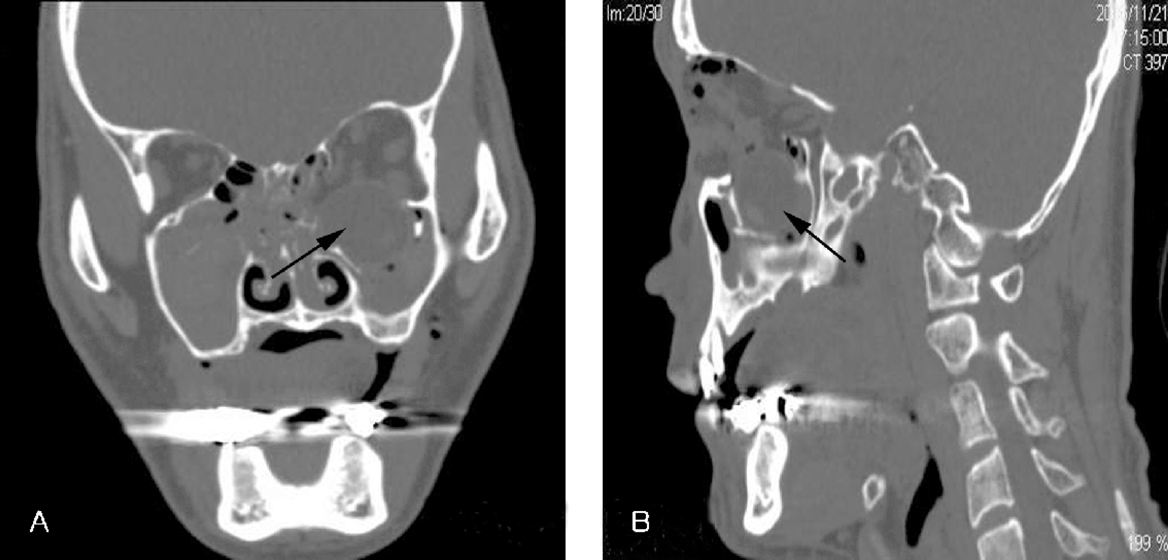 | Figure 2.(A) Preoperative coronal CT scan shows fracture of the left inferior and medial orbital wall and herniated eyeball (arrow) in the maxillary sinus. Large amount of hemorrhage was found within the ethmoid and maxillary sinuses. (B) Preoperative sagittal CT scan shows herniated eyeball(arrow) in the maxillary sinus. |
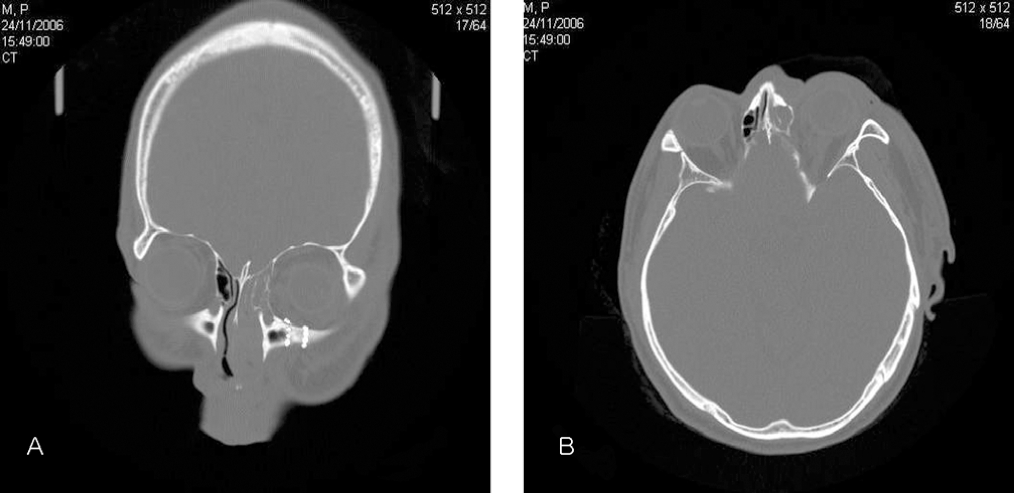 | Figure 4.(A) Postoperative CT shows the left globe within the orbit and part of the iliac bone. (B) Axial section of the left orbit shows normal location of the left globe. |

 XML Download