Abstract
Case summary
A 42-year-old woman presented with rapid enlarging mass, 15×12 mm in size at left upper eyelid. Orbit CT disclosed an enhanced, well-circumscribed preseptal lid mass. The histopathologic and immunohistochemical analyses after excisional biopsy were consistent with nodular fasciitis. There was no recurrence of the tumor after excision.
References
1. Spencer WH. Ophthalmic pathology: An atlas and textbook. 4th ed.Vol. 4. Philadelphia: WB Sauders;1996. p. 2380–2.
2. Enzinger FM, Weiss SW. Soft Tissue Tumors. 3rd ed.St. Louis: C.V. Mosby;1995. p. 165–76.
3. Alert DM, Jakobiec FA. Principles and practice of ophthalmology. 2nd ed.Philadelphia: WB Saunders;2000. p. 3450–1.
4. Recchia FM, Buckley EG, Townshend LM, Klintworth GK. Nodular fasciitis of the orbital rim in a pediatric patient. J Pediatr Ophthalmol Strabismus. 1997; 34:316–8.

5. Vestal KP, Bauer TW, Berlin AJ. Nodular fasciitis presenting as an eyelid mass. Ophthal Plast Reconstr Surg. 1990; 6:130–2.

6. Holds JB, Mamalis N, Anderson RL. Nodular fasciitis presenting as a rapidly enlarging episcleral mass in a 3-year-old. J Pediatr Ophthalmol Strabismus. 1990; 27:157–60.

7. Konwaler BE, Keasbey L, Kaplan L. Subcutaneous pseudosarcomatous fibromatosis (fasciitis). Am J Clin Pathol. 1955; 25:241–52.

8. Bernstein KE, Lattes R. Nodular (pseudosarcomatous) fasciitis, a nonrecurrent lesion: Clinicopathologic study of 134 cases. Cancer. 1982; 49:1668–78.

10. de Paula SA, Cruz AA, de Alencar VM, et al. Nodular fasciitis presenting as a large mass in the upper eyelid. Ophthal Plast Reconstr Surg. 2006; 22:494–5.

11. Price EB Jr, Silliphant WM, Shuman R. Nodular fasciitis: a clinicopathological analysis of 65 cases. Am J Clin Pathol. 1961; 35:122–36.
12. Shimizu S, Hashimoto H, Enjoji M. Nodular fasciitis: an analysis of 250 patients. Pathology. 1983; 16:161–6.

13. Stone DU, Chodosh J. Epibulbar nodular fasciitis associated with floppy eyelids. Cornea. 2005; 24:361–2.

15. Velagaleti GV, Tapper JK, Panova NE, et al. Cytogenetic findings in a case of nodular fasciitis of subclavicular region. Cancer Genet Cytogenet. 2003; 141:160–3.

16. Font RL, Zimmerman LE. Nodular fasciitis of the eye and adnexa. Arch Ophthalmol. 1966; 75:475–81.

17. Shields JA, Shields CL, Christian C, Eagle RC. Orbital nodular fasciitis simulating a dermoid cyst in an 8-month-old child. Ophthal Plast Reconstr Surg. 2001; 17:144–8.

18. Toledo AS, Rodriguez J, Cuasay NS, et al. Nodular fasciitis of the facial region: CT characteristics. J Comput Assist Tomogr. 1988; 12:898–9.
19. Sutton D. Textbook of radiology and imaging. 7th ed.Vol. 2. Edinburgh: Churchill Livingstone;2003. p. 1580–4.
20. Hymas DC, Mamalis N, Pratt DV, et al. Nodular fasciitis of the lower eyelid in a pediatric patient. Ophthal Plast Reconstr Surg. 1999; 15:139–42.

21. Kaw YT, Cuesta RA. Nodular fasciitis of the orbit diagnosed by fine needle aspiration cytology. A case report. Acta Cytol. 1993; 37:957–60.
22. Meffert JJ, Kennard CD, Davis TL, Quinn BD. Intradermal nodular fasciitis presenting as an eyelid mass. Int J Dermatol. 1996; 35:548–52.

23. Montgomery EA, Meis JM. Nodular fasciitis. Its morphologic spectrum and immunohistochemical profile. Am J Surg Pathol. 1991; 15:942–8.
24. Lee SK, Kwon SY. Nodular fasciitis of the face diagnosed by US-guided core needle biopsy: a case report. J Korean Radiol Soc. 2006; 55:551–5.

25. Choi MH, Jeon J, Son SW, et al. A case of recurrent nodular fasciitis. Korean J Dermatol. 2006; 44:1457–9.
26. Lee MW, Choi JH, Sung KJ, et al. Nodular fasciitis, review of 16 cases. Korean J Dermatol. 2001; 39:1–6.
Figure 1.
Axial orbit CT imaging disclosed an enhancing, well-circumscribed, preseptal mass in the left upper eyelid before surgery.

Figure 2.
Intraoperative photograph showed a solid mass contiguous with the dermis. The tumor was abutting from the periosteum of the lateral orbit at its superotemporal aspect.

Figure 4.
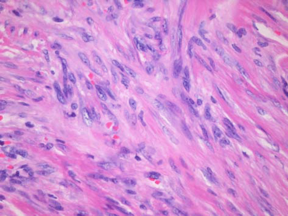
Histopathologic section showed interlacing bundles of spindle shaped cells compatible with immature fibroblasts. Small capillaries were abundant and were associated with extravasated red blood cells. (hematoxylin-eosin stain, ×40)


 PDF
PDF ePub
ePub Citation
 Print
Print


 XML Download