

 PDF
PDF ePub
ePub Citation
 Print
Print


Abstract
Purpose
To compare the long-term clinical results of one-piece Acrysof (SA60AT) hydrophobic acrylic intraocular lens (IOL) implantation compared with implantation of three-piece Acrysof (MA60BM) hydrophobic acrylic IOL.
Methods
We retrospectively analyzed each 50 eyes of 50 patients underwent MA60BM or SA60AT IOL implantation and followed for at least 6 months.
Results
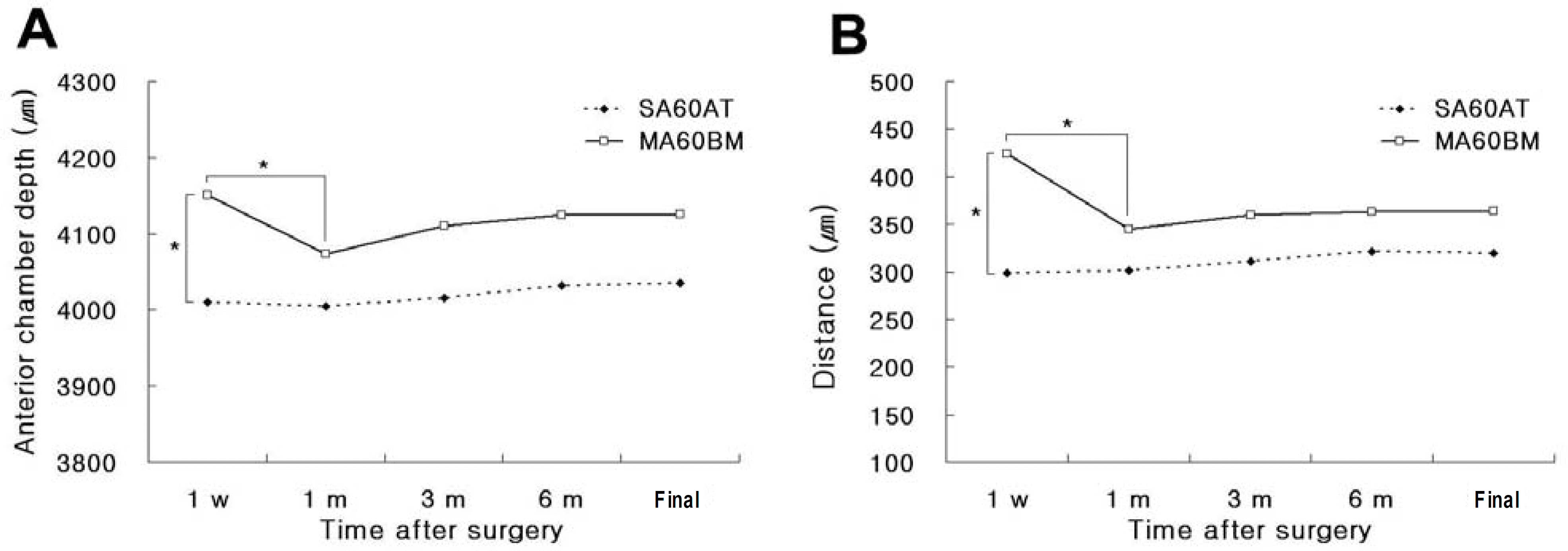
Final visual acuity of 0.5 or better was 38 eyes (76%) and 0.8 or better was 20 eyes (40%) in the SA60AT group. In the MA60BM group, it was 41 eyes (82%) and 23 eyes (46%) respectively. There were no significantly differences in predictability, intraocular pressure, endothelial cell density, astigmatism, and incidence of posterior capsule opacification between the two groups. Spherical equivalent at postoperative 1 week was −0.50±0.95D in SA60AT group and was −0.31±0.88D in MA60BM group (P=0.04). However, there was no significant difference between the two groups during follow up period. In MA60BM group, anterior chamber depth (P=0.02) and distance between iris and IOL (P=0.04) reduced significantly during the first postoperative month.
Go to : 
References
1. Kohnen S, Ferrer A, Brauweiler P. Visual function in pseudophakic eyes with poly (methyl methacrylate), silicone, and acrylic intraocular lenses. J Cataract Refract Surg. 1996; 22:1303–7.
2. Hayashi K, Hayashi H, Nakao F, Fumihiko H. Reduction in the area of the anterior capsule opening after poltmethymethacrylate, silicone, and soft acrylic intraocular lens implantation. Am J Ophthalmol. 1997; 123:441–7.
3. Magual E, Garcia J, Elvira JC, Hueso JR. Clinical results of AcrySof intraocular lens implantation. J Cataract Refract Surg. 1998; 24:114–7.
4. Oshika T, Suzuki Y, Kizaki H, Yaguchi S. Two year clinical study of a soft acrylic intraocular lens. J Cataract Refract Surg. 1996; 22:104–9.

5. Ursell PG, Spalton DJ, Pande MV, et al. Relationship between intraocular lens biomaterials and posterior capsule opacification. J Cataract Refract Surg. 1998; 24:352–6.
6. Cha YD, Oh SH, Lee DH. Comparative assessment of clinical results in various acrylate IOLs. J Korean Ophthalmol Soc. 2006; 47:740–7.
7. Choi JA, Kim TI, Tchah HW. Aberration change in pseudophakia with three types of acryl intraocular lens. J Korean Ophthalmol Soc. 2004; 45:405–12.
8. Oh SH, Kim JK, Lee DH. The clinical results of hydrophobic single-piece acrylic intraocular lenses after cataract surgery. J Korean Ophthalmol Soc. 2004; 45:2007–13.
9. Seo JH, Kim KB, Seo JB. A clinical comparison of Acrysof with hydrophilic acrylic IOLs. J Korean Ophthalmol Soc. 2001; 42:266–71.
10. Nishi O, Nishi K. Effect of the optic size of a single-piece acrylic intraocular lens on posterior capsule opacification. J Cataract Refract Surg. 2003; 29:348–53.
11. Caporossi A, Casprini F, Tosi GM, Baiocchi S. Preliminary results of cataract extraction with implantation of a single-piece AcrySof intraocular lens. J Cataract Refract Surg. 2002; 28:652–5.
12. Meacock WR, Spalton DJ, Boyce JF, Jose RM. Effecct of optic size on posterior capsule opacification: 5.5mm versus 6.0 mm AcrySof intraocular lenses. J Cataract Refract Surg. 2001; 27:1194–8.
13. Mian SI, Fahim K, Marcovitch A, et al. Nd:YAG capsulotomy rates after use of the AcrySof acrylic three piece and one piece intraocular lenses. Br J Ophthalmol. 2005; 89:1453–7.
14. Sacu S, Findl O, Menapace R, et al. Comparison of posterior capsule opacification between the 1-piece and 3-piece Acrysof intraocular lenses: two-year results of a randomized trial. Ophthalmology. 2004; 111:1840–6.
15. Bender LE, Nimsgern C, Jose R, et al. Effect of 1-piece and 3-piece AcrySof intraocular lenses on the development of posterior capsule opacification after cataract surgery. J Cataract Refract Surg. 2004; 786–9.
16. Byeun SH, Kim WS. Comparision of the three different hydrophilic acrylic intraocular lenses. J Korean Ophthalmol Soc. 2006; 47:1561–7.
17. Wirtitsch MG, Findle O, Menapace R, et al. Effect of haptic design on change in axial lens position after cataract surgery. J Cataract Refract Surg. 2004; 30:45–51.
18. Seong MC, Kim MJ, Choi CY, Tchah HW. Clinical results of single-piece hydrophilic IOL after cataract surgery. J Korean Ophthalmol Soc. 2006; 47:1394–400.
19. Lane SS, Burgi P, Milios GS, et al. Comparison of the biomechanical behavior of foldable intraocular lenses. J Cataract Refract Surg. 2004; 30:2397–402.
20. Nejima R, Miyata K, Honbou M, et al. A prospective, randomised comparison of single and three piece acrylic foldable intraocular lenses. Br J Ophthalmol. 2004; 88:727–8.
21. Cho HJ, Woo JM, Yang KJ. Ultrasound biomicroscopic dimensions of the anterior chamber in angle closure glaucoma patients. Korean J Ophthalmol. 2002; 16:20–5.
22. Yoon KC, Park SW, Song BY. The role of ultrasound biomicroscopy in operation for limbal dermoid. J Korean Ophthalmol Soc. 2004; 45:364–9.
23. Spaeth GL, Azuara-Blanco A, Araujo SV, Augsburger JJ. Intraobserver and interobserver agreement in evaluating the anterior chamber angle configuration by ultrasound biomicroscopy. J Glaucoma. 1997; 6:13–7.
24. Choi HY, Jun RM, Choi KR. Ultrasound Biomicroscopy (UBM plus, model P45, Paradigm). Intraobserver reproducibility and agreement of measurements. J Korean Ophthalmol Soc. 2003; 44:1112–7.
25. Oh DE, Jun RM, Choi KR. Quantified values of anterior segment in normal adult koreans using ultrasound biomicroscopy. J Korean Ophthalmol Soc. 2004; 45:251–8.
26. Sorenson AL, Holladay JT, Kim T, et al. Ultrasonographic measurement of induced myopia associated with capsular bag distention syndrome. Ophthalmology. 2000; 107:902–8.
27. Manabe S, Oh H, Amino K, et al. Ultrasound biomicroscopic analysis of posterior chamber intraocular lenses with transscleral sulcus suture. Ophthalmology. 2000; 107:2172–8.
28. Erickson P. Effects of intraocular lens position errors on postoperative refractive error. J Cataract Refract Surg. 1990; 16:305–11.
29. Petternel V, Menapace R, Findl O, et al. Effect of optic edge design and haptic angulation on postoperative intraocular lens position change. J Cataract Refract Surg. 2004; 30:52–7.
Go to :
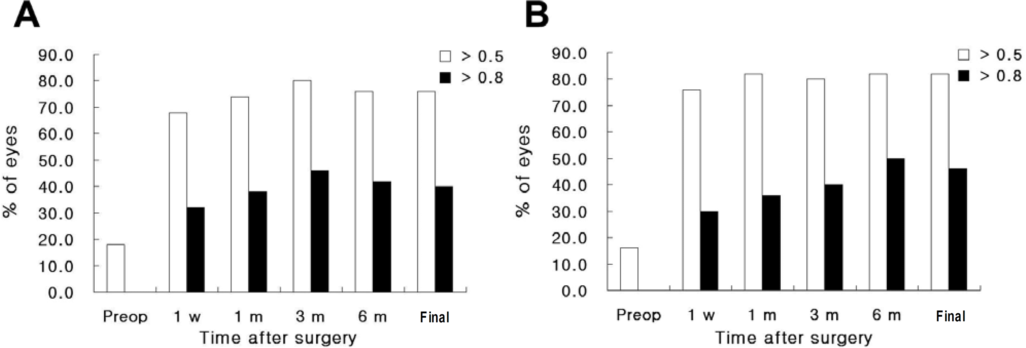 | Figure 1.Changes of preoperative and postoperative uncorrected visual acuity between SA60AT (A) and MA60BM (B) groups. |
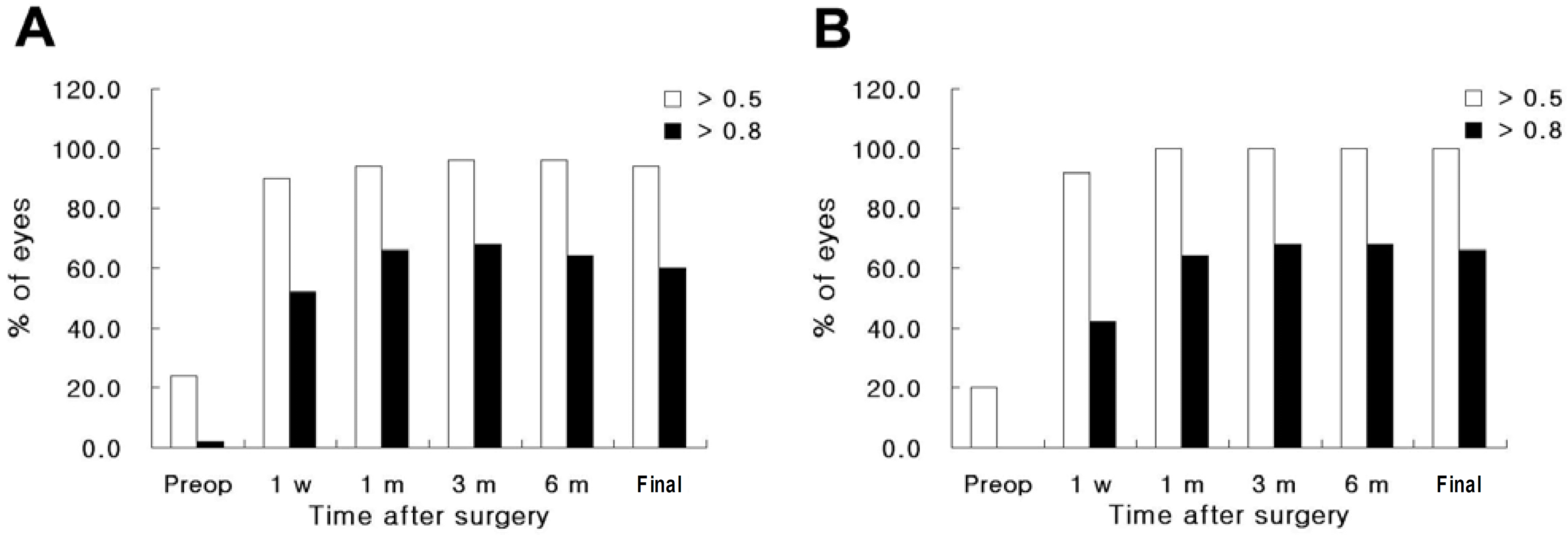 | Figure 2.Changes of preoperative and postoperative best corrected visual acuity between SA60AT (A) and MA60BM (B) groups. |
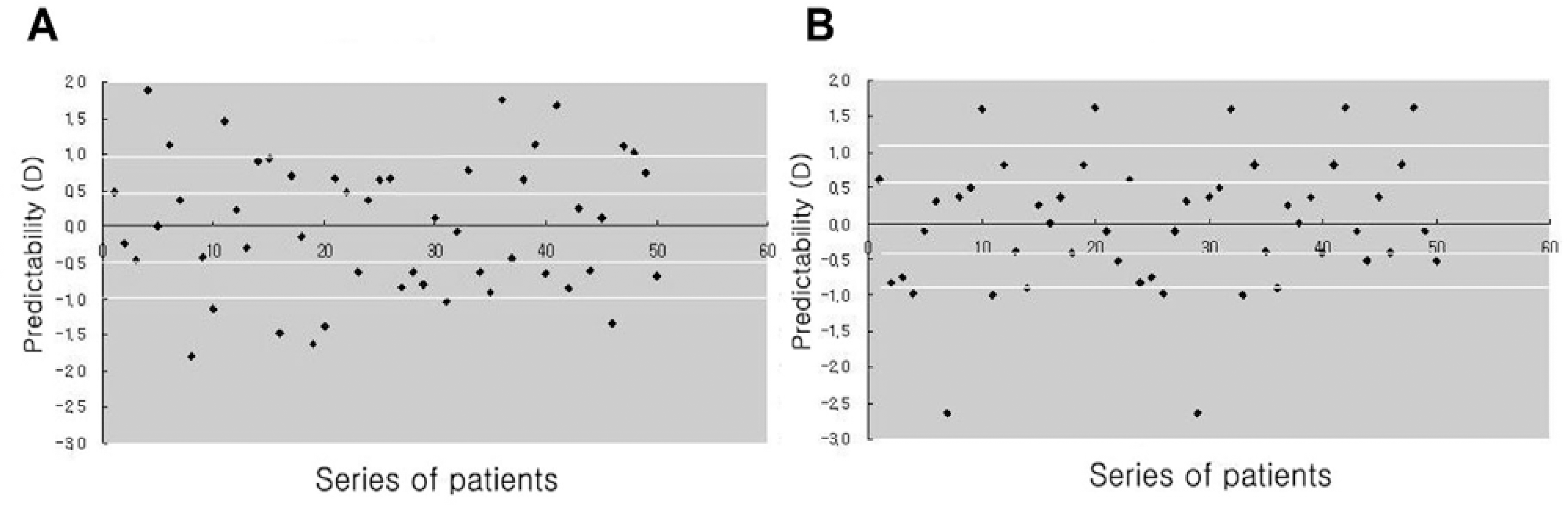 | Figure 3.Predictability of the manifest refraction with SA60AT (A) and MA60BM (B) at the final follow-up. |
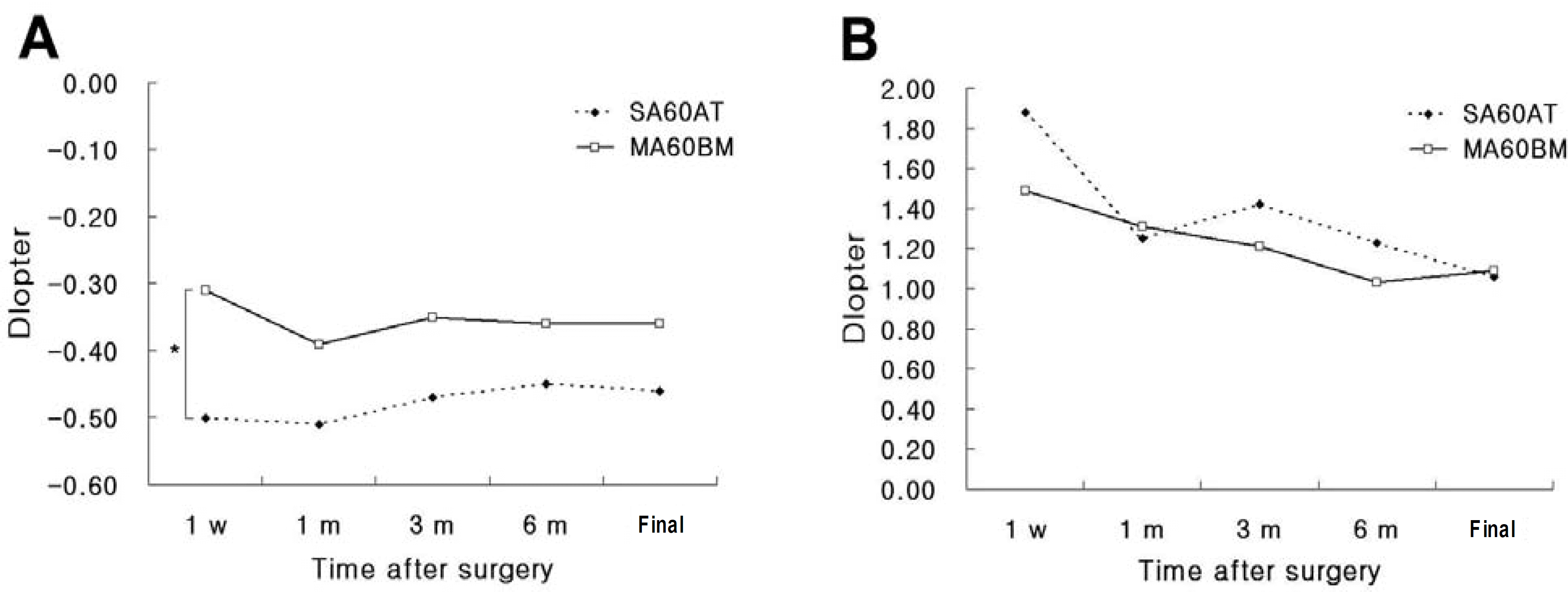 | Figure 4.Changes of the postoperative spherical equivalent (A) and astigmatism (B) (*: P<0.05). |
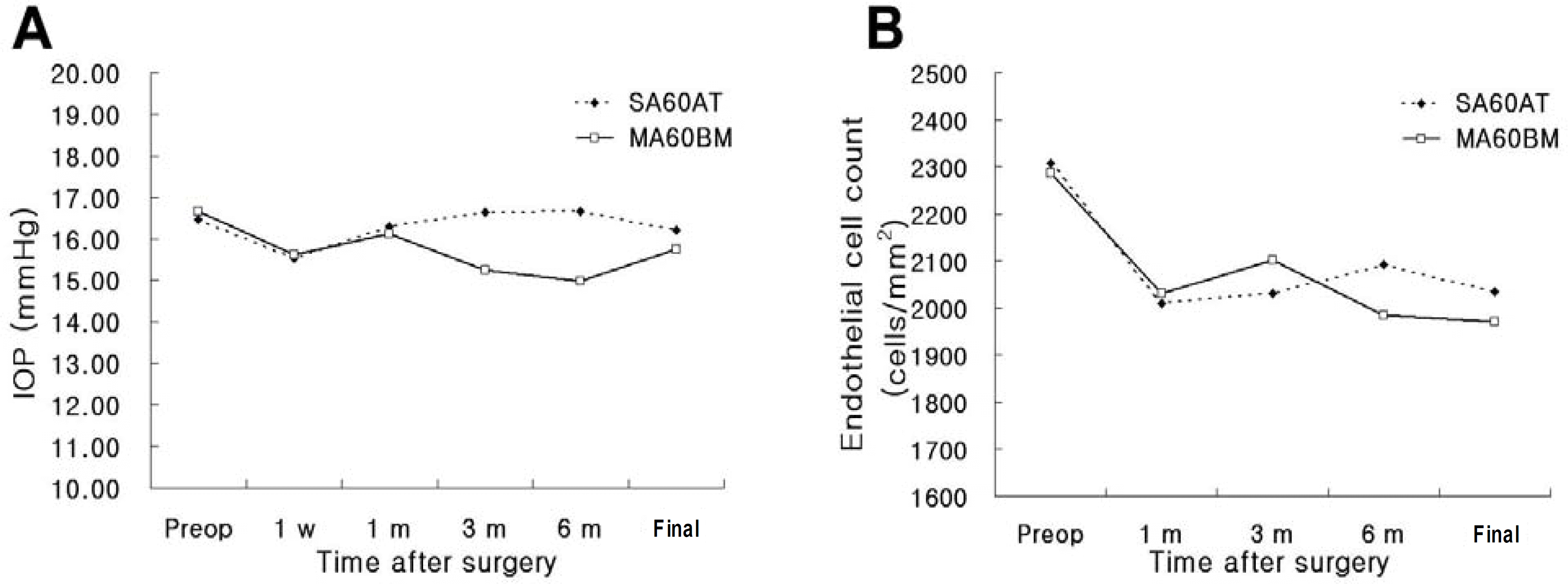 | Figure 5.Changes of the postoperative intraocular pressure (A) and endothelial cell count (B). |
 XML Download