

 PDF
PDF ePub
ePub Citation
 Print
Print


Abstract
Purpose
This is a report on the long-term follow-up of patients who had received black diaphragm intraocular lens implantation after traumatic aniridia.
Methods
This is a retrospective study on the long-term follow-up for more than 50 months of six patients who had received black diaphragm intraocular lens implantation after traumatic aniridia.
Results
An improvement in visual acuity of more than two letters, according to the Snellan chart, occurred in three out of six eyes that were available for follow-up studies after an average period of 7 years and 8 months (50-115 months) after the operation, and no visual acuity decline groups were present. Early complications of black diaphragm intraocular lens implantation consisted of cystoid macular edema and transient intraocular pressure rise. The complications late occurred included bullous keratopathy in four eyes, glaucoma in three eyes, and exotropia in three eyes. We performed penetrating keratoplasty in two of the four eyes suffering from bullous keratopathy, and an Ahmed valve implantation in two of the three eyes suffering from glaucoma. There were signs of reduced visual acuity in four eyes due to complications of the surgery.
Conclusions
The authors of this report recommend care in use of black diaphragm intraocular lens implantation, by studies done with an average follow-up period of 7 years and 8 months post operatively. Also reported were severe complications with this treatment such as bullous keratopathy and glaucoma.
References
1. Sundmacher R, Reinhard T, Althaus C. Black diaphragm intraocular lens for correction of aniridia. Ger J Ophthalmol. 1994; 25:180–5.
2. Suh DS, Roh JH, Kim SD. Clinical Experience with Black-Diaphragm Intraocular Lens in Traumatic (Postoperative) Aniridia and Aphakia. J Korean Ophthalmol Soc. 1998; 39:1183–91.
3. Park SJ, Kim HT, Kim SM, Chung SK. Implatation of black diaphragm intraocular lens in cataract surgery with congenital aniridia. J Korean Ophthalmol Soc. 1998; 39:1748–54.
4. Reinhard T, Engelhardt S, Sundmacher R. Black diaphragm aniridia intraocular lens for congenital aniridia: Long-term follow-up. J Cataract Refract Surg. 2000; 26:375–81.

5. Liu GJ, Okisaka S, Mizukawa A, Momose A. Histopathological study of pseudophakic bullous keratopathy developing after anterior chamber of iris-supported intraocular lens implant. Jpn J ophthalmol. 1993; 37:414–25.
6. Funderburgh JL, Hevelone ND, Roth MR, et al. Decorin and biglycan of normal and pathologic human corneas. Invest Ophthalmol Vis Sci. 1998; 39:1957–64.
7. Kenyon KR. The synthesis of basement membrane by the cornea epithelium in bullous keratopathy. Invest Ophthalmol. 1969; 8:156–68.
8. Vote BJ, Tranos P, Bunce C, et al. Long-term outcome of combined pars plana vitrectomy and scleral fixated sutured posterior chamber intraocular lens implatation. Am J Ophthalmol. 2006; 141:308–12.
9. Walton DS. Aniridia with glaucoma. Chandler PA, Grant WM, editors. Glaucoma. Philadelphia: Lea and Febiger;1986. 1:chap. 39.
10. Sidikaro Y, Von Noorden GK. Observations in sensory heterotropia. J Pediatr Ophthalmol. 1982; 19:12–9.
Figure 1.
Postoperative photographs. (A) Case 1; Postoperative 15 months fluorescent angiography reveal hyperfluorescent leakage at macular. (B) Case 1; After penetrating keratoplasty, slit lamp photography appearance. (C) Case 3; Last follow-up slit lamp photography appearance. Inferior corneal opacity was observed due to bullous keratopathy. (D) Case 4; Last follow-up slit lamp photography appearance of implantation of the black-diaphragm intraocular lens.
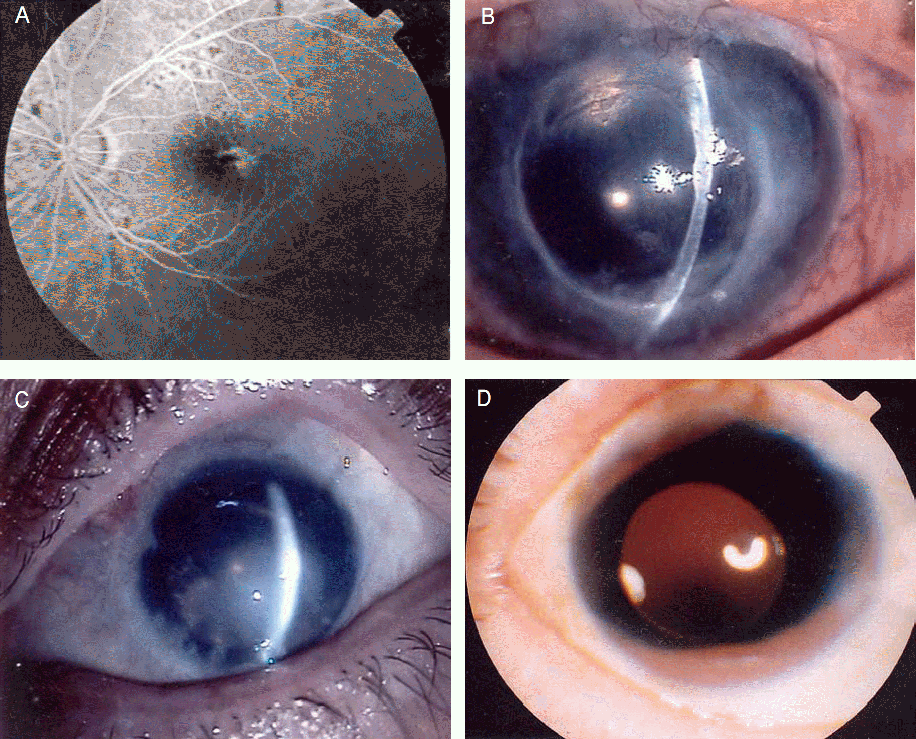
Figure 2.
The black diaphragm IOL, type 67 G, black polymethylmethacrylate diaphragm fixed tightly to the optic zone.
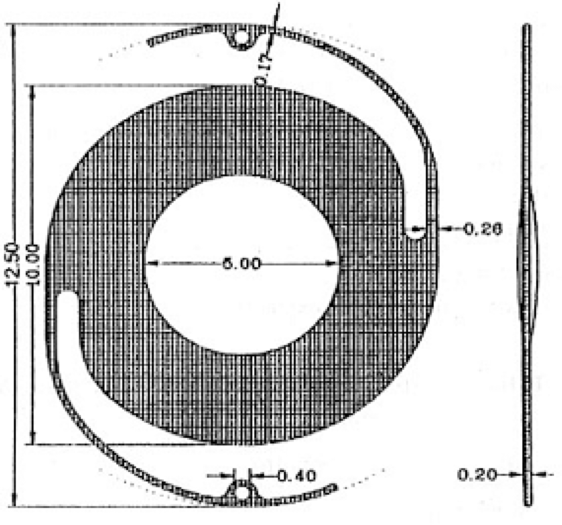
Table 1.
Patient's data before black diaphragm IOL implantation, complications, visual acuity outcome, and follow-up periods after scleral fixed black-diaphragm IOL implantation
 XML Download