

 PDF
PDF ePub
ePub Citation
 Print
Print


Abstract
Purpose
To investigate quantitative and morphometric endothelial changes in phakic eyes implanted with an iris-fixed phakic intraocular lens in Korean patients.
Methods
We prospectively examined the endothelial cell density (ECD), ECD loss (ECL), coefficient variations (CV), and frequency of Hexagons (6A) in 46 phakic eyes implanted with an iris-fixed phakic intraocular lens. The effect of anterior chamber depth (ACD) and diopter of iris-fixed phakic intraocular lens on ECD was statistically analyzed.
Results
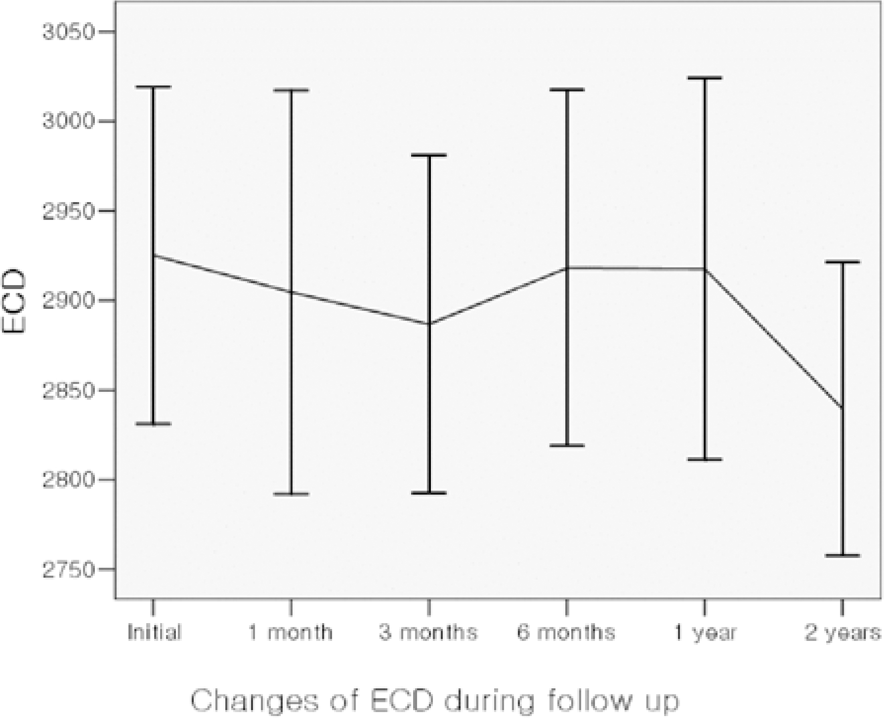
Preoperative mean ECD was 2923±257 cells/mm2. ECL at postoperative 1, 3, 6, 12, and 24 monthswas 0.71%, 0.88%, 0.70%, 0.64%, and 2.34%. It was significant only in the month 24 results. The mean CV value at the same time was 0.343, 0.342, 0.334, 0.329, 0.326, and 0.338. The change was significant at the 12th month. The mean 6A value at the same time was 58.2%, 57.8%, 59.7%, 59.6%, 60.1%, and 58.8%. There was no significant change in 6A value. Preoperative ACD and diopter of iris-fixed phakic intraocular lens didn't affect the ECD change.
Conclusions
This two-year follow-up study of changes in endothelial cells after implantation iris-fixated phakic intraocular lens showed statistically significant decreases in the cells, which is not very significant considering the physiologic reduction rate of the cells. Thses decreases showed a tendency to be stabilized with recovery of morphological changes. Further long-term follow-up is needed to determine its long-lasting effect.
Go to : 
References
1. Heitzmann J, Binder PS, Kassar BS, Nordan LT. The correction of high myopia using excimer laser. Arch Ophthalmol. 1993; 111:1627–34.
2. Kim TG, Joo CK. 2 cases of corneal ectasia detected after LASIK. J Korean Ophthalmol Soc. 1999; 40:846–9.
3. Geggel HS, Talley AR. Delayed onset keratectasia following laser in situkeratomileusis. J Cataract Refract Surg. 1999; 25:582–6.
4. Holladay JT, Dudeja KR, Chang J. Functional vision and corneal changes after laser in situkeratomileusis determined by contrast sensitivity, glare test and corneal topography. J Cataract Refract Surg. 1999; 25:663–9.
5. Fechner PU. Iris claw lens (German). Klin Monatsbl Augenheilkd. 1987; 191:26–9.
6. Fechner PU, Worst JF. A new concave intraocular lens for the correction of high myopia. European Journal of Implant and Refractive Surgery. 1989; 1:41–3.
7. Fechner PU, Strobel J, Wichmann W. Correction of myopia by implantation of a concave Worst-iris claw lens into phakic eyes. Refract Corneal Surg. 1991; 7:286–98.

8. Landesz M, Worst JG, Siertsema JV, van Rij G. Correction of high myopia with the Worst myopia claw intraocular lens. J Refract Surg. 1995; 11:16–25.
9. Izak MG, Izak A. Inflammatory reaction associated to Artisan phakic refractive IOL implantation. Budo CJR, editor. The ARTISAN Lens. 1st ed.Panama: Highlights of Ophthalmology;2004. 1:chap. 14.
10. Maloney RK, Nguyen LH, John ME. Artisan phakic intraocular lens for myopia. Short-term results of a prospective, multicenter study. Ophthalmology. 2002; 109:1631–41.
11. Sanders DR, Doney K, Poco M, et al. United States food and drug administration clinical trial of the implatable collamer lens(ICL) for moderate to high myopia. Three-year follow-up. Ophthalmology. 2004; 111:1683–92.
12. Sarikkola AU, Sen HN, Uusitalo RJ, Laatikainen L. Traumatic cataract and other adverse events with implantable contact lens. J Cataract Refract Surg. 2005; 31:511–24.
13. Assetto V, Benedetti S, Pesando P. Collamer intraocular contact lens to correct high myopia. J Cataract Refract Surg. 1996; 22:551–6.
14. Smallman D, Probst L, Rafuse PE. Pupillary block glaucoma secondary to posterior chamber phakicintraocular lens implantation for high myopia. J Cataract Refract Surg. 2004; 30:905–7.
15. Kodjikian L, Gain P, Donate D, et al. Malignant glaucoma induced by a phakic posterior chamber intraocular lens for myopia. J Cataract Refract Surg. 2002; 28:2217–21.
16. Perez-Santonja JJ, Iradier MT, Sanz-Iglesias L, et al. Endothelial changes in phakic eyes with anterior chamber intraocular lenses to correct high myopia. J Cataract Refract Surg. 1996; 22:1017–22.
17. de Souza RF, Forseto A, Nose R. Anterior chamber intraocular lens for high myopia: five year results. J Cataract Refract Surg. 2001; 27:1248–53.
18. Alio JL, de la Hoz F, Perez-Santonja JJ, et al. Phakic anterior chamber lenses for the correction of myopia: a 7-year cumulative analysis of complications in 263 cases. Ophthalmology. 1999; 106:458–66.
19. Perez-Santonja JJ, Alio JL, Jimenez-Alfaro I, Zato MA. Surgical correction of severe myopia with an angle-supported phakic intraocular lens. J Cataract Refract Surg. 2000; 26:1288–302.
20. Asano-Kato N, Toda I, Hori-Komai Y, et al. Experience with the Artisan phakic intraocular lens in Asian eyes. J Cataract Refract Surg. 2005; 31:910–5.
21. Landesz M, van Rij G, Luyten G. Iris-claw phakic intraocular lens for high myopia. J Refract Surg. 2001; 17:634–40.
22. Benedetti S, Casamenti V, Marcaccio L, et al. Correction of myopia of 7 to 24 diopters with the Artisan phakic intraocular lens: two-year follow-up. J Refract Surg. 2005; 21:116–26.
23. Kim HC, Lee SY, Shim CB. The clinical results of iris-fixated phakic IOL. J Korean Ophthalmol Soc. 2005; 46:353–9.
24. Benedetti S, Casamenti V, Benedetti M. Long-term endothelial changes in phakiceyes after Artisan intraocular lens implantation to correct myopia Five-year study. J Cataract Refract Surg. 2007; 33:784–90.
25. Menezo JL, Cisneros AL, Salvador VR. Endothelial study of iris-claw phakic lens: Four year follow-up. J Cataract Refract Surg. 1998; 24:1039–49.
26. Lee ES, Cho YJ, Kim EK. Short-term changes in corneal endothelium after iris-fixated phakic intraocular lens insertion. J Korean Ophthalmol Soc. 2005; 46:410–5.
27. Budo CJR. Manufacture. Budo CJR, editor. The ARTISAN Lens. 1st ed.Panama: Highlights of Ophthalmology;2004. 1:chap. 22.
28. Pop M, Payette Y. Initial results of endothelial cell counts after Artisan lens for phakic eyes. An evaluation of the united states food and drug administration Ophtec study. Ophthalmology. 2004; 111:309–17.
29. Tahzib NG, Nuijts RM, Wu WY, Budo CJ. Long-term Study of Artisan Phakic Intraocular Lens Implantation for the Correction of Moderate to High Myopia. Ophthalmology. 2007; 114:1133–42.
30. Bourne WM, Nelson LR, Hodge DO. Central corneal endothelial cell changes over a ten-year period. Invest Ophthalmol Vis Sci. 1997; 38:779–82.
31. Waring GO III, Bourne WM, Edelhauser HF, Kenyon KR. The corneal endothelium; normal and pathologic structure and function. Ophthalmology. 1982; 89:531–90.
32. Odenthal MTP, Gan IM, Oosting J, et al. Long-term changes in corneal endothelial morphology after discontinuation of low gas-permeable contact lens wear. Cornea. 2005; 24:32–8.
33. Ohno K, Nelson LR, McLaren JW, et al. Comparison of recording systems and analysis methods in specular microscopy. Cornea. 1999; 18:416–23.
34. Doughty MJ, Mller A, Zaman ML. Assessment of the reliability of human corneal endothelial cell density estimates using a non contact specular microscope. Cornea. 2000; 19:148–58.
Go to :
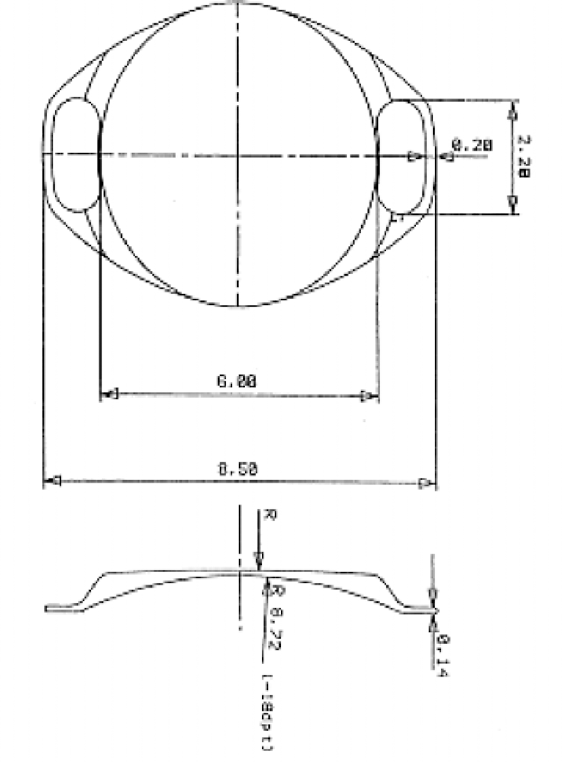 | Figure 1.Diagram of the 6-mm Artisan lens (Ophtec BV, Groningen, Netherlands) with relevant dimensions. The haptics lie in the pupillary planes, whereas the optic vaults anterior to the pupil to prevent iris chafing. |
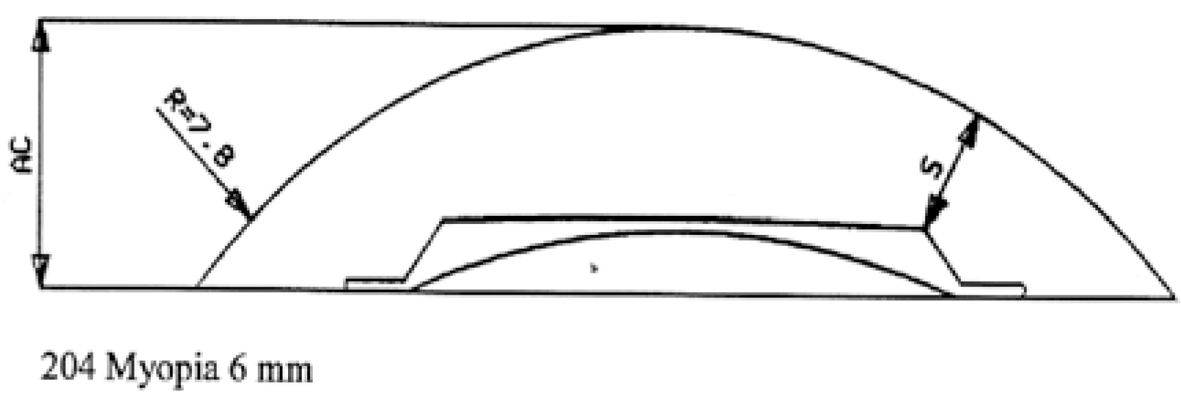 | Figure 2.Critical distance (S). At least, more than 1 mmis recommended for preventing iris rubbing or IOL movement. |
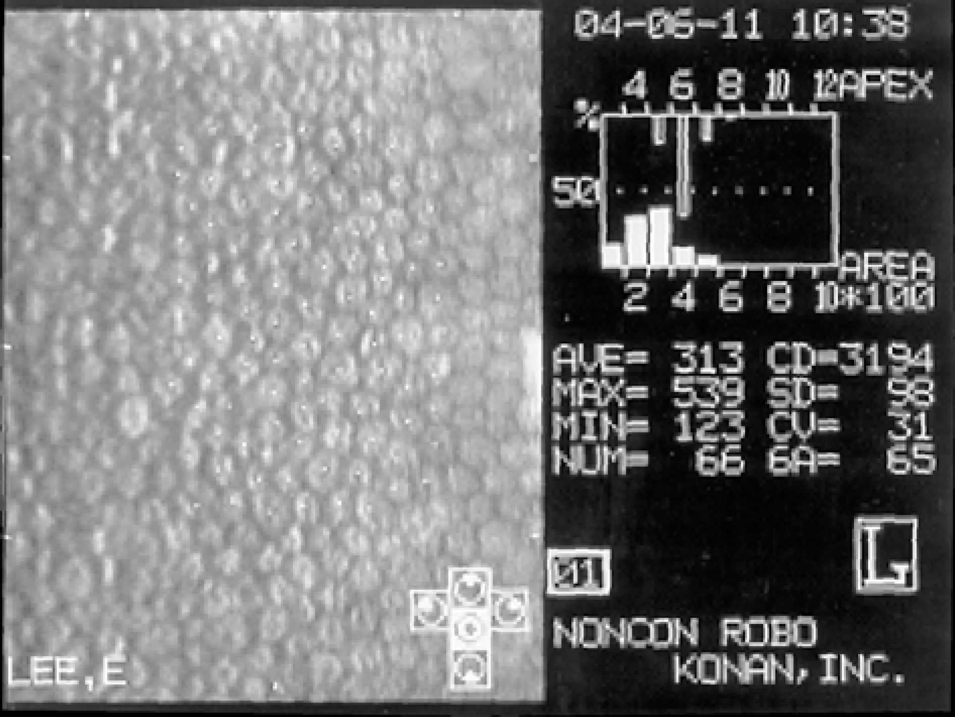 | Figure 3.Center method in calculating endothelial cell density. After 100 cell dotting, endothelial cell density is calculated automatically. |
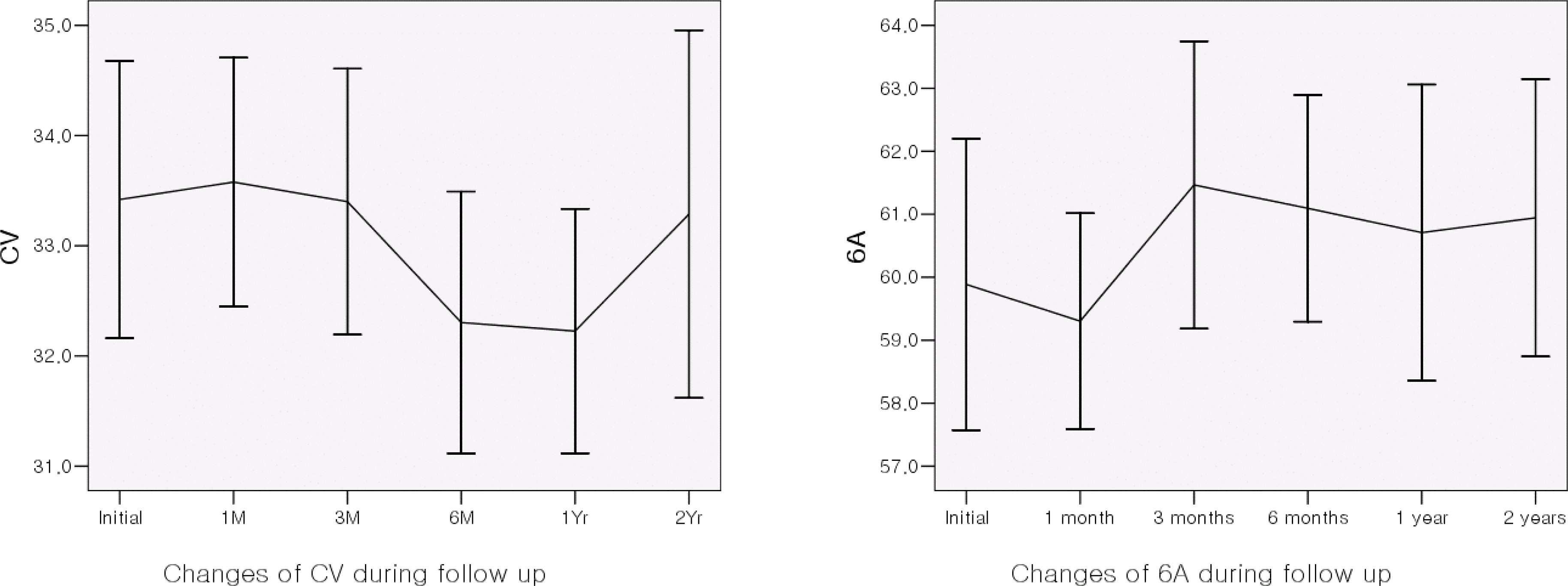 | Figure 5.Morphologic changes in coefficient variation (CV) and frequency (6A) of hexagons (%) at two-year follow-up. |
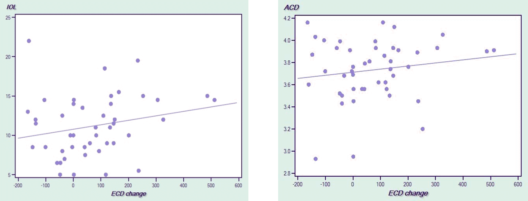 | Figure 6.Scatter plot showing the relationship between anterior chamber depth (ACD) and endothelial cell density chage (ECD change) after 24months and between diopter of intraocular lens (IOL) and endothelial cell density change (ECD change) after 24 months. |
Table 1.
Patient characteristics
Table 2.
Endothelial cell density (ECD) changes
| ECD | Changes from Baseline (%) P-value | ||
|---|---|---|---|
| Pre-operation | 2923±257 | ||
| 1st month | 2885±312 | −0.71 | 0.453 |
| 3rd month | 2888±260 | −0.88 | 0.320 |
| 6th month | 2908±275 | −0.70 | 0.239 |
| 12th month | 2921±296 | −0.64 | 0.474 |
| 24th month | 2854±254 | −2.34 | 0.004* |
Table 3.
Morphologic changes of endothelial cells; Coefficient variation (CV) in cell area
| CV | Changes from Baseline (%) | P-value | |
|---|---|---|---|
| 1st month | 0.342±0.034 | 0.11 | 0.945 |
| 3rd month | 0.334±0.032 | −0.33 | 0.880 |
| 6th month | 0.329±0.040 | −3.83 | 0.091 |
| 12th month | 0.326±0.031 | −4.94 | 0.005* |
| 24th month | 0.338±0.043 | −1.58 | 0.453 |
 XML Download