Abstract
Purpose
To evaluate the efficacy of conjunctivo-limbal autograft after wide excision of primary and recurrent pterygia.
Methods
Twenty-one eyes of 18 patients with primary pterygium and 18 eyes of 18 patients with recurrent pteygium underwent conjunctivo-limbal autograft after wide excision of pterygium. All patients underwent follow-up for more than six months. Recurrence rates and complications were evaluated.
Results
With a minimum of six months of follow-up, fibrovascular tissue in the excised area, not invading the cornea, was noted in one eye (5.6%) in the recurrent pterygium group but no further surgical interventions for the cosmetic problem were needed. One eye (4.8%) showed wound dehiscence, three eyes (14.3%) showed subgraft hemorrhage, and one eye (4.8%) showed subconjunctival fibrosis at the donor site in the primary pterygium group, while two eyes (11.1%) showed subgraft hemorrhage, and one eye (5.6%) showed Tenon's Capsule granuloma at the donor site in the recurrent pterygium group.
Go to : 
References
1. Dushku N, John MK, Schultz GS, Reid TW. Pterygia pathogenesis: corneal invasion by matrix metalloproteinase expressing altered limbal epithelial basal cells. Arch Ophthalmol. 2001; 119:695–706.
2. Sakoonwatanyoo P, Tan DT, Smith DR. Expression of p63 in pterygium and normal conjunctiva. Cornea. 2004; 23:67–70.

3. Solomon A, Grueterich M, Li DQ, et al. Overexpression of Insulin-like growth factor-binding protein-2 in pterygium body fibroblasts. Invest Ophthalmol Vis Sci. 2003; 44:573–80.

4. Di Girolamo N, McCluskey P, Lloyd A, et al. Expression of MMPs and TIMPs in human pterygia and cultured pterygium epithelial cells. Invest Ophthalmol Vis Sci. 2000; 41:671–9.
5. Li DQ, Lee SB, Gunja-Smith Z, Liu Y, et al. Overexpression of collagenase (MMP-1) and stromelysin (MMP-3) by pterygium head fibroblasts. Arch Ophthalmol. 2001; 119:71–80.
6. Dushku N, Hatcher SL, Albert DM, Reid TW. p53 expression and relation to human papillomavirus infection in pingueculae, pterygia, and limbal tumors. Arch Ophthalmol. 1999; 117:1593–9.

7. Threlfall TJ, English DR. Sun exposure and pterygium of the eye: a dose-response curve. Am J Ophthalmol. 1999; 128:280–7.

8. Tsai YY, Cheng YW, Lee H, et al. Oxidative DNA damage in pterygium. Mol Vis. 2005; 11:71–5.
9. Jurgenliemk-Schulz IM, Hartman LJ, Roesink JM, et al. Prevention of pterygium recurrence by postoperative single-dose beta-irradiation: a prospective randomized clinical double-blind trial. Int J Radiat Oncol Biol Phys. 2004; 59:1138–47.
10. Liddy BS, Morgan JF. Triethylene thiophosphoramide (thio-tepa) and pterygium. Am J Ophthalmol. 1966; 61:888–90.
11. Segev F, Jaeger-Roshu S, Gefen-Carmi N, Assia EI. Combined mitomycin C application and free flap conjunctival autograft in pterygium surgery. Cornea. 2003; 22:598–603.

12. Sharma A, Gupta A, Ram J, Gupta A. Low-dose intraoperative mitomycin-C versus conjunctival autograft in primary pterygium surgery: long term follow-up. Ophthalmic Surg Lasers. 2000; 31:301–7.

13. Kenyon KR, Wagoner MD, Hettinger ME. Conjunctival autograft transplantation for advanced and recurrent pterygium. Ophthalmology. 1985; 92:1461–70.

14. Dadeya S, Malik KP, Gullian BP. Pterygium surgery: conjunctival rotation autograft versus conjunctival autograft. Ophthalmic Surg Lasers. 2002; 33:269–74.

15. Ahn DG, Auh SJ, Choi YS. The clinical results of limbal conjunctival autograft transplantation with intraoperative mitomycin c application for recurrent pterygia. J Korean Ophthalmol Soc. 1999; 40:2443–9.
16. Tan DT, Chee SP, Dear KB, et al. Effect of pterygium morphology on pterygium recurrence in a controlled trial comparing conjunctival autografting with bare sclera excision. Arch Ophthalmol. 1997; 115:1235–40.

17. Guler M, Sobaci G, Ilker S, et al. Limbal-conjunctival autograft transplantation in cases with recurrent pterygium. Acta Ophthalmol (Copenh). 1994; 72:721–6.
18. Al Fayez MF. Limbal versus conjunctival autograft transplantation for advanced and recurrent pterygium. Ophthalmology. 2002; 109:1752–5.

19. Gris O, Guell JL, del Campo Z. Limbal-conjunctival autograft transplantation for the treatment of recurrent pterygium. Ophthalmology. 2000; 107:270–3.

20. Allan BD, Short P, Crawford GJ, et al. Pterygium excision with conjunctival autografting: an effective and safe technique. Br J Ophthalmol. 1993; 77:698–701.

21. Oh TH, Choi KY, Yoon BJ. The effect of conjunctival autograft for recurrent pterygium. J Korean Ophthalmol Soc. 1994; 35:1335–9.
22. Shimazaki J, Yang HY, Tsubota K. Limbal autograft transplantation for recurrent and advanced pterygia. Ophthalmic Surg Lasers. 1996; 27:917–23.

23. Ti SE, Tseng SC. Management of primary and recurrent pterygium using amniotic membrane transplantation. Curr Opin Ophthalmol. 2002; 13:204–12.

24. Solomon A, Pires RT, Tseng SC. Amniotic membrane transplantation after extensive removal of primary and recurrent pterygia. Ophthalmology. 2001; 108:449–60.

25. Ma DH, See LC, Hwang YS, Wang SF. Comparison of amniotic membrane graft alone or combined with intraoperative mitomycin C to prevent recurrence after excision of recurrent pterygia. Cornea. 2005; 24:141–50.

26. Prabhasawat P, Barton K, Burkett G, Tseng SC. Comparison of conjunctival autografts, amniotic membrane grafts, and primary closure for pterygium excision. Ophthalmology. 1997; 104:974–85.

27. Koh YM, Kim JY, Ji NC. A comparative study of recurrence rate in bilateral pterygium surgery: conjunctival autograft transplantation versus bare scleral. J Korean Ophthalmol Soc. 2001; 42:1543–8.
28. Cho JW, Chung SH, Seo KY, Kim EK. Conjunctival mini-flap technique and conjunctival autotransplantation in rterygium surgery. J Korean Ophthalmol Soc. 2005; 46:1471–7.
29. Mutlu FM, Sobaci G, Tatar T, Yildirim E. A comparative study of recurrent pterygium surgery: limbal conjunctival autograft transplantation versus mitomycin C with conjunctival flap. Ophthalmology. 1999; 106:817–21.

30. Ti SE, Chee SP, Dear KB, Tan DT. Analysis of variation in success rates in conjunctival autografting for primary and recurrent pterygium. Br J Ophthalmol. 2000; 84:385–9.

31. Oh TH, Choi KY, Yoon BJ. The effect of conjunctival autograft for recurrent pterygium. J Korean Ophthalmol Soc. 1994; 35:1335–9.
32. Kim YS, Kim JH, Byun YJ. Limbal-conjunctival autograft transplantation for the treatment of primary pterygium. J Korean Ophthalmol Soc. 1999; 40:1804–10.
Go to : 
 | Figure 1.Classification of pterygium. (A) Grade T1 (atrophic) episcleral vessels are unobscured. (B) Grade T2 (intermediate) episcleral vessels are partially obscured. (C) Grade T3 (fleshy) episcleral vessels are totally obscured. |
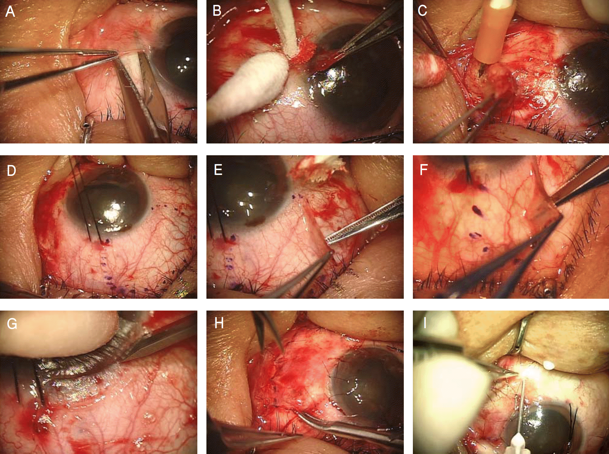
 | Figure 2.Photographs illustrating the surgical technique of conjunctivo-limbal autograft. (A) excision and blunt dissection of pterygium from the cornea and sclera by using conjunctival scissors. (B) remnant pterygium of the cornea was dissected off by corneal forceps. (C) wide excision of subconjunctival Tenon's tissue was done by the Ellman cautery. (D) the donor site was marked with a gentian violet. (E-F) conjunctival graft is dissected with conjunctival scissors leaving the underlying Tenon's capsule intact. (G) the limbus is dissected with No. 64 Beaver blade. (H) the conjunctivo-limbal graft is transferred and secured with multiple interrupted sutures. (I) triamcinolon was injected |
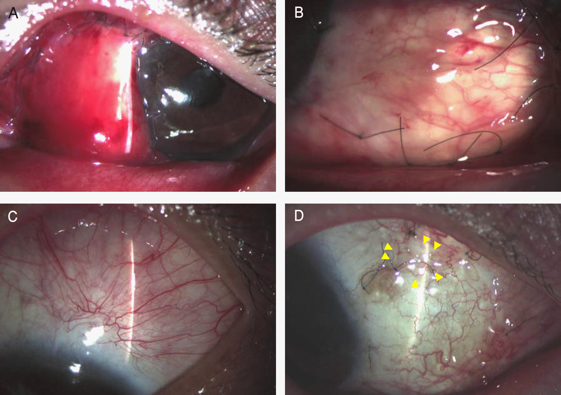
 | Figure 3.Grading of recurrence after pterygium surgery. (A) Grade 0, normal appearance of the operated site. (B) Grade 1, fine episcleral vessels in the excised area (C) Grade 2, fibrovascular tissue in the excised area, reaching to the limbus, but not invading the cornea (conjunctival recurrence). (D) Grade 3, fibrovascular tissue invading the cornea (corneal recurrence) |
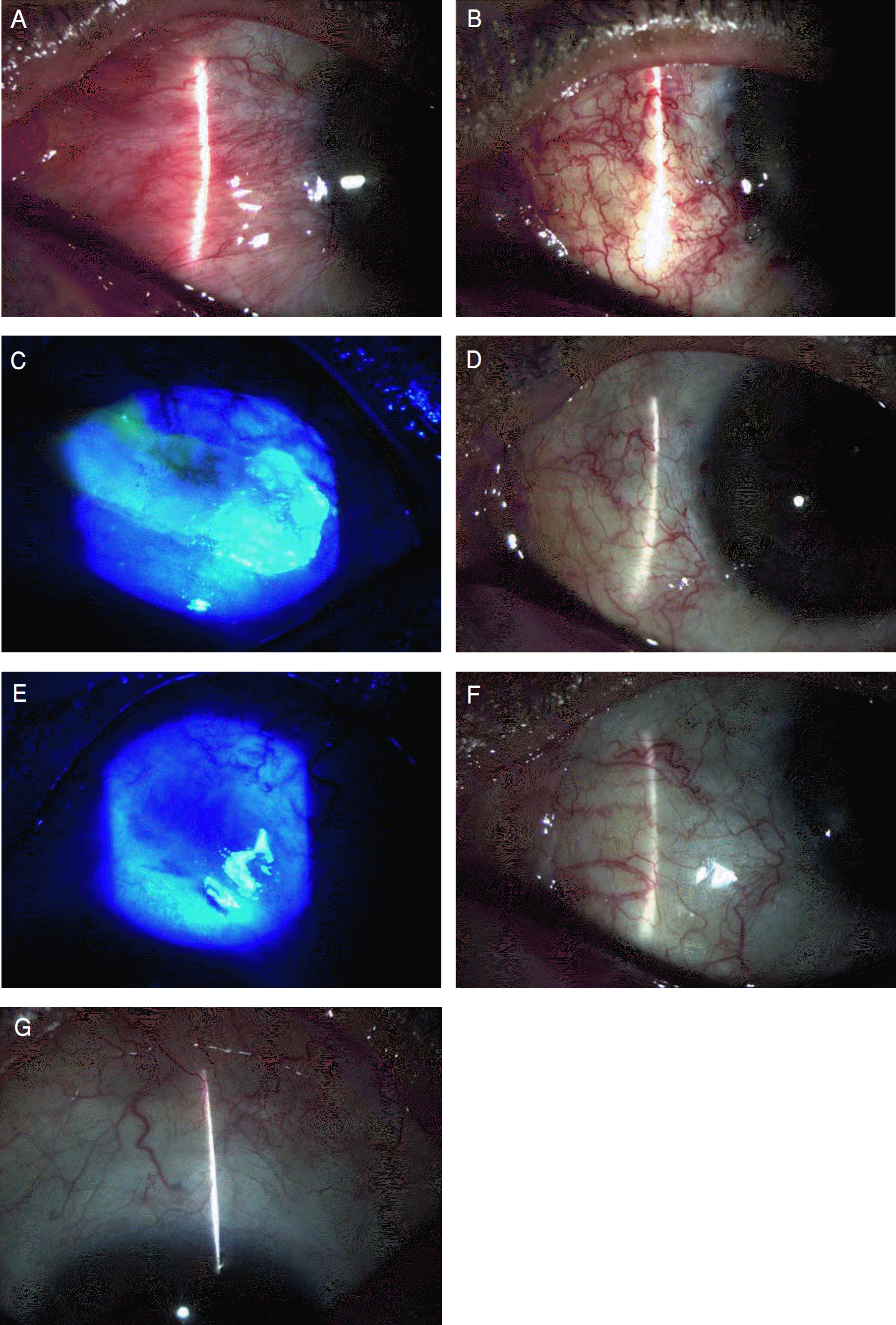
 | Figure 4.(A) The left eye before surgery has a large and fleshy recurrent pterygium. (B-C) At 2 days after surgery. The well anchored conjunctivo-limbal autograft was seen B. Note epithelial defect at donor site under fluorescein staining C. (D-E) At 3 weeks after surgery. The surgical wound is healed D and complete epithelization at donor site under fluorescein staining is seen E. (F-G) At 6 months after surgery. Normal appearance (Grade 0) is seen F and there is no scarring or excessive vascularization at donor site G. |


 PDF
PDF ePub
ePub Citation
 Print
Print


 XML Download