

 PDF
PDF ePub
ePub Citation
 Print
Print


Abstract
Purpose
We evaluated the effect of amniotic membrane transplantation with fibrin glue after redundant conjunctival resection.
Methods
After enchelon-shaped resection of inferior redundant conjunctiva, an amniotic membrane was transplanted using fibrin glue in symptomatic conjunctivochalasis. Conjunctiva was fixed to the episclera with 10-0 nylon.
Results
The subjects were 18 eyes of 9 patients (two males=4 eyes, seven females=14 eyes) with an average age of 67.9±7.7 years (range: 54-79 years). Mean operation time was 35.6±5.6 minutes/eye (range: 30-45 minutes/eye) and mean numbers of sutures were 8±0.76 (range: 7-9). The mean follow-up period was 10.4±3 months (range: 6-14 months) and the mean time to full epithelization was 14.5±3.4 days (range: 11-20 days). At postoperative days 1-2, the lower tear meniscus was reconstructed. There were no recurrences or any postoperative complications observed.
Conclusions
Amniotic membrane transplantation after redundant conjunctival resection in conjunctivochalasis resulted in reconstruction of the lower tear meniscus with stable conjunctival surface and marked improvement of subjective symptoms. Operation time and early postoperative irritation symptoms could be reduced with the use of fibrin glue.
Go to : 
References
1. Pinkerton OD. Bulbar conjunctivo-chalasis. Arch Ophthalmol. 1972; 88:532.

2. Liu D. Conjunctivochalasis. A cause of tearing and its management. Ophthal Plast Reconst Surg. 1986; 2:25–8.
3. Byon DS, Song CH, Shim JK, et al. Conjunctivochalasis as a cause of epiphora and its histopathological findings. J Korean Ophthalmol Soc. 1996; 37:400–4.
4. Bosniak SL, Smith BC. Conjunctivochalasis. Adv Ophthalmic Plast Reconstr Surg. 1984; 3:153–5.
5. Hughes WL. Conjunctivochalasis. Am J Ophthalmol. 1942; 25:48–51.
6. Meller D, Tseng SC. Conjunctivochalasis: literature review and possible pathophysiology. Surv Ophthalmol. 1998; 43:225–32.
7. Yokoi N, Komuro A, Nishii M, et al. Clinical impact of conjunctivochalasis on the ocular surface. Cornea. 2005; 24:S24–31.
8. Tseng SC, Prabhasawat P, Lee SH. Amniotic membrane transplantation for conjunctival surface reconstruction. Am J Ophthalmol. 1997; 124:765–74.
9. Meller D, Tseng SC. Reconstruction of amniotic membrane for conjunctival and corneal surface reconstruction. Ophthalmologe. 1998; 95:805–13.
10. Meller D, Maskin SL, Pires RT, Tseng SC. Amniotic membrane transplantation for symptomatic conjunctivochalasis refractory to medical treatments. Cornea. 2000; 19:796–803.
11. Kruse FE, Meller D. Amniotic membrane transplantation for reconstruction of the ocular surface. Ophthalmologe. 2001; 98:801–10.
12. Cohen RA, McDonald MB. Fixation of conjunctival autografts with an organic tissue adhesive. Arch Ophthalmol. 1993; 111:1167–8.
13. Hick S, Demers PE, Brunette I, et al. Amniotic membrane transplantation and fibrin glue in the management of corneal ulcers and perforations: A review of 33 cases. Cornea. 2005; 24:369–77.
14. Koranyi G, Seregard S, Kopp ED. Cut and paste: a no suture, small incision approach to pterygium surgery. Br J Ophthalmol. 2004; 88:911–4.
15. Uy HS, Reyes JM, Flores JD, Lim-Bon-Siong R. Comparison of fibrin glue and sutures for attaching conjunctival autografts after pterygium excision. Ophthalmology. 2005; 112:667–71.
16. Sharma A, Kaur R, Kumar S, et al. Fibrin glue versus N-butyl-2-cyanoacrylate in corneal perforations. Ophthalmology. 2003; 110:291–8.
17. Koranyi G, Seregard S, Kopp ED. The cut-and-paste method for primary pterygium surgery: long-term follow-up. Acta Ophthalmol Scand. 2005; 83:293–301.
18. Pfister RR, Sommers CI. Fibrin sealant in corneal stem cell transplantation. Cornea. 2005; 24:593–8.
19. Hoh H, Schirra F, Kienecker C, Ruprecht KW. Lidparrallele konjunktivale Falten(LIPCOF) sind ein sicheres diagnostisches Zeichen des trockenen Auges. Ophthalmologe. 1995; 92:802–8.
20. Watanabe A, Yokoi N, Kinoshita S, et al. Clinicopathologic study of conjunctivochalasis. Cornea. 2004; 23:294–8.
21. Li DQ, Meller D, Liu Y, Tseng SC. Overexpression of MMP-1 and MMP-3 by cultured conjunctivochalasis fibroblasts. Invest Ophthalmol Vis Sci. 2000; 41:404–10.
22. Meller D, Li DQ, Tseng SC. Regulation of collagenase, stromelysin, and gelatinase B in human conjunctival and conjunctivochalasis fibroblasts by interleukin-1β and tumor necrosis factor-α. Invest Ophthalmol Vs Sci. 2000; 41:2922–9.
23. Ko SM, Kim MK, Kim JC. The role of mast cell in hyperlaxity of conjunctiva. J Korean Ophthalmol Soc. 1997; 38:949–55.
24. Otaka I, Kyu N. A new surgical technique for management of conjunctivochalasis. Am J Ophthalmol. 2000; 129:385–7.
25. Oh SJ, Byon DS. Treatment of conjunctivochalasis using bipolar cautery. J Korean Ophthalmol Soc. 1999; 40:707–11.
26. Kim HH, Shin DS, Lee KW. Effects of cauterization with suturing in treatment of conjunctivochalasis; 4 Cases. J Korean Ophthalmol Soc. 2006; 47:843–6.
Go to :
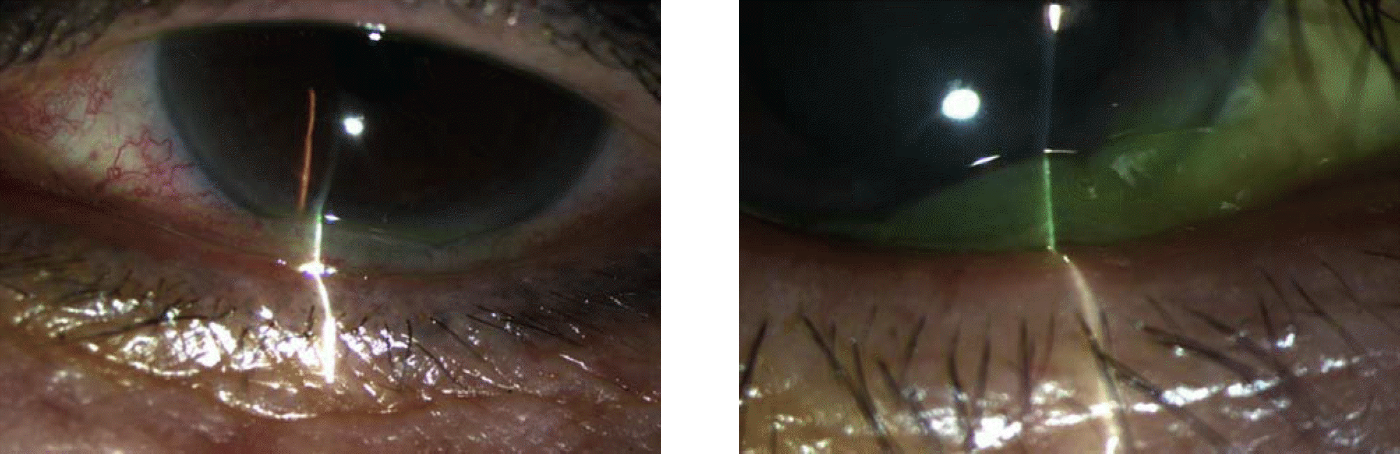
 | Figure 2.(A) Conjunctivochalasis at ordinary condition. (B) Worsening of conjunctivochalasis after vigorous blinking. |
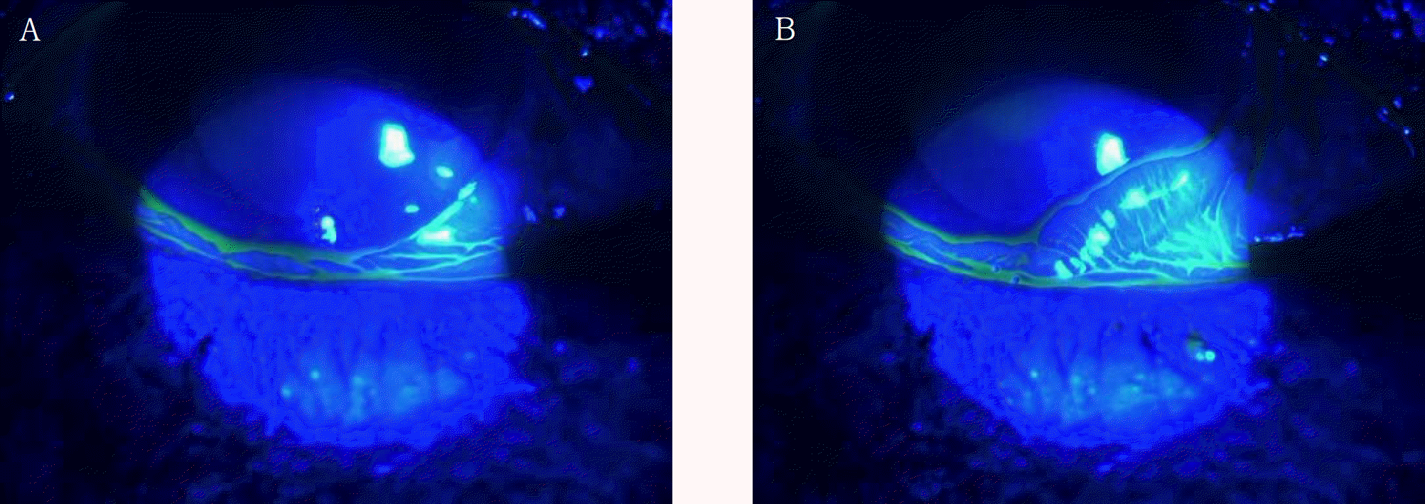
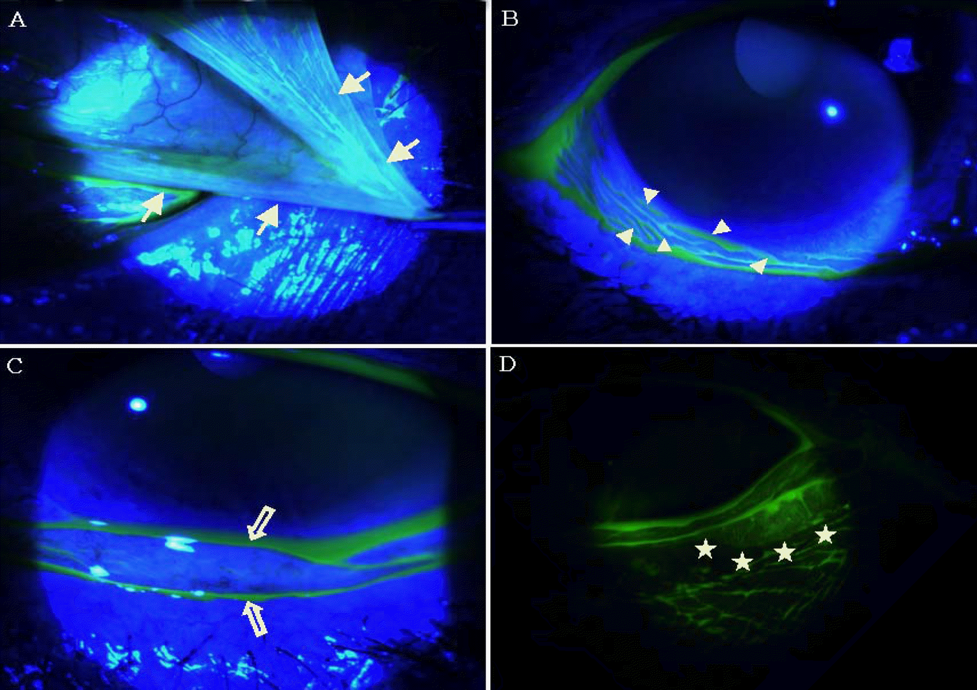 | Figure 3.Objective signs of conjunctivochalasis. (A) Redundancy of conjunctiva. (B) Ectopic tear meniscus. (C) Interruption of lower tear meniscus. (D) Anterior migration of the mucocutaneous junction. |
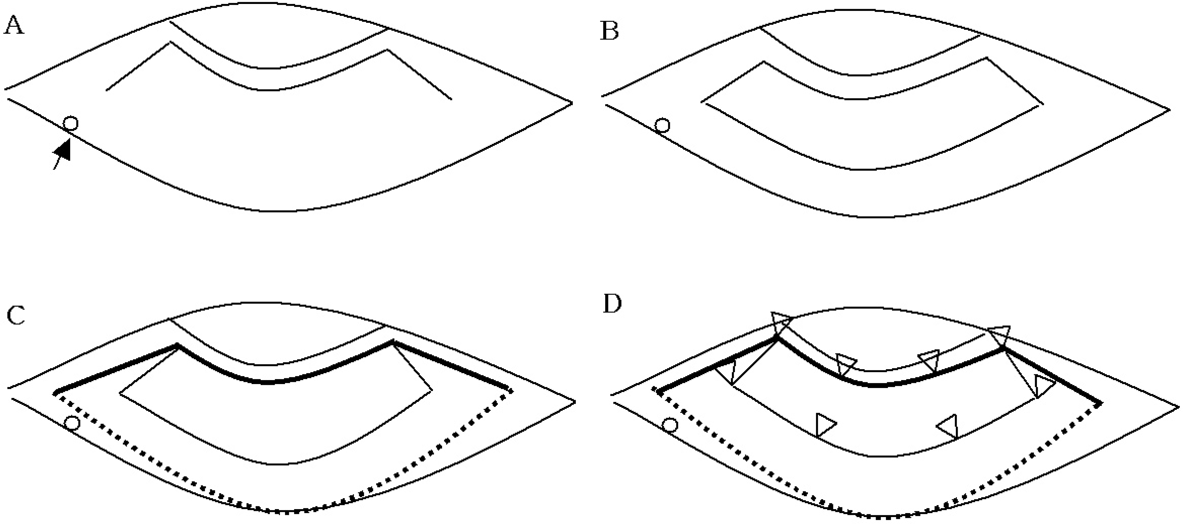 | Figure 4.Schematic presentation of conjunctivochalasis surgery. (A) The conjunctival incision 1mm from the limbus (black arrow: lower punctum). (B) Enchelon-shaped resection of redundant conjunctiva. (C) Covering of amniotic membrane leaving 2 mm excess margin in length and width. (D) Conjunctiva fixed to the episclera with interrupted 10-0 nylon sutures. |
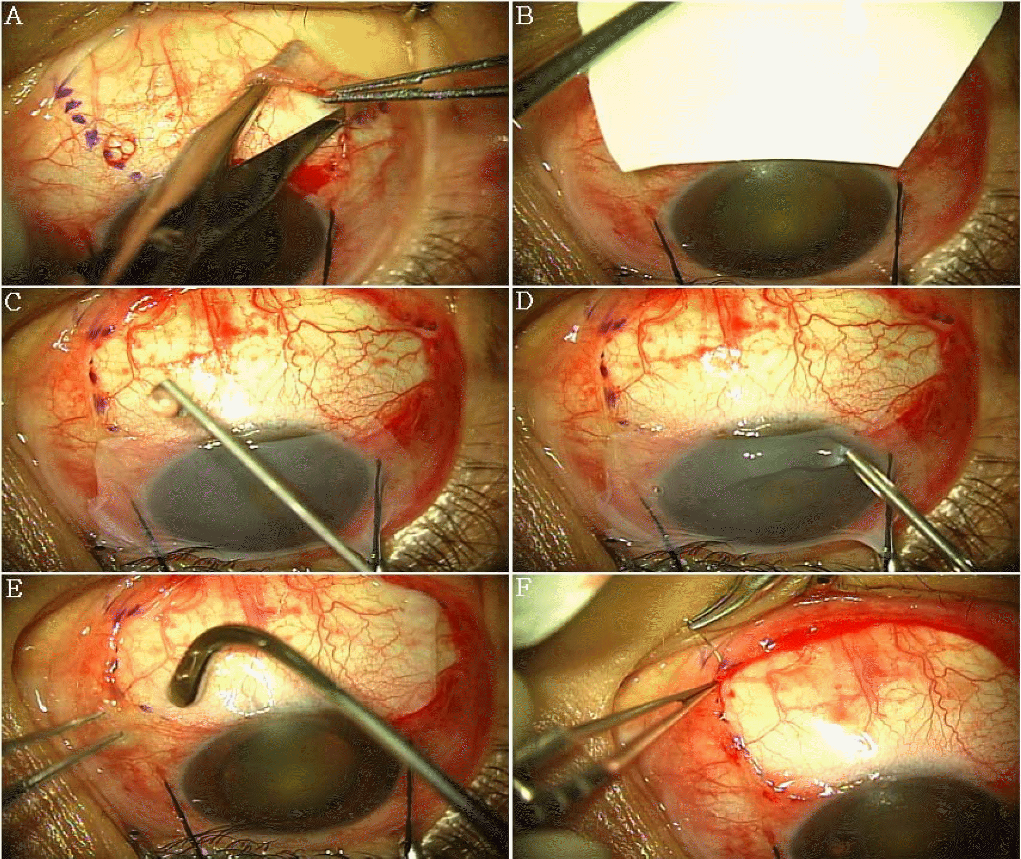 | Figure 5.(A) An enchelon-shaped resection was performed to remove inferior redundant conjunctiva. (B) The amniotic membrane was peeled off the carrier paper from the storage medium. (C) Thrombin solution was placed on the exposed sclera. (D) A drop of fibrinogen solution was applied on stromal side of amniotic membrane. (E) The graft was immediately transferred onto the exposed sclera and rapidly smoothered with a muscle hook. (F) The surrounding conjunctival edge was then secured to the episclera by interrupted sutures with 10-0 nylon. |
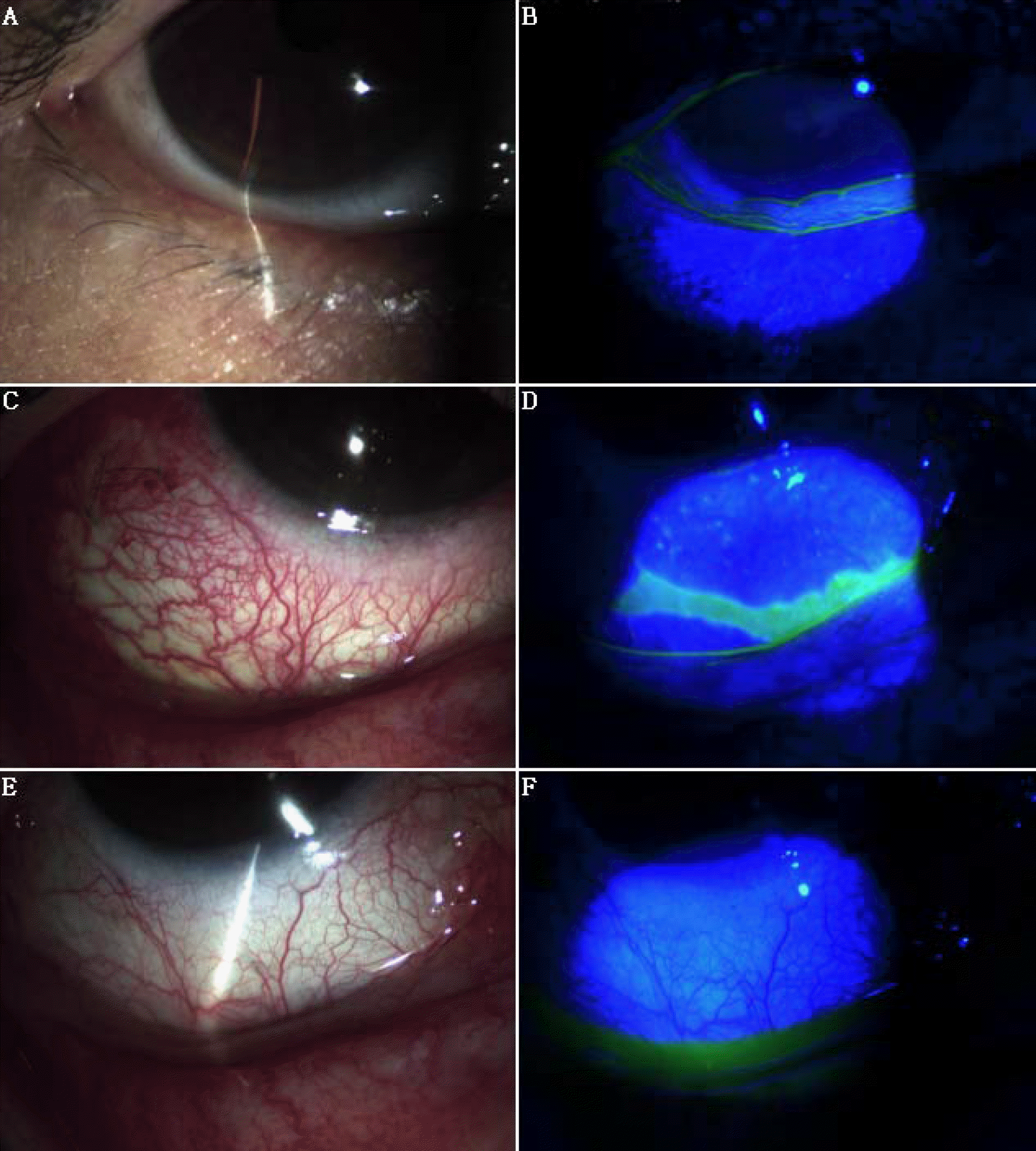 | Figure 6.(A, B) Preoperative appearance showed the redundant conjunctival tissue and interruption of lower tear meniscus. (C, D) One week after operation, the size of epithelial defect decreased. (E, F) One month after operation, smooth, quiet, non-inflamed conjunctival surface and complete epithelization is found. |
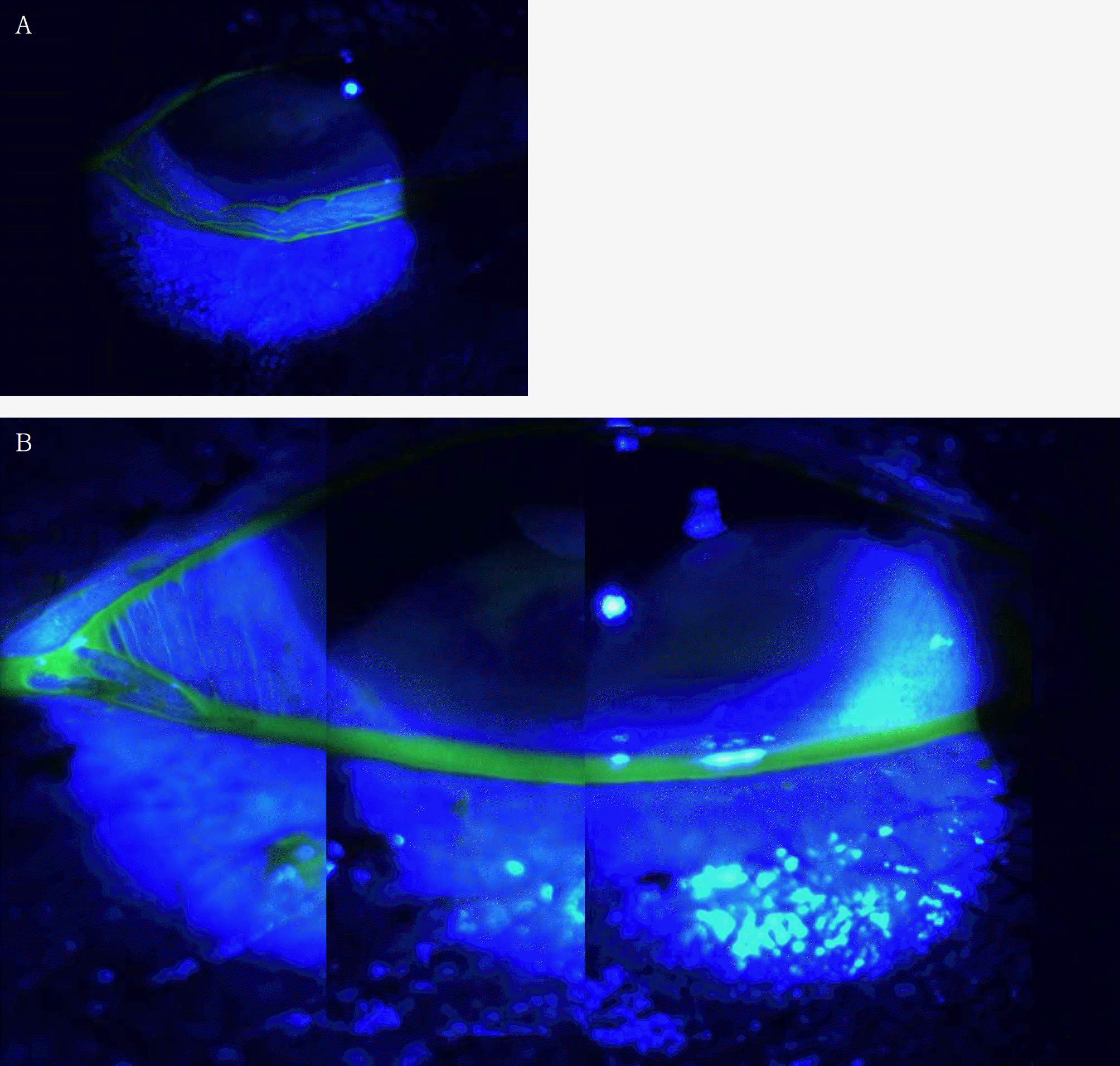 | Fig. 7.(A) Preoperative photograph shows interruption of the lower tear meniscus. (B) Three months after operation, panoramic picture of representative example of a patient with conjunctivochalasis. There is no evidence of redundant conjunctiva between the cornea and lower lid, and a completely reconstructed lower tear meniscus is found. |
Table 1.
Grading of conjunctivochalasis (LIOCF)19
Table 2.
Symptoms before and after surgery
| Before | Resolved | ‡ RI | Improved | Unchanged | Aggravated | |
|---|---|---|---|---|---|---|
| * FBS(eyes) | 18 | 6 | 8 | 4 | 0 | 0 |
| † MD(eyes) | 18 | 4 | 6 | 8 | 0 | 0 |
Table 3.
Schirmer I test and tear break-up time before and after surgery
| Before | 1 month | 3 months | 6 months | |
|---|---|---|---|---|
| Schirmer I test (mm/5 min) | 14.44±3.50 | 15.78±4.41 (P=0.488) | 15.89±4.31 (P=0.447) | 17.33±4.69 (P=0.158) |
| TBUT* (seconds) | 5.22±2.28 | 5.89±1.97 (P=0.516) | 6.33±1.41 (P=0.232) | 5.89±1.62 (P=0.484) |
 XML Download