

 PDF
PDF ePub
ePub Citation
 Print
Print


Abstract
Purpose
To describe the clinical course of young patients with central retinal vein occlusion (CRVO).
Methods
We reviewed the records of patients 50 years or younger who presented with CRVO and who were followed up for at least 6 months.
Results
The average age of the patients was 37.7 years, and the mean follow-up time was 26.3 months. Participants included 27 patients with CRVO younger than 50 years among the total 393 patients with CRVO, and the sex distribution was nearly equal; 13 patients were male and 14 patients were female. Associated systemic diseases were hypertension (8 patients), diabetes (3 patients), stroke (3 patients), and myocardial infarction (1 patient). Of the 17 patients who had no systemic disease, 8 patients had hypercholesterolemia or another hematologic abnormalities. Of the 18 eyes with non-ischemic CRVO, 10 eyes (55.6%) showed improved final visual acuities (2 or more lines). Of the 9 eyes with ischemic CRVO, only 1 eye (11.1%) showed improved visual acuity.
Conclusions
Hematological examination and treatment with systemic evaluation were recommended to younger patients with CRVO. The classification of retinal ischemia of young patients as well as that of older patients could be useful for the follow-up of patients and the assessment of its progression in the future.
Go to : 
References
1. The central vein occlusion study group Baseline and early natural history report. Arch Ophthalmol. 1993; 111:1087–95.
2. The eye disease case-control study group Risk factors for central retinal vein occlusion. Arch Ophthalmol. 1996; 114:545–54.
3. Gutman FA. Evaluation of a patient with central retinal vein occlusion. Ophthalmology. 1983; 90:481–3.

4. Fong AC, Schatz H. Central retinal vein occlusion in young adults. Surv Ophthalmol. 1993; 37:393–417.
5. Fong AC, Schatz H, McDonald HR. . Central retinal vein occlusion in young adult (papillophlebitis). Retina. 1992; 12:3–11.
6. Klien BA, Olwin JH. A survey of the pathogenesis of retinal venous occlusion. Emphasis upon choice of therapy and an analysis of the therapeutic results in fifty-three patients. AMA Arch Ophthalmol. 1956; 56:207–47.
7. Klien BA. Sidelights on retinal venous occlusion. Am J Ophthalmol. 1966; 61:25–36.
8. Green WR, Chan CC, Hutchins GM, Terry JM. Central retinal vein occlusion: a prospective histopathologic study of 29 eyes in 28 cases. Retina. 1981; 1:27–55.
9. Lahey JM, Tunc M, Kearney J. . Laboratory evaluation of hypercoagulable state in patients with central retinal vein occlusion who are less than 56 years of age. Ophthalmology. 2002; 109:126–31.
10. Hayreh SS, Zimmerman MB, Podhajsky P. Hematologic abnormalities associated with various type of retinal vein occlusion. Graefes Arch Clin Exp Ophthalmol. 2002; 240:180–96.
11. Adamczuk YP, Varela LI, Martinuzzo ME. . Central retinal vein occlusion and thrombophilia risk factors. Blood Coagul Fibrinolysis. 2002; 13:623–6.
12. Bandello F, Viganò D'Angelo S, Parlavecchia M. . Hypercoagulability and high lipoprotein (a) levels in patients with central retinal vein occlusion. Thromb Haemost. 1994. 72:p. 39–43.
13. Ministry of Welfare Report on 2005 national health and nutrition survey. Seoul: Ministry of Health and Welfare;2007; 103–30.
14. Walters RF, Spalton DJ. Central retinal vein occlusion in people aged 40 years or less: a review of 17 patients. Br J Ophthalmol. 1990; 74:30–5.
15. Magargal LE, Gonder JR, Maher V. Central retinal vein obstruction in the young adult. Trans Pa Acad Ophthalmol Otolaryngol. 1985; 37:148–53.
16. Priluck IA, Robertson DM, Hollenhorst RW. Long-term follow-up of occlusion of the central retinal vein in young adults. Am J Ophthalmol. 1980; 90:190–202.
17. The Central Vein Occlusion Study Group Natural history and clinical management of central retinal vein occlusion. Arch Ophthalmol. 1997; 115:486–91.
18. Chen V, Moisseiev J, Treister G. Severe ischemic process in a young man with CRVO. Metabol Pediatr Sys Ophthalmol. 1988; 11:67–9.
19. Magargal LE, Donoso LA, Sanborn GE. Retinal ischemia and risk of neovascularization following central retinal vein destruction. Ophthamology. 1982; 89:1241–5.
Go to :


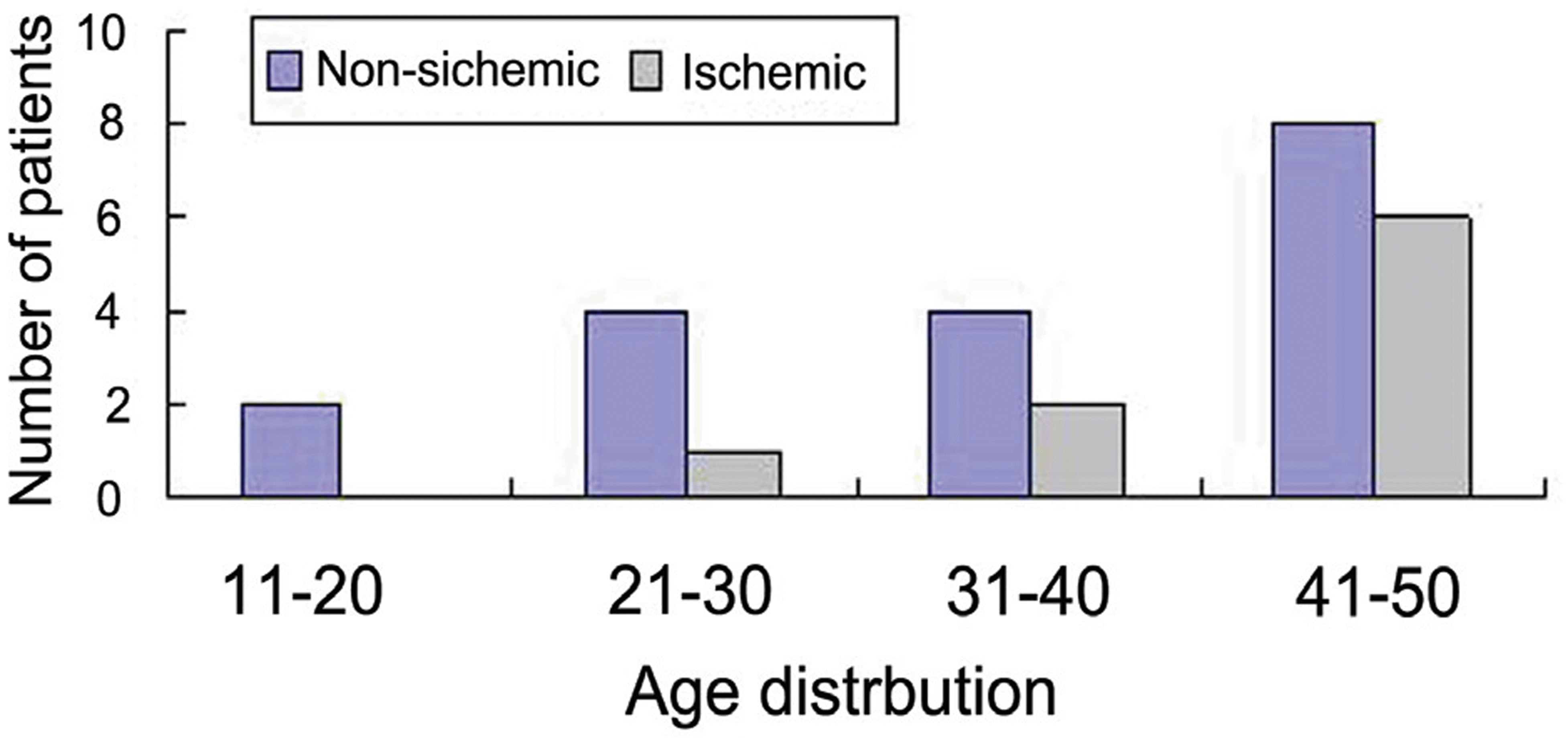 | Figure 3. Age distribution of patients with central retinal vein occlusion less than 50 years of age. |
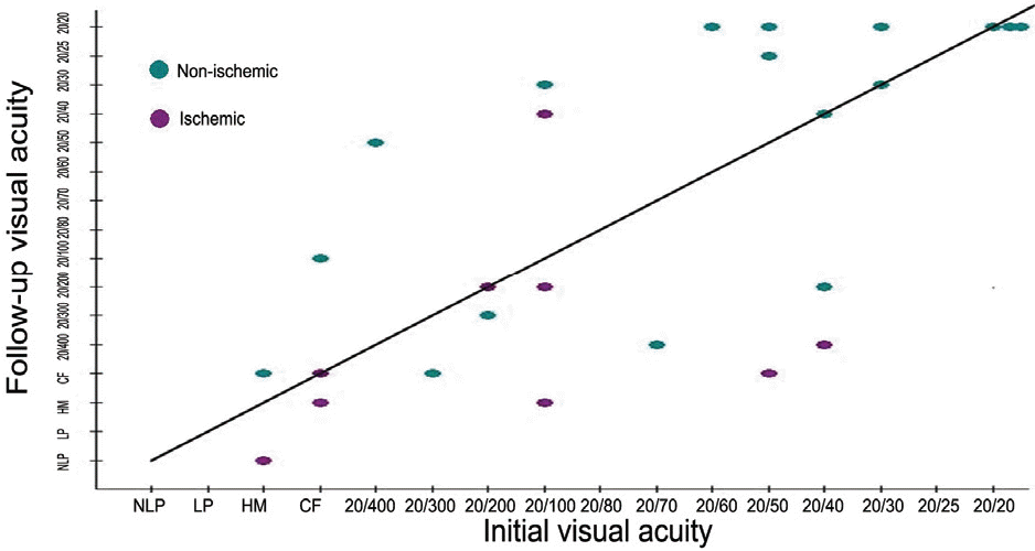
Table 1.
Systemic risk factors of young adults with central retinal vein occlusion
| CRVO∗ (%) | Total (%) | |||
|---|---|---|---|---|
| Ischemic | Non-ischemic | |||
| Risk factors (+) | CV† disease (Stroke & MI‡) | 2 (22.2) | 2 (11.1) | 4 (14.8) |
| HTN | 2 (22.2) | 6 (33.3) | 8 (29.6) | |
| DM § | 3 (33.3) | 0 | 3 (11.1) | |
| IIOP§ | 1 (11.1) | 0 | 1 (3.70) | |
| Risk factors (-) | 5 (55.5) | 12 (66.6) | 17 (62.9) | |
Table 2.
Laboratory studies of young adults with central retinal vein occlusion
| Risk factor∗ (+) | Risk factor (-) | Total | |
|---|---|---|---|
| Hematocrit | 2 (7.40%) | 0 | 2 (7.40%) |
| Hypercholesterolemia | 3 (11.1%) | 6 (22.2%) | 9 (33.3%) |
| ESR | 0 | 1 (3.70%) | 1 (3.70%) |
| AST/ALT | 0 | 1 (3.70%) | 1 (3.70%) |
| Total | 5 (18.5%) | 8 (29.6%) | 13 (48.1%) |
 XML Download