

 PDF
PDF ePub
ePub Citation
 Print
Print


Abstract
Purpose
To retrospectively evaluate the clinical results of insertion of a hydrophilic acrylic plate posterior chamber intraocular lens (ThinOptX) after bimanual microincision phacoemulsification.
Methods
Thirty-four eyes of 30 patients who underwent bimanual phacoemulsification and ThinOptX implantation through a 2.0 mm incision between July 2004 and May 2006 were followed-up for more than 12 months. We examined best corrected visual acuity (BCVA), refractive errors, corneal endothelial cell density, halo and contrast sensitivity, posterior capsule opacification (PCO), and intraoperative and postoperative complications.
Results
The preoperative mean logMAR BCVA was 0.43±0.24, and the postoperative BCVA was 0.04±0.09 after 6 months and 0.14±0.12 after 12 months. The preoperative corneal endothelial cell density was 2562±347.90 cells/mm2, and decreased to 2241±294.88 (cells/mm2) at 12 months postoperative. Postoperative contrast sensitivity at 6 months was increased in both photopic and mesopic condition. Halo was noted in all examined eyes. A PCO of 29% was evaluated at 6 months postoperative and a PCO of 52% was evaluated at 12 months postoperative.
Conclusions
Bimanual phacoemulsification and ThinOptX implantation through a 2.0 mm incision resulted in good initial visual outcome and correction of refractive errors. However, problems such as halo and PCO associated with IOL design were noted. Therefore, further evaluation and correction of the IOL are needed.
References
1. Prakash P, Kasaby HE, Aggrarwal RK, Humfrey S. Microincision bimanual phacoemulsification and ThinOptX® implantation through a 1.70mm incision. Eye. 2007; 21:177–82.
2. Paul T, Braga-Mele R. Bimanual microincisional phacoemulsifi cation: the future of cataract surgery? Curr Opin Ophthalmol. 2005; 16:2–7.
3. Shearing SP, Relyea RL, Louiza A. . Routine phacoemulsification through a one-millimetre non-sutured incision. Cataract. 1985; 2:6–11.
4. Kim HJ, Kim JH, Lee DH. Endothelial cell damage in microincision cataract surgery and coaxial phacoemulsification. J Korean Ophthalmol Soc. 2007; 48:19–26.
5. Dogru M, Honda R, Omoto M. . Early visual results with the rollable ThinOptx intraocular lens. J Cataract Refract Surg. 2004; 30:558–5.

6. Wejde G, Kugelberg M, Zetterstrom C. Posterior capsule opacification: comparison of 3 intraocular lenses of different materials and design. J Cataract Reftract Surg. 2003; 29:1556–9.
7. Kellman CD. Phaco-emulsification and aspiration; a new technique of cataract removal; a preliminary report. Am J Ophthalmol. 1967; 64:23–35.
8. Cavallini GM, Campi L, Masini C. . Bimanual microphacoemulsification vs coaxial miniphacoemulsification: Prospective study. J Cataract Refract Surg. 2007; 33:387–92.
9. Cinhüseyinoglu N, Celik L, Yaman A. . Microincisional cataract surgery and Thinoptx rollable intraocular lens implantation. Graefes Arch Clin Exp Ophthalmol. 2006; 244:802–7.
10. Jung HW, Kim IC. Posterior capsular opacification and Nd:YAG laser capsulotomy in 811B, SI40NB, MA60BM intraocular lens. J Korean Ophthalmol Soc. 2003; 44:1072–8.
11. Heatley CJ, Spalton DJ, Kumar AJ. . Comparison of posterior capsule opacification rates between hydrophilic and hydrophobic single-piece acrylic intraocular lenses. J Cataract Refract Surg. 2005; 31:718–24.
12. Mencucci R, Ponchietti C, Virgili G. . Corneal endothelial damage after cataract surgery: Microincision versus standard technique. J Cataract Refract Surg. 2006; 32:1351–4.
13. Crema AS, Walsh A, Yamane Y, Nosé W. Comparative study of coaxial phacoemulsification and microincision cataract surgery. One-year follow-up. J Cataract Refract Surg. 2007; 33:1014–8.
14. Alió J, Rodríguez-Prats JL, Galal A, Ramzy M. Outcomes of microincision cataract surgery versus coaxial phacoemulsification. Ophthalmology. 2005; 112:1997–2003.
15. Wilczynski M, Drobniewski I, Synder A, Omulecki W. Evaluation of early corneal endothelial cell loss in bimanual microincision cataract surgery (MICS) in comparison with standard phacoemulsification. Eur J Ophthalmol. 2006; 16:798–803.
16. Mencucci R, Ponchietti C, Virgili G. . Corneal endothelial damage after cataract surgery: microincision versus standard technique. J Cataract Refract Surg. 2006; 32:1351–4.
17. Bordeianu CD. Thinoptix implant the last border in modern cataract surgery Ofthalmologia. 2005; 49:58–66.
18. Paik JS, Kim MJ, Park SH, Joo CK. Contrast sensitivity and glare of different edge designed intraocular lenses. J Korean Ophthalmol Soc. 2007; 48:259–65.
Figure 2
. Distant visual acuity after ThinOptX implantation. There is statistically significant difference of BCVA between postoperative 6 months and 12 months (* p=0.02).
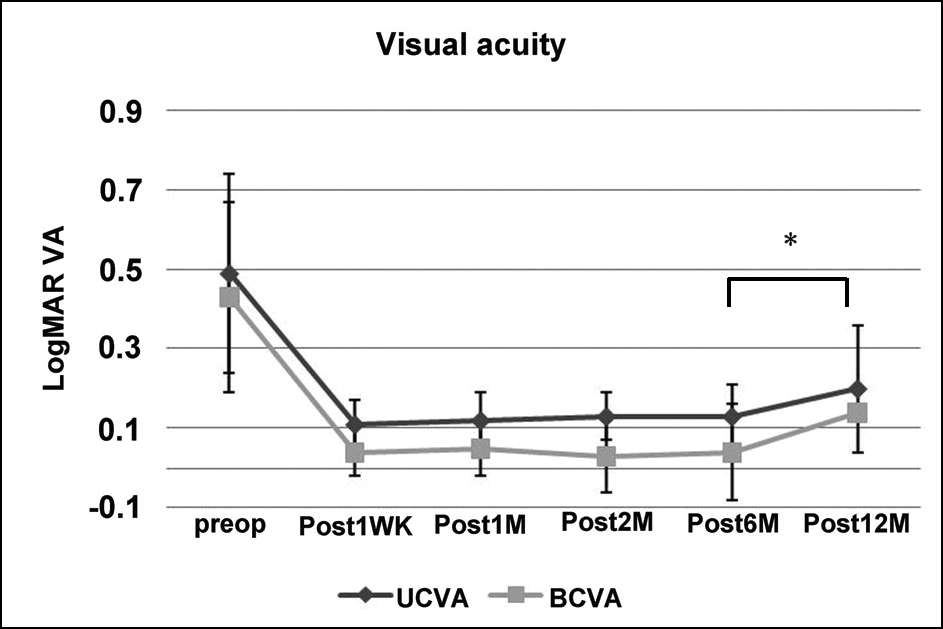
Figure 3
. Change of endothelial cell density in the patients who was inserted ThinOptX. There is statistically significant difference between preoperative endothelial cell density and postoperative endothelial density (* p<0.01).
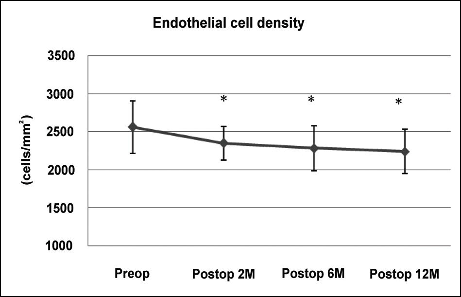
Figure 4
Contrast sensitivity in the patients on whom ThinOptX at photopic was inserted and mesopic conditions.
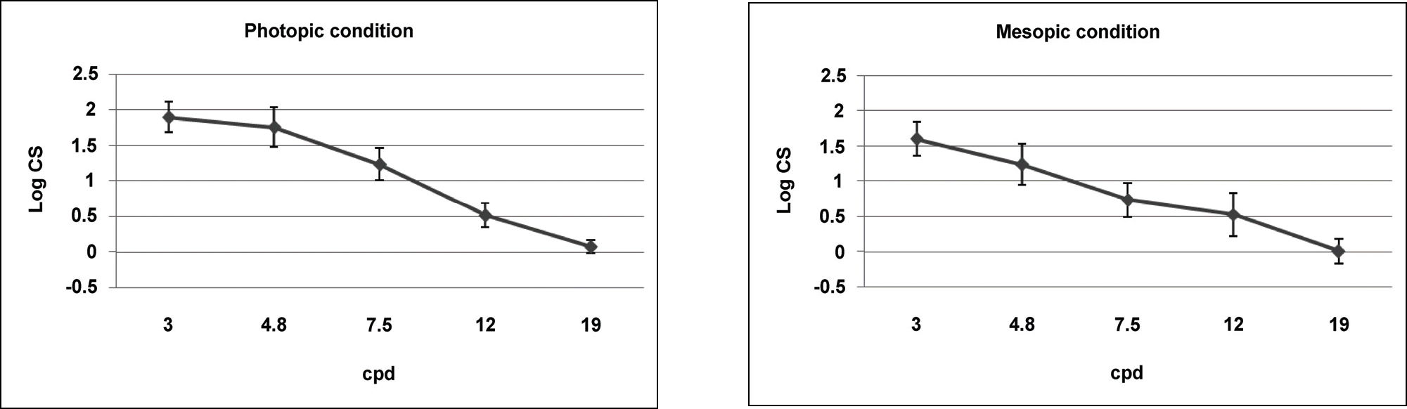
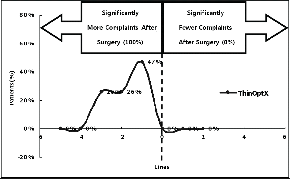

Figure 7.
Site of anterior capsulotomy seen at the inferior margin of continuous curvilinear capsulorrhexis.

Table 1.
Grading of posterior capsular opacification
Table 2.
Characteristics of inserted ThinOptX IOL patients
Table 3.
Test results of contrast sensitivity (Log CS) at photopic condition (100 cd/m2) and mesopic condition (30 cd/m2)
| Contrast sensitivity at various cycles per degree (cpd) | |||||
|---|---|---|---|---|---|
| 3 | 4.8 | 7.5 | 12 | 19 | |
| Photopic condition∗ | 1.90±0.21 | 1.76±0.28 | 1.24±0.23 | 0.52±0.17 | 0.08±0.09 |
| Mesopic condition† | 1.60±0.24 | 1.24±0.29 | 0.74±0.24 | 0.53±0.30 | 0.01±0.18 |
 XML Download