Abstract
Purpose
To evaluate the effects of cheap tinted contact lenses on corneal swelling and ocular surface inflammation, compared to hydrogel and silicone hydrogel contact lenses.
Methods
Forty eyes of 20 New Zealand white rabbits were randomly assigned to 4 groups. Two types of tinted contact lenses, hydrogel lenses, and silicone hydrogel lenses were each applied to 10 rabbit eyes. Corneal thickness and tear lactate dehydrogenase (LDH) activity were measured at 1 and 4 days after contact lens wear, and the inflammation of ocular surface was scored at 4 days after contact lens wear. The internal surface of the cheap tinted lens was examined with a scanning electron microscope to compare the surface quality between the tinted and non-tinted area.
Results
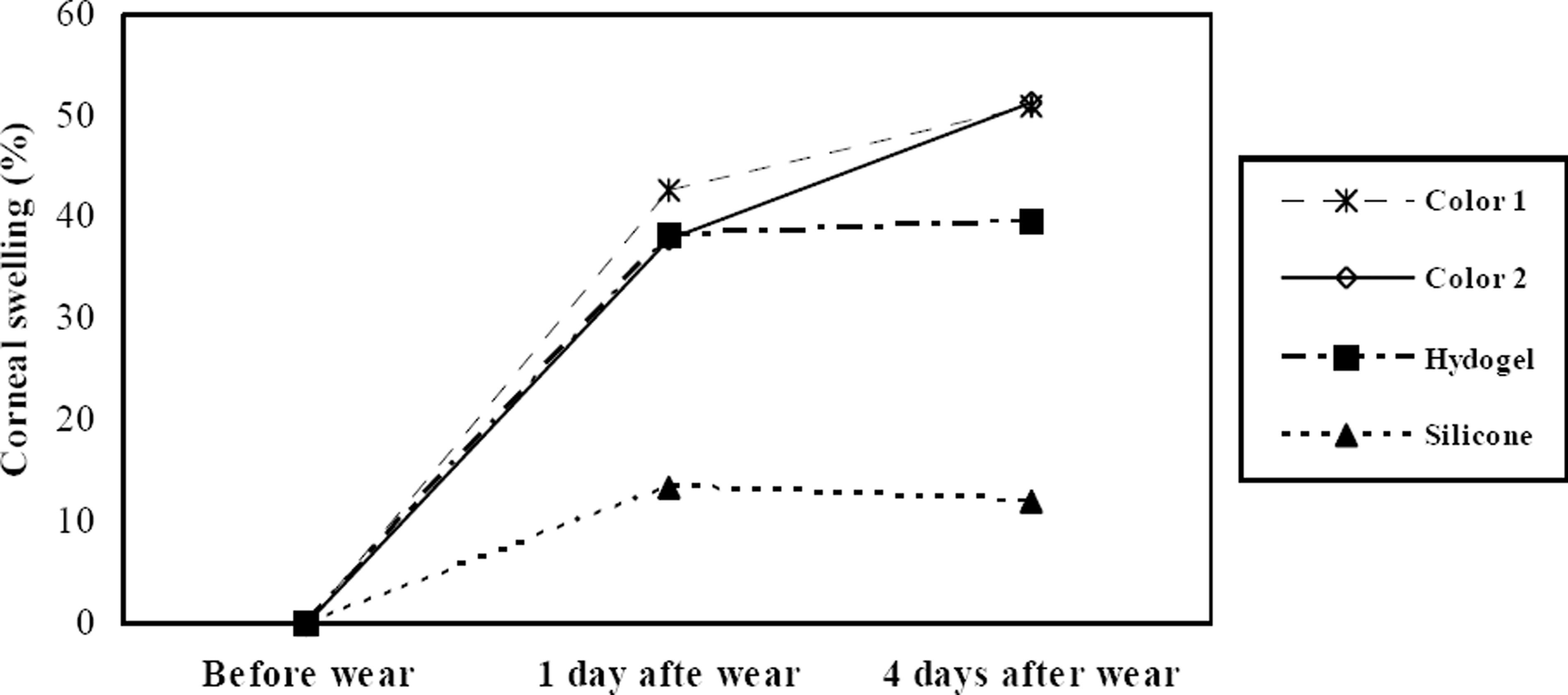
Although the corneal swelling of the silicone hydrogel lens group was significantly lower than the other 3 lens groups after contact lens wear ( p<0.01), the common hydrogel lens group was not different from the 2 tinted contact lens groups ( p>0.1). Tear LDH activity at 1 and 4 days after contact lens wear showed no significant difference among the 4 groups ( p>0.29). The scores of ocular surface inflammation in the 2 tinted contact lens groups were greater than the hydrogel and silicone hydrogel lens groups ( p=0.03). The scanning electron microscope revealed the internal surface of the tinted area in the tinted contact lens was coarse and irregular though the surface of the non-tinted area was relatively smooth.
Conclusions
Regarding corneal swelling and tear LDH activity, the cheap tinted contact lenses used in Korea were not significantly different from the common hydrogel contact lenses. However, tinted contact lenses showed a greater tendency to provoke ocular surface inflammation than other lenses. The coarse and irregular surface of the tinted area in the tinted contact lens appears to play a role in provoking severe ocular surface inflammation.
Go to : 
References
1. Park YM, Hahn TW, Choi SH. . Acanthamoeba keratitis related to cosmetic contact lenses. J Korean Ophthalmol Soc. 2007; 48:991–4.
2. Conners MS, Stoltz RA, Webb SC. . A closed eye contact lens model of corneal inflammation. Part1: Increased synthesis of cytochrome P450 arachidonic acid metabolites. Invest Ophthalmol Vis Sci. 1995; 36:828–40.
3. Cunha MC, Thomassen TS, Cohen EJ. . Complications associated with contact lens use. CLAO J. 1987; 13:107–11.
4. Liesegang TJ. Physiologic changes of the cornea with contact lens wear. CLAO J. 2002; 28:12–27.
5. Park YJ, Lee GJ, Park JJ. . The long-term effects of soft contact lens wear on corneal thickness, curvature and endothelium. J Korean Ophthalmol Soc. 2005; 46:945–53.
6. Lee WJ, Yoon GS, Shyn KH. Corneal complications in contact lens wearer. J Korean Ophthalmol Soc. 1996; 37:225–32.
7. Lee JH, Wee WR, Sohn JH. The effect of short-term wearing of disposable contact lenses on corneal topography. J Korean Ophthalmol Soc. 1994; 35:1020–6.
8. Bonanno JA. Contact lens induced corneal acidosis. CLAO J. 1996; 22:70–74.
9. Cannella A, Bonafini JA Jr. Polymer chemistry. Bennett ES, Weissman BA, editors. Clinical contact lens practice. Philadelphia: Lippincott Wiliams & Wilins;2005. chap. 10.
10. Korean contact lens study society Contact lens: Principles and practice. Seoul: Naeoe Haksool;2007. chap. 13.
11. Hamano H, Maeda N, Hamano T. . Corneal thickness change induced by dozing while wearing hydrogel and silicone hydrogel lenses. Eye Contact Lens. 2008; 34:56–60.

12. Fullard RJ, Carney LG. Use of tear enzyme activities to assess the corneal response to contact lens wear. Acta Ophthalmol. 1986; 64:216–20.

13. Kim MS, Park HS, Kim JH. Rigid gas permeable contact lens induced corneal hypoxia in rabbit. J Korean Ophthalmol Soc. 1997; 38:1936–46.
Go to : 
 | Figure 2.Photographs of corneal epithelial defects at 4 days after contact lens wear. (A) Large epithelial defect (grade 3) is shown in the color lens 1 group. (B) Small epithelial defect (grade 1) is observed in the hydrogel lens group. |
 | Figure 3.Scanning electron micrographs of the internal surface of a color contact lens (×100). (A) The surface of non-tinted area in the color lens is smooth. (B) The surface of tinted area is coarse and irregular. |
Table 1.
Characteristics of four soft contact lenses
| Group | Base Curve | Diameter | Thickness | Water content | Dk/t value∗ |
|---|---|---|---|---|---|
| Color lens 1 | 8.6 mm | 14 mm | 0.178 mm | 38 % | 13 |
| Color lens 2 | 8.4 mm | 14 mm | 0.088 mm | 35 % | 10 |
| Hydrogel lens | 8.5 mm | 14 mm | 0.134 mm | 38 % | 15 |
| Silicone hydrogel lens | 8.4 mm | 14 mm | 0.063 mm | 51 % | 52 |
Table 2.
Comparison of tear lactate dehydrogenase activities (U/L) among the four groups
| Group | Color lens 1 | Color lens 2 | Hydrogel lens | Silicone hydrogel lens | p value† |
|---|---|---|---|---|---|
| Before CL∗ wear | 255 (±236.5) U/L | 151 (±78.7) U/L | 203 (±178.5) U/L | 171 (±125.3) U/L | 0.528 |
| 1 day after CL wear | 506 (±492.5) U/L | 431 (±244.8) U/L | 334 (±196.7) U/L | 398 (±288.1) U/L | 0.743 |
| 4 days after CL wear | 354 (±132.1) U/L | 272 (±129.9) U/L | 306 (±174.2) U/L | 216 (±101.7) U/L | 0.163 |
| p value† | 0.240 | 0.003 | 0.279 | 0.031 |


 PDF
PDF ePub
ePub Citation
 Print
Print


 XML Download