

 PDF
PDF ePub
ePub Citation
 Print
Print


Abstract
Purpose
To compare the efficacy and safety of intraoperative laser indirect ophthalmoscopy and cryopexy for rhegmatogenous retinal detachment through the use.
Methods
We retrospectively analyze the clinical results of 60 patients (61 eyes) with rhegmatogenous retinal detachment. All eyes had an attached macula and were scheduled for conventional scleral buckling surgery with cryopexy (25 eyes) or laser indirect ophthalmoscopy (36 eyes) from March 2001 to August 2007. The visual acuities of the two groups at the first postoperative day, 1 week, 1 month, and 3 months were compared. Retinal reattachment, macular pucker, cystoid macular edema, and proliferative vitreoretinopathy were confirmed 3 months after surgery.
Results
The visual recovery was faster in patients who received laser indirect ophthalmoscopy (1 day, P=0.044; 1 week, P=0.017). During the follow-up period, anatomical failure in the laser indirect ophthalmoscopy group did not develop, but there were two anatomical failures in the cryopexy group. In addition, 1 PVR was detected in the cryopexy group. However, neither group exhibited macular pucker or cystoid macular edema. Postoperative additional laser photocoagulation was performed on 7 eyes (19.4%) in thelaser indirect ophthalmoscopy group and 2 eyes (4.0%) in the cryopexy group.
Go to : 
References
1. Chignell A. Retinal detachment surgery without cryotherapy. Trans Ophthalmol Soc U K. 1997; 97:30–2.

2. Fetkenhour CL, Hauch TL. Scleral buckling without thermal adhesion. Am J Ophthalmol. 1980; 89:662–6.
3. Sabates NR, Sabates FN, Sabates R, et al. Macular detachment after retinal detachment surgery. Am J Ophthalmol. 1989; 108:22–9.
4. Appiah AP, Hirose T. Secondary causes of premacular fibrosis. Ophthalmology. 1989; 96:389–92.
5. Lobes LA Jr, Grand G. Incidence of cystoid macular edema following scleral buckling procedure. Arch Ophthalmol. 1980; 98:1230–2.
6. Lobes LA Jr, Burton TC. The incidence of macular pucker after retinal detachment surgery. Am J Ophthalmol. 1978; 85:72–7.
7. Bonnet M, Guenoun S. Surgical risk factors for severe postoperative proliferative vitreoretinopathy (PVR) in retinal detachment with grade B PVR. Grafes Arch Clin Exp Ophthalmol. 1995; 233:789–91.
8. Machemer R, Aaberg TM, Freeman HM, et al. An updated classification of retinal detachment with proliferative vitreo- retinopathy. Am J Ophthalmol. 1991; 112:159–65.
9. Arrindel EL, Wu JC, Wolf MD, et al. High resolution magnetic resonance imaging evaluation of blood-retinal barrier integrity following transscleral diode laser treatment. Arch Ophthalmol. 1995; 113:96–102.
10. Barnes SD, LoRusso FJ. Does transscleral diode laser retinopexy enhance dispersion of viable retinal pigment epithelial cells? Invest Ophthalmol Vis Sci. 1998; 39:s108.
11. Sato Y, Berkowitz BA, Wilson CA, De Juan E Jr. Blood- retinal barrier breakdown caused by diode versus argon laser endophotocoagulation. Arch Ophthalmol. 1992; 110:277–81.
12. McHugh DA, Schwartz S, Hamilton PA, et al. Diode laser contact transscleral retinal photocoagulation: a clinical study. Br J Ophthalmol. 1995; 79:1083–7.
13. Haller JA, Blair N, de Juan E Jr, et al. Transscleral diode laser retinopexy in retinal detachment surgery: results of a multicenter trial. Retina. 1998; 18:399–404.
14. Jennings T, Fuller T, Vukich JA, et al. Transscleral contact retinal photocoagulation with an 810nm semiconductor diode laser. Ophthalmic Surg. 1990; 21:492–6.
15. Arrindel EL, Wu JC, Wolf MD, et al. MRI evaluation of blood-retinal barrier following transconjunctival diode laser photocoagulation and retinal cryotherapy. Invest Ophthalmol Vis Sci. 1992; 33:S1125.
16. Steel DH, West J, Campbell WG. A randomized controlled study of the use of transscleral diode laser and cryotherapy in the management of rhegmatogenous retinal detachment. Retina. 2000; 20:346–57.
17. Veckeneer M, Van Overdam K, Bouwens D, et al. Rando- mized clinical trial of cryotherapy versus laser photo- coagulation for retinopexy in conventional retinal detachment surgery. Am J Ophthalmol. 2001; 32:343–7.
18. Cowley M, Con Wey BP, Campochiaro PA, et al. Clinical risk factors for proliferative vitreoretinopathy. Arch Ophthalmol. 1989; 107:1147–51.
19. Grizzard WS, Hilton GF, Hammer ME, Taren D. A multi- variate analysis of anatomic success of retinal detachments treated with scleral buckling. Grafes Arch Clin Exp Ophthalmol. 1994; 232:1–7.
20. Tseng W, Cortez RT, Ramirez G, et al. Prevalence and risk factors for proliferative vitreoretinopathy in eyes with rhegmatogenous retinal detachment but no previous vitreo- retinal surgery. Am J Ophthalmol. 2004; 137:1105–15.
21. Avitabile T, Bartolotta G, Torrisi B, Reibaldi A. A randomized prospective study of rhegmatogenous retinal detachment cases treated with cryopexy versus frequency-doubled Nd:YAG laser-retinopexy during episcleral surgery. Retina. 2004; 24:878–82.
22. Mietz H, Hiemann K. Onset and recurrence of proliferative vitreoretinopathy in various vitreoretinal disease. Br J Ophthal- mol. 1995; 79:874–7.
23. Burton RL, Cairns JD, Campbell WG, et al. Needle drainage of subretinal fluid. A randomized clinical trial. Retina. 1993; 13:13–6.
24. Salicone A, Smiddy WE, Venkatraman A, Feuer W. Visual recovery after scleral buckling procedure for retinal detachment. Ophthalmology. 2006; 113:1734–42.
25. Benner JD, Huang M, Morse LS, et al. Comparison of photocoagulation with argon, krypton, and diode laser indirect ophthalmoscopes in rabbit eyes. Ophthalmology. 1992; 99:1554–63.
26. Lee SH, Chang MH, Choi WS. Clinical analysis of laser photocoagulation following scleral buckling for the treatment of rhegmatogenous retinal detachment. J Korean Ophthalmol Soc. 1999; 40:3111–6.
Go to :
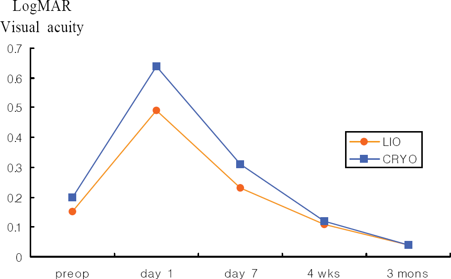 | Figure 1.Visual recovery. LIO=laser indirect ophthalmoscopy; CRYO=Cryopexy; logMAR VA=logarithm of minimal angle of resolution visual acuity; preop=before surgery; wks=weeks; mons=months. |
Table 1.
Preoperative and perioperative features of two study groups with rhegmatogenous retinal detachment
| Characteristics | LIO* group (n=36) | Cryo† group (n=25 | ) P value | Test type |
|---|---|---|---|---|
| Mean age, yr (mean±SD | ) 31.92±11.27 | 36.88±13.41 | 0.176 | Mann-Whitney test |
| Sex (M/F) | 17/19 | 12/13 | 0.952 | Pearson chi-square test |
| IOP‡(Mean±SD) | 15.31±2.69 | 16.08±2.75 | 0.220 | Mann-Whitney test |
| R.E§ (mean±SD) | -5.38±5.71 | -4.92±4.23 | 0.626 | Mann-Whitney test |
| P∏/PS#/AP# | 34/0/2 | 23/2/0 | 0.763 | Chi-square test (linear-by-linear association) |
Table 2.
LogMAR visual acuity before and after surgery (Mann-Whitney test, P<0.05)
| LIO* group (mean±SD) | Cryo† group (mean±SD) | P-value | |
|---|---|---|---|
| Before surgery | 0.15±0.17 | 0.20±0.31 | 0.967 |
| Day 1 | 0.49±0.46 | 0.64±0.34 | 0.044 |
| Day 7 | 0.23±0.28 | 0.31±0.21 | 0.017 |
| 4 weeks | 0.11±0.16 | 0.12±0.13 | 0.342 |
| 3 months | 0.04±0.10 | 0.04±0.07 | 0.369 |
 XML Download