

 PDF
PDF ePub
ePub Citation
 Print
Print


Abstract
Purpose
We present a case of choroidal osteoma and report a pattern of optical coherence tomography and multifocal electroretinography with a literature review.
Case summary
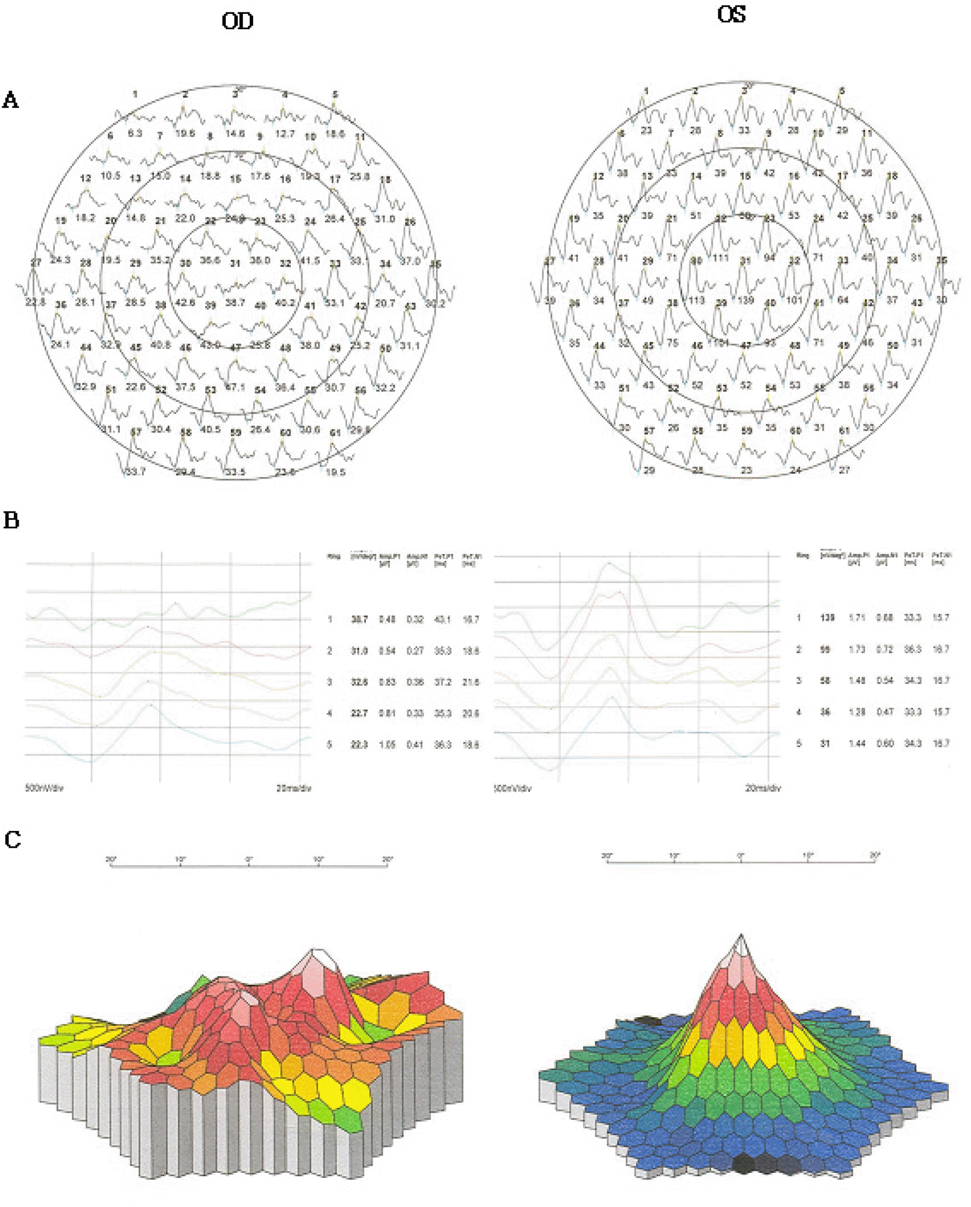
Using B-scan and CT, we performed OCT and MF-ERG in a patient diagnosed with choroidal osteoma. OCT revealed serous retinal detachment in the macula and a thick, irregular plate-like structure with high reflectivity in the choroidal mass lesion. MF-ERG demonstrated that Trace arrays show suppression of central and peripheral signals, especially in central lesions. Ring averages show reduced amplitudes in all locations, and 3D-topography of the response density shows marked suppression of the central signal and no foveal peak in comparison with the unaffected left eye.
References
1. Shields CL, Shields JA, Augsburger JJ. Choroidal osteoma. Surv Ophthalmol. 1988; 33:17–27.

2. Gass JD. New observations concerning choroidal osteomas. Int Ophthalmol. 1979; 2:71–84.
3. Gass JD, Guerry RK, Jack RL, Harris G. Choroidal osteoma. Arch Ophthalmol. 1978; 96:428–35.
4. Battaglia PM, Da PS, Toto L, et al. Photodynamic therapy for choroidal neovascularization associated with choroidal osteoma. Retina. 2001; 2:660–1.
5. Kida Y, Shibuya Y, Oguni M, et al. Choroidal osteoma in infant. Am J Ophthalmol. 1997; 124:119–20.
6. Mizota A, Tanabe R, Adachi-Usami E. Rapid enlargement of choroidal osteoma in a 3-year-old girl. Arch Ophthalmol. 1998; 116:1128–9.
7. Noble KG. Bilateral choroidal osteoma in three siblings. Am J Ophthalmol. 1990; 109:656–60.
8. Kayazawa F, Shimamoto S. Choroidal osteoma. Two cases in Japanese women. Ann Opthalmol. 1981; 13:1053–6.
9. Joffe L, Shields JA, Fitzgerald JR. Osseous choristoma of the choroid. Arch Ophthalmol. 1978; 96:1809–12.
10. Sin DH, Oum BS. Choroidal osteoma: Two cases report. J Korean Ophthalmol Soc. 1993; 34:264–71.
11. Grand MG, Burgess DB, Singerman LJ, Ramsey J. Choroidal osteoma. Treatment of associated subretinal neovascular membranes. Retina. 1984; 4:84–9.
12. Hoffman ME, Sorr EM. Photocoagulation of subretinal neovascularization associated choroidal osteoma. Arch Ophthalmol. 1987; 105:998–9.
13. Morrison DL, Magargal LE, Ehrlich DR, et al. Review of choroidal osteoma: Successful krypton red laser photocoagulation of an associated subretinal neovascular membrane involving the fovea. Ophthalmol Surg. 1987; 18:299–303.
14. Baum MD, Pilkerton AR, Berler DK, Kramer KK. Choroidal osteoma. Ann Ophthalmol. 1979; 11:1849–51.
15. Shelds JA, Shields CL. Osseous tumors of the uvea: Intraocular tumors: a text and atlas. 1st ed.Vol. 1. Philadephia: WB Saunders;1992. p. 261–72.
16. DePotter P, Shields JA, Shields CL, Rao VM. Magnetic resonance imaging in choroidal osteoma. Retina. 1992; 11:221–3.
17. Fukasawa A, Iijima H. Optical coherence tomography of choroidal osteoma. Am J Ophthalmol. 2002; 133:419–21.
18. Ide T, Ohguro N, Hayashi A, et al. Optical coherence tomography patterns of choroidal osteoma. Am J Ophthalmol. 2000; 130:131–4.
19. Yoon KC, Jeong JH, Seo MS. Choroidal osteoma. J Korean Ophthalmol Soc. 1997; 38:167–71.
Figure 1.
(A) Fundus photograph of the right eye shows large, well-demarcated, round, yellow-white lesion involving the peripapillary area and macula. (B) Early fluorescein angiogram of the right eye demonstrates hyperfluorescence due to window defect corresponding to choroidal lesion. (C) Late fluorescein angiogram of the right eye demonstrates hyperfluorescence without leakage.
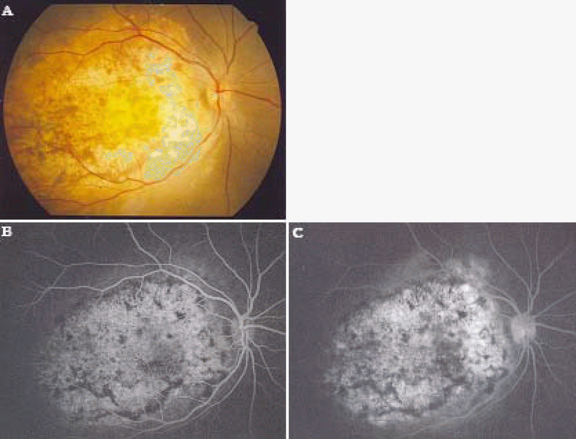
Figure 2.
(A) A-scan ultrasonography demonstrates the high-intensity echo spike corresponding to the anterior surface of choroidal osteoma. (B) B-scan ultrasonography demonstrates the slightly elevated, highly reflective choroidal mass and posterior acoustic shadowing. (C), (D) Computed tomography shows dense choroidal lesion having the density of normal bone.

Figure 3.
(A) The visual field of the right eye shows a defect relatively small compared to lesion size. (B) The visual field of the left eye. (C) Optical coherence tomography of the right eye corresponding to the scanning line shown in the fundus photograph shows serous retinal detachment in the macula and thick, irregular plate-like structure with high reflectivity in choroidal mass lesion. (D) Optical coherence tomography of the left eye.
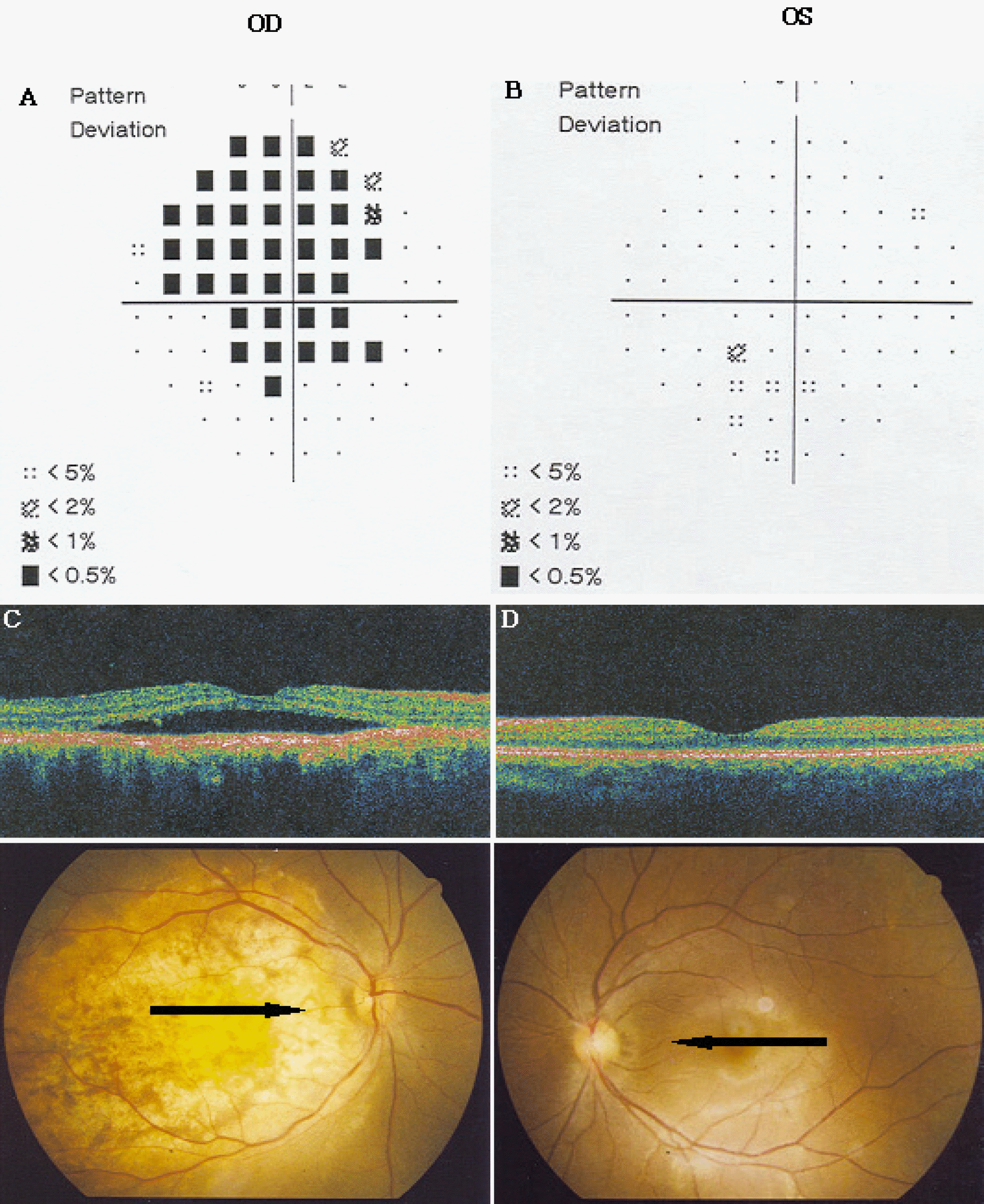
Figure 4.
Multifocal electroretinography. (A) Trace arrays of the right eye show suppression of central and peripheral signal especially in the central lesion compared with unaffected left eye. (B) Ring averages of the right eye show reduced amplitude in all locations in comparison with unaffected left eye. (C) 3D-Topography of the response density in the right eye shows marked suppression of the central signal and no foveal peak in comparison with unaffected left eye.
 XML Download