

 PDF
PDF ePub
ePub Citation
 Print
Print


Abstract
Purpose
To prospectively investigate the change of clinical manifestations after 1 year of administration of anthocyanoside (Tagen-F®) to patients with NPDR-associated macular edema.
Methods
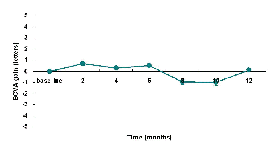
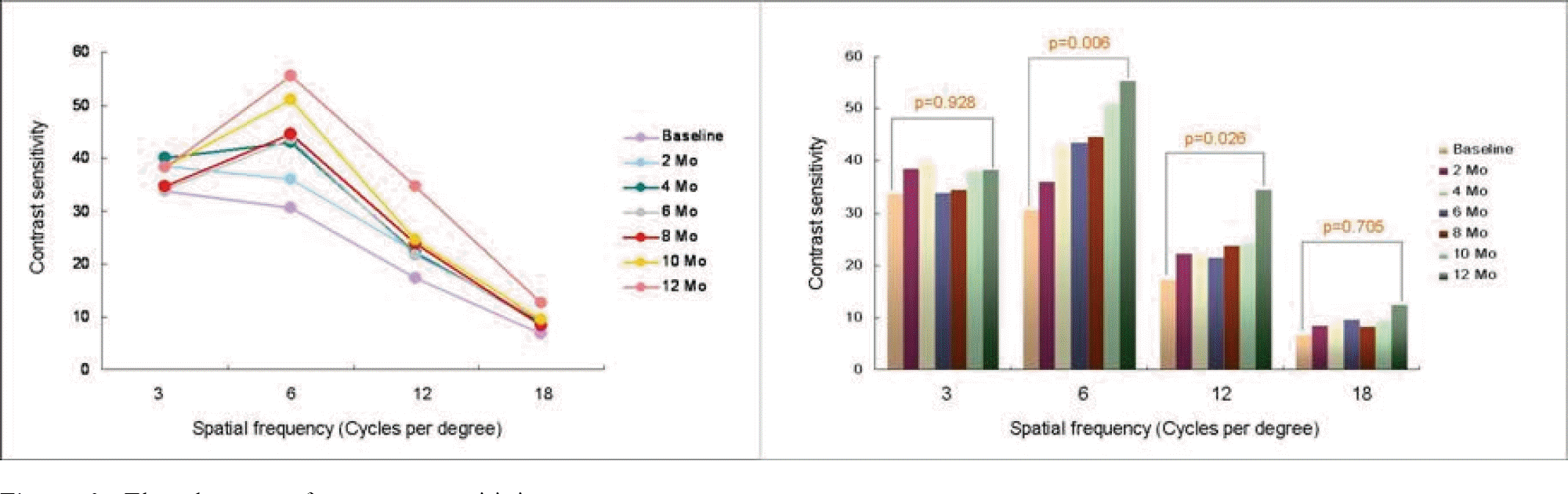
One hundred seventy-five eyes in 88 patients were enrolled in this study, at 5 centers, from March, 2005 to October, 2005. Patients took 3 capsules of Vaccinium myrtillus extract (170 mg/capsule, Tagen-F®, Kukje pharmaceutical) per day. The primary endpoints were corrected visual acuity and contrast sensitivity which were checked at 2 months following the beginning of treatment [East 1]. The secondary endpoints were the number of hard exudates, microaneurysms, leaking points and the changes of foveal thickness. These were examined at the beginning of, 6 months after, and 12 months after treatment.
Results
Corrected visual acuity showed no significant changes during 12 months. Contrast sensitivity improved gradually, especially in 12, 16 cycles per degree [East 2]. There was no statistically significant changes in the numbers of hard exudates, microaneurysms, and leaking points. Foveal thickness measured by OCT was maintained and there was no aggravation of macular edema.
Go to : 
References
1. Amos AF, McCarthy DJ, Zimmet P.The rising global burden of diabetes and its complications: estimates and projections to the year 2010. Diabetic Med. 1997; 14:S1–85.

2. Aiello LP, Gardner TW, King GL. . Diabetic retinopathy. Diabetes Care. 1998; 21:143–56.
3. Brownlee M. Biochemistry and molecular cell biology of diabetic complications. Nature. 2001; 414:813–20.
4. Yamagishi S, Imaizumi T. Diabetic vascular complications: pathophysiology, biochemical basis and potential therapeutic strategy. Curr Pharm Des. 2005; 11:2279–99.
5. Nishikawa T, Edelstein D, Du XL. . Normalizing mitochondrial superoxide production blocks three pathways of hyperglycaemic damage. Nature. 2000; 404:787–90.
6. Feldman EL. Oxidative stress and diabetic neuropathy: a new understanding of an old problem. J Clin Invest. 2003; 111:431.
7. Vessby J, Basu S, Mohsen R. . Oxidative stress and antioxidant status in type 1 diabetes mellitus. J Intern Med. 2002; 251:69–76.
8. Marra G, Cotroneo P, Pitocco D. . Early increase of oxidative stress and reduced antioxidant defenses in patients with uncomplicated type 1 diabetes: a case for gender difference. Diabetes Care. 2002; 25:370–5.
9. Reunanen A, Knekt P, Aaran RK, Aromaa A. Serum antioxidants and risk of non-insulin dependent diabetes mellitus. Eur J Clin Nutr. 1998; 52:89–93.
10. Rahimi R, Nikfar S, Larijani B, Abdollahi M. A review on the role of antioxidants in the management of diabetes and its complications. Biomed Pharmacother. 2005; 59:365–73.
11. Busciala A, Bosisio E. Vascular effects of wine ployphenols. Cardiovas Res. 2004; 63:593–602.
12. Amano S, Yamagishi S, Kato N. . Sorbitol dehydrogenase overexpression potentiates glucose toxicity to cultured retinal pericytes. Biochem Biophys Res Commun. 2002; 299:183–8.
13. Lekakis J, Rallidis LS, Andreadou I. . Polyphenolic compounds from red grapes acutely improve endothelial function in patients with coronary heart disease. Eur J Cardiovasc Prev Rehabil. 2005; 12:596–600.
14. Burns J, Gardner PT, O'Neil J. . Relationship among antioxidant activity, vasodilation capacity and phenolic contents of red wines. J Agric Food Chem. 2000; 48:220–30.
15. Jindra LF, Zemon V. Contrast sensitivity testing: A more complete assessment of vision. J Cataract Refract Surg. 1989; 14:141–8.
16. Kim EA, Koo YJ, Han YB. Contrast sensitivity changes in patients with diabetic retinopathy. J Korean Ophthalmol Soc. 1995; 36:1523–8.
17. Arend O, Remky A, Evans D. . Contrast sensitivity loss is coupled with capillary dropout in patients with diabetes. Invest Ophthalmol Vis Sci. 1997; 38:1819–24.
18. Klein R, Meuer SM, Moss SE, Klein BE. Retinal microaneurysm counts and 10-year progression of diabetic retinopathy. Arch Ophthalmol. 1995; 113:1386–91.
Go to :

 XML Download