Abstract
Purpose
To determine the preoperative factors of different types of diabetic macular edema (DME) classified using Optical Coherence Tomography (OCT) and to evaluate the short-term therapeutic effects and pattern changes of intravitreal triamcinolone acetonide injection (IVTA).
Methods
Seventy-seven eyes of 60 patients, who had been previously diagnosed with DME through fundoscopy and fluorescein angiography, were enrolled, and each patient was classified as one of three DME types according to his/her OCT features: Type 1, diffuse retinal thickening; Type 2, cystoid macular edema; and Type 3, serous macular detachment. We compared age, sex, the duration of diabetes mellitus (DM), and decreased visual acuity (VA). We analyzed VA, intraocular pressure (IOP), foveal thickness (FT), total macular volume (TMV), and pattern changes that occurred between pre-operation and 1 month post-operation.
Results
The duration of DM was short in Type 3 DME patients. There were no differences in age or the duration of decreased VA. Pre-operative VA was higher in Type 1 than in Type 2 or 3 patients. FT and TMV increased in thickness from Type 1 through Type 3. VA after IVTA improved in Types 2 and 3. FT and TMV after IVTA decreased in each type. However, the extent of the changes in Types 2 and 3 was greater than that in Type 1. Seventy-four percent of Type 2 and 83% of Type 3 changed to Type 1 after IVTA.
Go to : 
References
1. Klein R, Klein BE, Moss SE. . The Wisconsin epidemiologic study of diabetic retinopathy IV: Diabetic macular ede ma. Ophthalmology. 1984; 91:1464–74.
2. Johnson RN, Howard SH, McDonald R, Ai E. Ryan SJ, editor. Fluorescein angiography: basic principles and interpretation. Retina, revised. St. Louis: Mosby;2001. v. 2:p. chap. 55.

3. Early Treatment Diabetic Retinopathy Study Research Group Early Treatment Diabetic Retinopathy Study report number 1: Photocoagulation for diabetic macular edema. Arch Ophthalmol. 1985; 103:1796–806.
4. Shahidi M, Ogura Y, Blair NP. . Retinal thickness analysis of quantitative assessment of diabetic macular edema. Arch Ophthalmol. 1991; 109:1115–9.
5. Oshima Y, Emi K, Yamanishi S, Motokura M. Quantitative assessment of macular thickness in normal subjects and patients with diabetic retinopathy by scanning retinal thickness analyser. Br J Ophthalmol. 1999; 83:54–61.

6. Zambarakji HJ, Amoaku WM, Vernon SA. Volumetric analysis of early macular edema with the Heidelberg Retina Tomograph in diabetic retinopathy. Ophthalmology. 1998; 105:1051–9.
7. Hee MR, Izatt JA, Swanson EA. . Optical coherence tomography of the human retina. Arch Ophthalmol. 1995; 113:325–32.

8. Hee MR, Puliafito CA, Wong C. . Quantitative assessment of macular edema with optical coherence tomography. Arch Ophthalmol. 1995; 113:1019–29.

9. Konno S, Akiba J, Yoshida A. Retinal thickness measurements with optical coherence tomography and the scanning retinal thickness analyzer. Retina. 2001; 21:57–61.

10. Otani T, Kishi S, Maruyama Y. Patterns of diabetic macular edema with optical coherence tomography. Am J Ophthalmol. 1999; 127:688–93.

11. Kang SW, Park CY, Ham DI. The correlation between fluorescein angio and optical coherence tomographic features in clinically significant diabetic macular edema. Am J Ophthalmol. 2004; 137:313–22.
12. Kim DH, Kim SH, Kim HW, Yoon IH. Risk factors for diffuse diabetic macular edema as classified by optical coherence tomography. J Korean Ophthalmol Soc. 2006; 47:548–55.
13. Jonas JB, Sofker A. Intraocular injection of crystalline cortisone as adjunctive treatment of diabetic macular edema. Am J Ophthalmol. 2001; 132:425–7.

14. Martidis A, Duker JS, Greenberg PB. . Intravitreal triamcinolone for refractory diabetic macular edema. Ophthal mology. 2002; 109:920–7.

15. Ip MS. Intravitreal injection of triamcinolone: an emerging treatment for diabetic macular edema. Diabetes Care. 2004; 27:1794–7.
16. Brasil OF, Smith SD, Galor A. . Predictive factors for short-term visual outcome after intravitreal triamcinolone acetonide injection for diabetic macular oedema: an optical coherence tomography study. Br J Ophthalmol. 2007; 91:761–5.

17. Kim YG, Yu SY, Kwak HW. The effect of intravitreal triamcinolone acetonide injection according to the diabetic macular edema type. J Korean Ophthalmol Soc. 2005; 46:84–9.
18. Nussenblatt RB, Kaufman SC, Palestine AG. . Macular thickening and visual acuity. Measurement in patients with cystoid macular edema. Ophthalmology. 1987; 94:1134–9.

19. Kim BY, Smith SD, Kaiser PK. Optical coherence tomography patterns of diabetic macular edema. Am J Ophthalmol. 2006; 142:405–12.
20. Ferris FL III, Patz A.Macular edema. A complication of diabetic retinopathy. Surv Ophthalmol. 1984; 28:S452–61.

21. Antcliff RJ, Marshall J. The pathogenesis of edema in diabetic maculopathy. Semin Ophthalmol. 1999; 14:223–32.

23. Lobo C, Bernardes R, Faria de Abreu Jr, Cunha-Vaz JG. Novel imaging techniques for diabetic macular edema. Doc Ophthalmol. 1999; 97:341–7.

24. Yannoff M, Fine BS, Brucker AG, Eagle RC Jr. Pathology of human cystoid macular edema. Surv Ophthalmol. 1984; 28:S505–11.
25. Fine BS, Brucker AJ. Macular dema Macular edema and cystoid macular edema. Am J Ophthalmol. 1981; 92:466–81.
26. Kaiser Pk, Riemann CD, Sears JE, Lewis H. Macular traction detachment and diabetic macular edema associated with posterior hyaloidal traction. Am J Ophthalmol. 2001; 131:44–9.

27. Yamaguchi Y, Otani T, Kishi S. Resolution of diabetic cystoid macular edema associated with spontaneous vitreofoveal separation. Am J Ophthalmol. 2003; 135:116–8.

28. Iida T, Yannuzzi LA, Spaide RF. . Cystoid macular degeneration in chronic central serous chorioretinopathy. Retina. 2003; 23:1–7.

29. Sabates NR, Sabates FN, Sabates R. . Macular changes after retinal detachment surgery. Am J Ophthalmol. 1989; 108:22–9.

30. Alkuraya H, Kangave D, Abu El-Asrar AM. The correlation between optical coherence tomographic features and severity of retinopathy, macular thickness and visual acuity in diabetic macular edema. Int Ophthalmol. 2005; 26:93–9.

31. Cho HY, Lee JH. The correlation between visual acuity and patterns of diabetic macular edema in OCT images. J Korean Ophthalmol Soc. 2003; 44:2028–34.
32. Jonas JB, Martus P, Degenring RF. . Predictive factors for visual acuity after intravitreal triamcinolone treatment for diabetic macular edema. Arch Ophthalmol. 2005; 123:1338–43.

33. Mason JO 3rd, Somaiya MD, Singh RJ. Intravitreal concentration and clearance of triamcinolone acetonide in non-vitrectomized hyman eyes. Retina. 2004; 24:900–4.
34. Kang SW, Sa HS, Cho HY, Kim JI. Macular grid photocoagulation after intravitreal triamcinolone aceonide for diffuse diabetic macular edema. Arch Ophthalmol. 2006; 124:653–8.
Go to : 
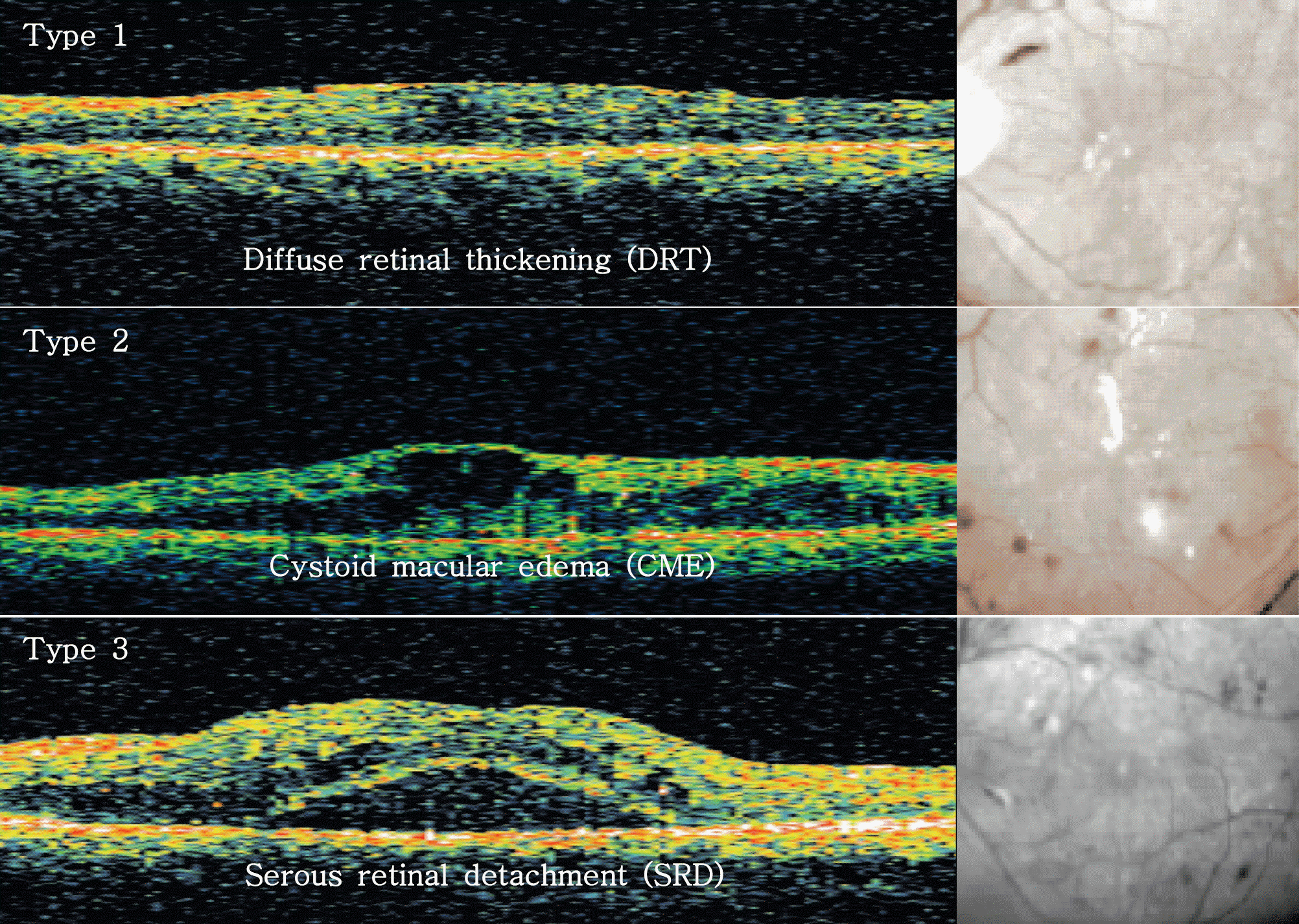 | Figure 1.The classification of clinically significant diabetic macular edema by OCT features. Type 1 (top): the thickening of the fovea with homogeneous optical reflectivity throughout the whole layer of the retina. Type 2 (middle): the thickening of the fovea with markedly decreased optical reflectivity in the outer retinal layer. Type 3 (bottom): the foveolar detachment without vitreofoveal traction; OCT=optical coherence tomography. |
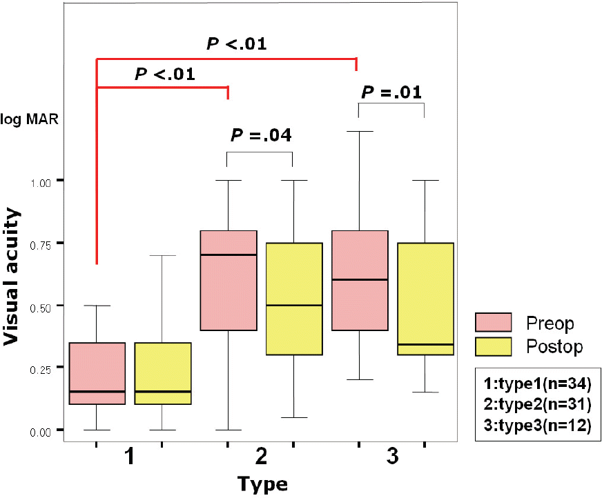 | Figure 2.An analysis of the VA before and after IVTA. Pre-operative VA was significantly higher in type 1 than in types 2 and 3 (Kruskal-Wallis test= p<.01, with p<.05 regarded as significant). VA after IVTA improved significantly in types 2 and 3 (Wilcoxon’s signed rank test, p=.04, p=.01, with p<.05 regarded as significant); VA=visual acuity; IVTA=intravitreal triamcinolone acetonide injection; log MAR=the logarithm of the minimum angle of resolution. |
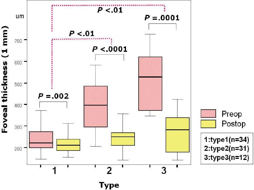 | Figure 3.An analysis of the foveal thickness before and after IVTA. Foveal thickness significantly decreased in every types (Wilcoxon’s signed rank test, p=.002, p<.0001, p=.0001, with p<.05 regarded as significant). But the extent of changes in Types 2 and 3 was greater than in Type 1 (Kruskal-Wallis test p<.01, with p<.05 regarded as significant). |
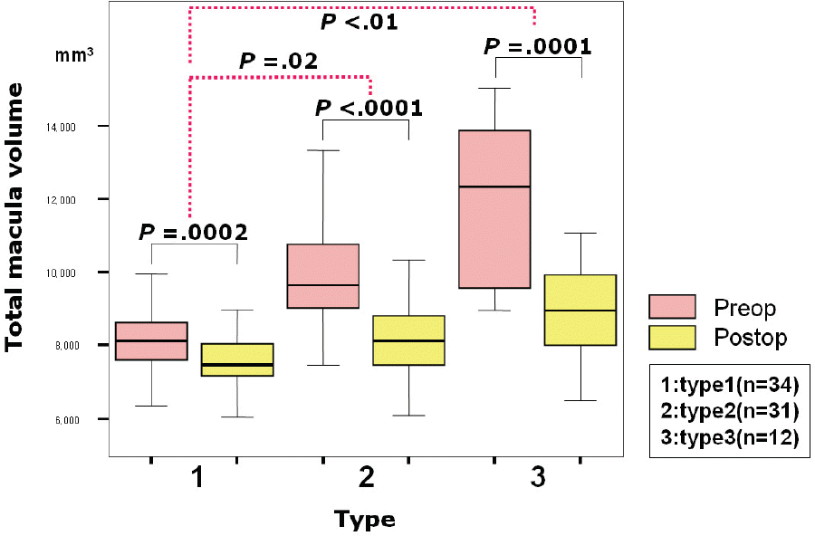 | Figure 4.An analysis of total macular volume before and after IVTA. Total macular volume was significantly decreased in every types (Wilcoxon’s signed rank test, p=.0002, p<.0001, p=.0001, with p<.05 regarded as significant). But the extent of changes in Types 2 and 3 was greater than in Type 1 (Kruskal-Wallis test p<.01, with p<.05 regarded as significant). |
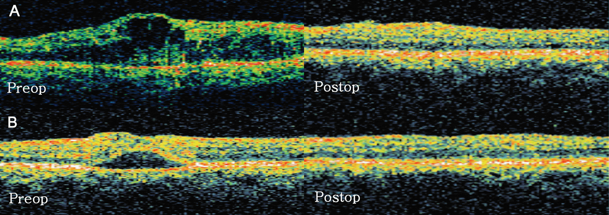 | Figure 5.DME pattern changes before and after IVTA injection at 1 month. The OCT feature changed to diffuse thickened macula (Type 1) in (A) 74% of cystoid macular edema (Type 2), (B) 83% of serous retinal detachment (Type 3); DME=diabetic macular edema; OCT=optical coherence tomography. |
Table 1.
Basic characteristics of eyes with different OCT types
| Type1 | Type2 | Type3 | |
|---|---|---|---|
| No of eyes | 34 | 31 | 12 |
| Age (yr) | 52.1 | 55.3 | 53.7 |
| Sex (M:F) | 22:12 | 21:10 | 7:5 |
| DM duration (yr) | 10.67±0.63 | 14.21±8.13 | 4.14±3.61* |
| Decreased VA duration (yr) | 0.47±0.69 | 0.71±1.18 | 0.60±0.38 |
| Preoperative VA (log MAR) | 0.23±0.21* | 0.62±0.30 | 0.62±0.31 |
| IOP (mmHg) | 15.7±4.49 | 16.1±12.2 | 15.6±3.63 |
| Foveal thickness (µm) | 242.1±64.2* | 395.9±117.5* | 511.8±127.2* |
| Macular volume (mm3) | 8.2±1.23* | 9.7±1.41* | 12.0±2.26* |


 PDF
PDF ePub
ePub Citation
 Print
Print


 XML Download