Abstract
Purpose
To evaluate the clinical features of subretinal hemorrhage after photodynamic therapy in eyes with exudative age-related macular degeneration.
Methods
We retrospectively reviewed data for 267 eyes of 243 patients who had undergone PDT for the treatment of ARMD between January 2005 and December 2006. Best corrected visual acuity, fundus photography, fluorescein angiography, and ICG angiography were performed before and after treatment. We followed up the patients at 1 week, 1 month, and 3 months after treatment and at 3-month intervals thereafter.
Results
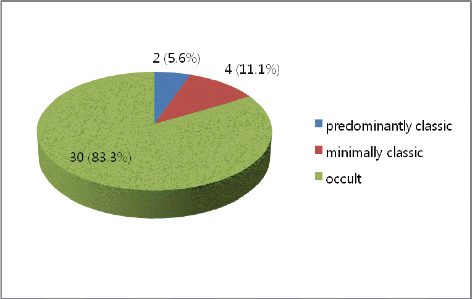
Postoperative subretinal hemorrhage was seen in 36 (13.4%) of 267 eyes. The pretreatment and post-treatment mean visual acuities were logMAR 0.80 and logMAR 1.05 respectively, representing a decrease of 2.05 lines. On FAG, two eyes were predominantly classic, four eyes were minimally classic, and 30 eyes were occult. The laser irradiation spot size was under 3,000 µm in one case and from 3,000 µm to 5,000 µm in 19 cases and over 5,000 µm in 16 eyes.
Go to : 
References
1. Blinder KJ, Blumenkranz MS, Bressler NM. . Verteporfin therapy of subfoveal choroidal neovascularization in pathologic myopia: 2-year results of a randomized clinical trial- VIP report No.3. Ophthalmology. 2003; 110:667–73.
2. Bressler NM. Treatment of Age-Related Macular Degeneration with Photodynamic Therapy (TAP) Study Group. Photo dynamic therapy of subfoveal choroidal neovascularization in age-related macular degeneration with verteporfin: two-year results of 2 randomized clinical trials-TAP report 2. Arch Ophthalmol. 2001; 119:198–207.
3. Verteporfin In Photodynamic Therapy Study Group. Verteporfin therapy of subfoveal choroidal neovascularization in age-related macular degeneration: two-year results of a randomized clinical trial including lesions with occult with no classic choroidal neovascularization-Verteporfin In Photo dynamic Therapy report 2. Am J Ophthalmol. 2001; 131:541–60.
4. Verteporfin In Photodynamic Therapy Study Group. Photo dynamic therapy of subfoveal choroidal neovascularization in pathologic myopia with verteporfin. 1-year results of a randomized clinical trial-VIP report no. 1. Ophthalmology. 2001; 108:841–52.
5. Treatment of Age-related Macular Degeneration with Photodynamic Therapy (TAP) Study Group and Verteporfin In Photodynamic Therapy (VIP) Study Group. Acute severe visual acuity decrease after photodynamic therapy with verteporfin: Case reports from randomized clinical trials- TAP and VIP Report No. 3. Am J Ophthalmol. 2004; 137:683–96.
6. Klein R, Klein BE, Linton KL. Prevalence of age related maculopathy. The Beaver Dam Eye Study. Ophthalmology. 1992; 99:933–43.
7. Klein R, Davis MD, Magli YL. . The Wisconsin age related maculopathy grading system. Ophthalmology. 1991; 98:1128–34.
8. Bressler SB, Bressler NM, Fine SL. . Natural course of choroidal neovascular membranes within the foveal avascular zone in senile macular degeneration. Am J Ophthalmol. 1982; 93:157–63.

9. Bressler NM, Bressler SB, Fine SL. Age-related macular degeneration. Surv Ophthalmol. 1988; 32:375–413.

10. Guyer DR, Fine SL, Maguine MG. . Subfoveal choroidal neovascular membranes in age-related macular degeneration. Visual prognosis in eyes with relatively good visual acuity. Arch Ophthalmol. 1986; 104:702–5.

11. Klein BE, Klein R. Cataract and macular degeneration in older Americans. Arch Ophthalmol. 1982; 100:571–3.
12. Hart PM, Chakraverthy RF, Mackenzie G. . Therapy for subfoveal choroidal neovascularization of age-related macular degeneration: results of follow up in a non-randomized study. Br J Ophthalmol. 1996; 80:1046–1050.
13. Hasan T, Gragoudas E. . Vascular targeting in photodynamic occlusion of subretinal vessels. Ophthalmology. 1994; 101:1953–61.

14. Miller JW, Walsh AW, Kramer M. . Photodynamic therapy of experimental chorioidal neovascularization using lipoprotein-derived benzoporphyrin. Arch Ophthalmol. 1995; 113:801–8.
15. Kramer M, Miller JW, Michaud N. . Liposomal benzoporphyrin derivative verteporfin photodynamic therapy. Selective treatment of choroidal neovascularization in monkeys. Ophthalmology. 1996; 103:427–38.

16. TAP Study Group. Photodynamic therapy of subfoveal choroidal neovascularization in age related macular degeneration with verteporfin. One year results of two randomized clinical trials-TAP report 1. Arch Ophthalmol. 1999; 117:1329–45.
17. Theodossiadis GP, Panagiotidis D, Georgalas IG. . Retinal hemorrhage after photodynamic therapy in patients with subfoveal choroidal neovascularization caused by age-related macular degeneration. Graefes Arch Clin Exp Ophthalmol. 2003; 241:13–8.

18. Do DV, Bressler NM, Bressler SB. Large submacular hemorrhages after verteporfin therapy. Am J Ophthalmol. 2004; 137:558–60.

19. Gelisken F, Inhoffen W. . Subfoveal hemorrhage after verteporfin photodynamic therapy in treatment of choroidal neovascularization. Graefes Arch Clin Exp Ophthalmol. 2005; 243:198–203.

20. Diaz-De-Durana-Santa-Coloma E, Fernandez-Ares ML, Iturralde-Errea D. . Submacular hemorrhage following photodynamic therapy in the treatment of choroidal neovascularization. Ophthalmic Surg Lasers Imaging. 2006; 37:278–83.
21. The Japanese Age-related Macular Degeneration Trial (JAT) Study Group. Japanese age-related macular degeneration trial: 1-year results of photodynamic therapy with verteporfin in Japanese patients with subfoveal choroidal neovascularization secondary to age-related macular degeneration. Am J Ophthalmol. 2003; 136:1049–61.
22. Kim JW, Kim HK, Kim HC. Photodynamic therapy for choroidal neovascularization caused by age-related macular degeneration. J Korean Ophthalmol Soc. 2002; 43:1435–43.
23. Michels S. Schmidt-Erfurth U. Sequence of early vascular events after photodynamic therapy. Invest Ophthalmol Vis Sci. 2003; 44:2147–54.
24. Michels S, Barbazetto I. Invest Ophthalmol Vis Sci. 2002; 43:830–41.
25. Ojima Y, Tsujikawa A, Otani A. . Recurrent bleeding after photodynamic therapy in polypoidal choroidal vasculopathy. Am J Ophthalmol. 2006; 141:958–60.

26. Hirami Y, Tsujikawa A, Otani A. . Hemorrhagic complications after photodynamic therapy for polypoidal choroidal vasculopathy. Retina. 2007; 27:335–41.

27. Harbin TS Jr. . Massive hemorrhage complicating age-related macular degeneration. Clinicopathologic correlation and role of anticoagulants. Ophth almology. 1986; 93:1581–92.
28. Ehrlich R, Rosenblatt I. . Photodynamic therapy for occult choroidal neovascularization with pigment epithelium detachment in age-related macular degeneration. Arch Ophthalmol. 2004; 122:453–9.
29. Han JW, Lee WK. Photodynamic therapy of choroidal neovasculariation associated with large serous pigment epithelial detachment. J Korean Ophthalmol Soc. 2004; 45:79–86.
30. Pece A, Isola V, Vadalà M, Calori G. aPhotodynamic therapy with verteporfin for choroidal neovascularization associated with retinal pigment epithelial detachment in age-related macular degeneration. Retina. 2007; 27:342–8.
31. Moorfields Macular Study Group. Retinal pigment epithelial detachments in the elderly: a controlled trial of argon laser photocoagulation. Br J Ophthalmol. 1982; 66:1–16.
Go to : 
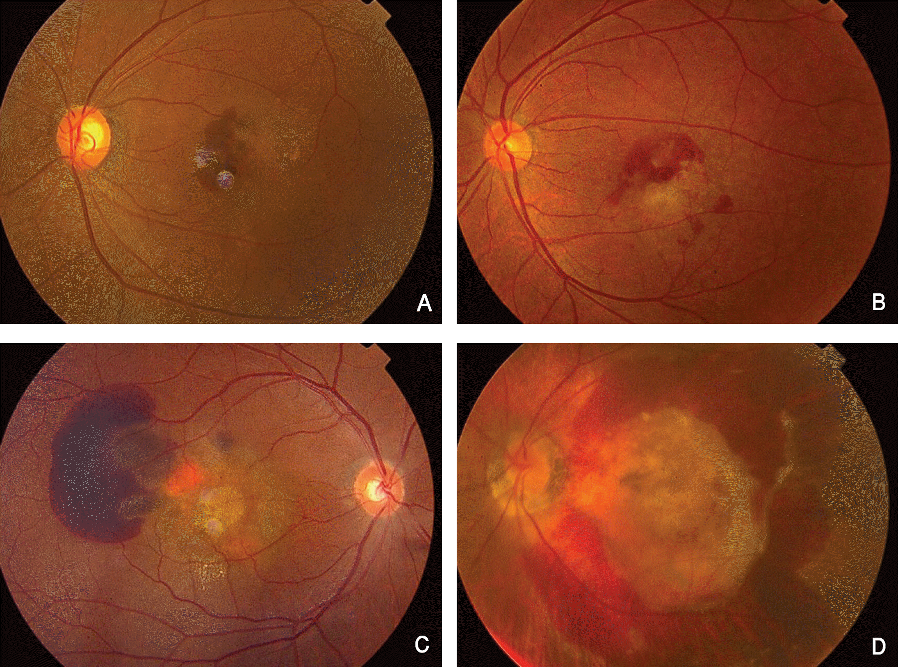
 | Figure 1.(A) Subretinal hemorrhage less than 2 disc diameter. (B) Subretinal hemorrhage more than 2 disc diameter. (C) Subretinal hemorrhage extends beyond the temporal vascular arcades. (D) Subretinal hemorrhage with vitreous hemorrhage. |
 | Figure 2.The fluorescein angiographic classification of age related macular degeneration which had subretinal hemorrhage after photodynamic therapy. |
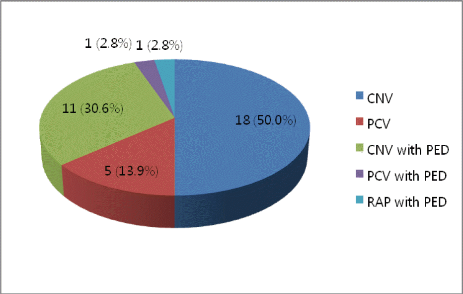
 | Figure 3.The indocyanine green angiographic classification of age related macular degeneration which had subretinal hemorrhage after photodynamic therapy. (CNV=choroidal neovascularization; PCV=polypoidal choroidal vasculopathy; RAP=retinal angiomatous proliferation; PED=pigment epithelial detachment) |
Table 1.
Demographics of the patients who had a retinal hemorrhage after photodynamic therapy
| Number of patients* | 36 (13.4%) |
|---|---|
| Age (years) | 63.12±5.2 (45-74) |
| Sex (M:F) | 25:11 |
| Preop BSCVA† (logMAR) | 0.80 |
| Postop BSCVA (logMAR) | 1.05 |
| Change of VA‡ (lines) | 2.05 |
Table 2.
Onset of hemorrhage after photodynamic therapy
| Onset of hemorrhage after PDT* | Eyes (percent) |
|---|---|
| <1 week | 9/36 (25.0%) |
| 1 week ≤ ~ < 1 month | 7/36 (19.4%) |
| ≥ 1 month | 20/36 (55.6%) |
Table 3.
Extent of hemorrhage after photodynamic therapy
| Extent of hemorrhage | Eye (percent) |
|---|---|
| < 2 disc diameter | 18/36 (50.0%) |
| ≥2 disc diameter | 9/36 (25.0%) |
| Massive hemorrhage* | 3/36 (8.3%) |
| Vitreous hemorrhage† | 6/36 (16.7%) |
Table 4.
Spot size of laser
| Spot size of laser (µm) | Eye (percent) |
|---|---|
| < 3,000 | 1/36 (2.7%) |
| 3,000 ≤ ~ < 5,000 | 19/36 (52.8%) |
| ≥ 5,000 | 16/36 (44.5%) |
Table 5.
Comparison of published retinal hemorrhage rate after photodynamic therapy of age related macular degeneration
| Author | Reference No | No. of treated eyes | Follow-up | Hemorrhage rate | Hemorrhage type |
|---|---|---|---|---|---|
| This study | 267 | 6~26 mo* | 36 (13.4%) | subretinal, vitreous | |
| TAP study group | 16 | 402 | 3 mo | 116 (29%) | subretinal, intraretinal |
| Theodossiadis | 17 | 215 | 48 hr† | 4 (1.86%) | extensive macular |
| Bressler | 18 | 55 | 2~3 mo | 5 (9%) | large submacular |
| Gelisken | 19 | 104 | 2 wk‡ | 23 (22%) | new subfoveal |
| Diaz | 20 | 504 | 34 mo | 8 (1.58%) | submacular |
| JAT study group | 21 | 64 | 12 mo | 1 (1.56%) | subretinal |
| Kim | 22 | 21 | 3~15 mo | 4 (19.0%) | subretinal, vitreous |
Table 6.
The relation between onset of hemorrhage and extent of hemorrhage after photodynamic therapy
| Onset of hemorrhage | Eyes (percent) | Extent of hemorrhage |
|---|---|---|
| <1 week | 9/36 (25.0%) | 7 eyes < 2DD |
| 2 eyes ≥ 2DD | ||
| 0 eye Massive hemorrhage* | ||
| 0 eye Vitreous hemorrhage† | ||
| 1 week ≤ ~ < 1 month | 7/36 (19.4%) | 5 eyes < 2DD |
| 2 eyes ≥ 2DD | ||
| 0 eye Massive hemorrhage* | ||
| 0 eye Vitreous hemorrhage† | ||
| ≥ 1 month | 20/36 (55.6%) | 6 eyes < 2DD |
| 5 eyes ≥ 2DD | ||
| 3 eyes Massive hemorrhage* | ||
| 6 eyes Vitreous hemorrhage† |


 PDF
PDF ePub
ePub Citation
 Print
Print


 XML Download