Abstract
Purpose
To compare surgically induced astigmatism (SIA) and some corneal higher order aberrations in patients who underwent microcoaxial cataract surgery (MCCS) or conventional cataract surgery.
Methods
A prospective randomized study included 60 eyes of 55 patients. Thirty eyes received MCCS using a 2.2mm clear corneal incision (group 1), and 30 eyes received conventional cataract surgery using a 2.8 mm clear corneal incision (group 2). SIA and corneal higher order aberrations were measured with a Keratometer (Humphrey, Zeiss) and i-Trace (Tracey Technologies) preoperatively, and at 1 and 3 months after cataract surgery. SIA was analyzed vectorially using the Alpins method.
Results
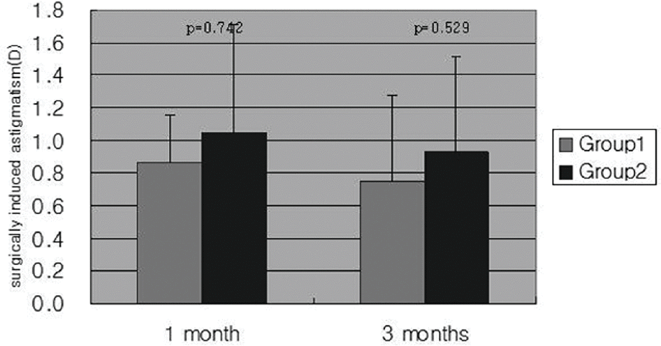
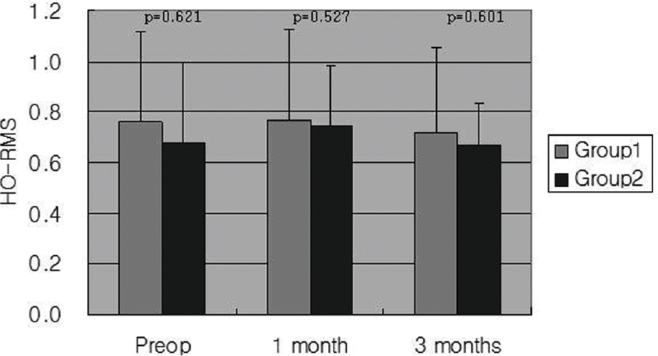
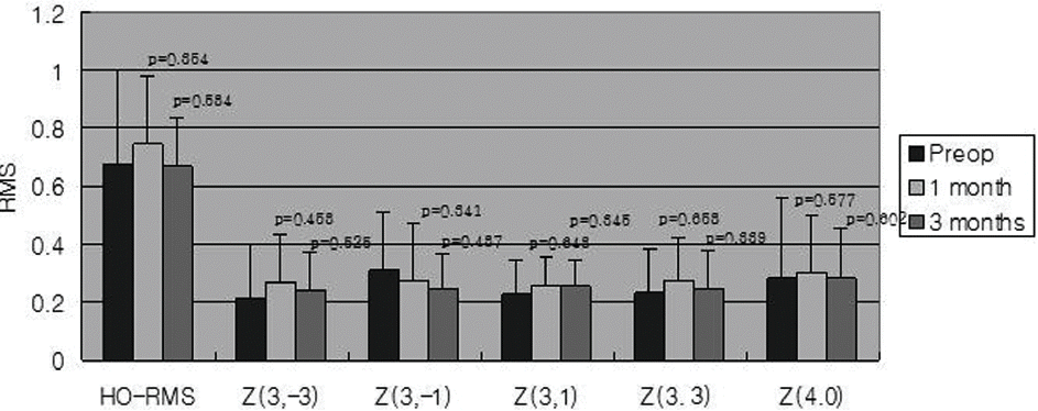
There was no significant difference in preoperative UCVA or BCVA between the two groups. At 1 month and 3 months after surgery, SIA in group 1 was less than that in group 2, but this difference was not significant. There was no statistically significant difference in postoperative change of corneal higher order aberrations in each group at 1 month or 3 months after surgery, and there was no statistically significant difference in corneal higher order aberrations between the two groups preoperatively, at 1 month, or 3 months after surgery.
References
1. Steiner GA, Binder PS, Parker WT, Perl T. The natural and modified course of post-cataract astigmatism. Ophthalmic Surg. 1982; 13:822–7.
2. Jaffe NS, Clayman HM. The pathophysiology of corneal astigmatism after cataract extraction. Trans Am Acad Ophthalmol Otolaryngol. 1975; 79:615–30.
3. Hu YJ, Lee KH, Joo CK. Comparison of surgically induced astigmatism between superior and temporal clear corneal incision in sutureless cataract surgery. J Korean Ophthalmol Soc. 1998; 39:495–500.
4. Kershner RM. Clear corneal cataract surgery and the correction of myopia, hyperopia, and astigmatism. Ophthalmology. 1997; 104:381–9.

5. Cravy TV. Calculation of the change in corneal astigmatism following cataract extraction. Ophthalmic Surg. 1979; 10:38–49.
6. Simsek S, Yasar T, Demirok A. . Effect of superior and temporal clear corneal incision on astigmatism after sutureless phacoemulsification. J Cataract Refract Surg. 1998; 24:515–8.
7. Long DA, Monica ML. A prospective evaluation of corneal curvature changes with 3.0- to 3.5-mm corneal tunnel phacoemulsification. Ophthalmology. 1996; 103:226–32.

8. Alió JL, Rodríguez-Prats JL, Gala A, Ramzy M. Outcomes of microincision cataract surgery versus coaxial phacoemulsification. Ophthalmology. 2005; 112:1997–2003.

9. Artal P, Berrio E, Guirao A, Piers P. Contribution of the cornea and internal surface to the change of ocular aberrations with age. J Opt Soc Am A Opt Image Sci. 2002; 19:137–43.
10. Kwon HL, Park HR, Koo BS. Kim TI. Higher-order aberrations in pseudophakia with different intraocular lenses. J Korean Ophthalmol Soc. 2005; 46:954–60.
11. Alpins NA. A new method of analyzing vectors for changes in astigmatism. J Cataract Refract Surg. 1993; 19:524–33.

12. Guirao A, Redondo M, Geraghty E. . Corneal optical aberrations and retinal image quality in patiens in whom monofocal intraocular lenses were implanted. Arch Ophthalmol. 2002; 120:1143–51.
14. Jiang Y, Le Q, Yang J, Lu Y. Changes in corneal astigmatism and higher order aberrations after clear corneal tunnel phacoemulsification guided by corneal topography. J Refract Surg. 2006; 22:S1083–8.

15. Jee DH, Lee PY, Joo CK. The comparison of astigmatism according to the incision size in cataract operation. J Korean Ophthalmol Soc. 2003; 44:594–8.
16. Ku HC, Kim HJ, Joo CK. The comparison of astigmatism according to the incision size in small incision cataract surgery. J Korean Ophthalmol Soc. 2005; 46:416–21.
17. Rodriguez P, Navarro R, González L, Hemández JL. Accuracy and reproducibility of Zywave, Tracey, and experimental aberrometers. J Refract Surg. 2004; 20:810–7.

18. Yao K, Tang X, Ye P. Corneal astigmatism, higher order aberrations, and optical quality after cataract surgery: microincision versus small incision. J Refract Surg. 2006; 22:S1079–82.

19. Kurz S, Krummenauer F, Gabriel P. . Biaxial micro incision versus coaxial small-incision clear corneal cataract surgery. Ophthalmology. 2006; 113:1818–26.
20. Cavallini GM, Campi L, Masini C. . Bimanual micro phacoemulsification versus coaxial miniphacoemulsification: prospective study. J Cataract Refract Surg. 2007; 33:387–92.
21. Osher RH. Microcoaxial phacoemulsification Part2: Clinical study. J Cataract Refract Surg. 2007; 33:408–12.
Figure 1.
Surgically induced astigmatism in the two groups. There is no statistically significant difference between the two groups ( p>0.05); Group 1=microcoaxial cataract surgery; Group 2=conventional cataract surgery.

Figure 2.
Corneal total higher order aberration in the two groups. There is no statistically significant difference between the two groups ( p>0.05); HO-RMS=corneal total higher order aberration root mean square (µm); Group 1= microcoaxial cataract surgery; Group 2=conventional cataract surgery.

Figure 3.
Corneal higher order aberrations in the microcoaxial cataract surgery group. There is no statistically significant difference among preoperative and postoperative 1 month, 3 months ( p>0.05); RMS=root mean square; HO-RMS=corneal total higher order aberration root mean square (µm); Z=Zernike coefficients of higher order aberration.

Figure 4.
Corneal higher order aberrations in the conventional cataract surgery group. There is no statistically significant difference among preoperative and postoperative 1 month and 3 months ( p>0.05); RMS=root mean square; HO-RMS=corneal total higher order aberration root mean square (µm); Z=Zernike coefficients of higher order aberration.

Table 1.
Demographics of patients
| Variables | Group 1 | Group 2 | P-value |
|---|---|---|---|
| No. of patients | 28 | 27 | - |
| No. of eyes | 30 | 30 | - |
| Sex (M/F) | 13/15 | 11/16 | 0.757 |
| Age (years) | |||
| mean | 70.6±9.2 | 68.8±7.6 | 0.754 |
| Preoperative | |||
| UCVA* | 0.35±0.22 | 0.32±0.33 | 0.724 |
| S.E.‡ | -3.12±0.21 | -2.89±0.19 | 0.342 |
| Postoperative 1month | |||
| S.E.‡ | -0.68±0.26 | -0.70±0.28 | 0.541 |
| Postoperative 3month | |||
| UCVA* | 0.74±0.26 | 0.76±0.27 | 0.654 |
| BCVA† | 0.87±0.33 | 0.85±0.20 | 0.682 |
| S.E.‡ | -0.69±0.21 | -0.80±0.12 | 0.242 |
Table 2.
Mean individual corneal higher order aberrations in each groups
|
|
Group 1 |
|
|
Group 2 |
|
|
|---|---|---|---|---|---|---|
| Preop | 1 month | 3 months | Preop | 1 month | 3 months | |
| * HO-RMS | 0.705±0.351 | 0.771±0.358 | 0.719±0.336 | 0.673±0.322 | 0.748±0.231 | 0.667±0.170 |
| † Z (3, -3) | 0.210±0.190 | 0.324±0.235 | 0.287±0.183 | 0.214±0.185 | 0.269±0.165 | 0.242±0.130 |
| † Z (3, -1) | 0.355±0.383 | 0.300±0.229 | 0.353±0.326 | 0.310±0.203 | 0.275±0.193 | 0.251±0.111 |
| † Z (3, 1) | 0.296±0.271 | 0.261±0.179 | 0.293±0.292 | 0.231±0.113 | 0.252±0.105 | 0.256±0.088 |
| † Z (3, 3) | 0.174±0.129 | 0.270±0.210 | 0.227±0.135 | 0.238±0.148 | 0.277±0.142 | 0.245±0.132 |
| † Z (4, 0) | 0.325±0.229 | 0.352±0.216 | 0.362±0.199 | 0.282±0.275 | 0.306±0.188 | 0.283±0.169 |


 PDF
PDF ePub
ePub Citation
 Print
Print


 XML Download