

 PDF
PDF ePub
ePub Citation
 Print
Print


Abstract
Purpose
To evaluate the clinical effect of photorefractive keratectomy (PRK) and laser assisted in situ keratomileusis (LASIK) on eyes with anisometropia due to residual astigmatism after cataract surgery.
Methods
We retrospectively reviewed the medical records of 11 eyes of 11 patients who had undergone cataract surgery from March 2002 to November 2005. PRK (2 eyes) and LASIK (9 eyes) was performed on 11 eyes with refractive myopic or mixed astigmatism over 1.5D after cataract surgery.
Result
Before laser surgery, the mean astigmatism was 3.23±1.02D and the mean spherical equivalent (SE) was -2.27±1.43D. Six months after laser surgery, the mean SE was 0.66±0.58D and the mean astigmatism was 0.73±0.39D. The changes in mean manifest SE and astigmatism were statistically significant between paired preoperative and postoperative values ( p<0.05). At 6 months after surgery, the mean uncorrected visual acuity and best corrected visual acuity significantly improved to 0.65±0.17 and 0.84±0.11, respectively. Three eyes (27.3%) developed mild haze and were treated without sequelae. There were no other complications.
Go to : 
References
1. O’Day DM. Management of cataract in adults. Quick reference guide for clinicians. The Cataract Management Guideline Panel of the Agency for Health Care Policy and Research. Arch Ophthalmol. 1993; 111:453–9.
2. Joyal H, Grégoire J, Faucher A. Photorefractive keratectomy to correct hyperopic shift after radial keratotomy. J Cataract Refract Surg. 2003; 29:1502–6.

3. Artola A, Ayala MJ, Claramonte P. . Photorefractive keratectomy for residual myopia after cataract surgery. J Cataract Refract Surg. 1999; 25:1456–60.
4. Trouman RC, Swinger C. Relaxing incision for control of postoperative astigmatism following keratoplasty. Ophthalmic Surg. 1980; 11:117–20.
5. Trouman RC. Corneal wedge resection and relaxing incisions for postkeratoplasty astigmatism. Int Ophthalmol Clin. 1993; 23:166–8.
6. Kim KS, Kim MS. The effect of PRK and LASIK for the correction of postkeratoplasty astigmatism. J Korean Ophthalmol Soc. 2004; 45:376–82.
7. Kang SH, Chung ES, Kim WJ. Three cases of LASIK for myopia and astigmatism after penetrating keratoplasty. J Korean Ophthalmol Soc. 2002; 43:2341–8.
8. Desai P. The national cataract surgery survey: II. Clinical outcomes. Eye. 1993; 7:489–94.
9. Norouzi H. Rahmati-Kamel M. Laser in situ keratomileusis for correction of induced astigmatism from cataract surgery. J Refract Surg. 2003; 19:416–24.
10. Salz JJ, Reader A. Lens implant exchanges for incorrect power: results of an informal survey. J Cataract Refract Surg. 1988; 14:221–4.
11. Jaffe NS, Clayman HM. The pathophysiology of corneal astigmatism after cataract extraction. Trans Am Acad Ophthalmol Otolaryngol. 1975; 79:615–30.
12. Hovding C, Natvik C, Sletteberg O. The refractive error after implantation of a posterior chamber intraocular lens: the accuracy of IOL power calculation in a hospital practice. Acta Ophthalmol. 1994; 72:612–6.
13. Erickson P. Effects of intraocular lens position errors on postoperative refractory error. J Cataract Refract Surg. 1990; 16:305–11.
14. Long DA, Monica ML. A prospective evaluation of corneal curvature changes with 3.0 to 3.5 mm corneal tunnel phacoemulsification. Ophthalmology. 1996; 103:226–32.
15. Kohnen T, Burkhard D, Jacobi KW. Comparison of the induced astigmatism after temporal clear corneal tunnel incisions of different sizes. J Cataract Refract Surg. 1995; 21:417–24.
16. Ayala MJ, Pérez-Santonja JJ, Artola A. . Laser in situ keratomileusis to correct residual myopia after cataract surgery. J Refract Surg. 2001; 17:12–6.
17. Park CY, Kim JH, Lee EH. . Transverse relaxing keratotomy for the correctionof astigmatism after cataract operation. J Korean Ophthalmol Soc. 1994; 35:485–90.
18. Waring GO, Lynn MJ, Nizam A. . Results of the prospective evaluation of radial keratotomy study five years after surgery. Ophthalmology. 1991; 98:1164–76.
19. Tielsch JM, Sommer A, Katz J. . Racial variations in the prevalence of primary open-angle glaucoma; the Baltimore Eye Survey. J Am Med Assoc. 1991; 266:369–74.
20. Gothard TW, Agapitos PJ, Bowers RA. . Four incision radial keratopathy for high myopia after penetrating keratoplasty. Refract Corneal Surg. 1993; 9:51–7.
21. sio R Jr, Wilson S. LASIK vs LASEK vs PRK: advantages and indications. Semin Ophthalmol. 2003; 18:2–10.
22. Yoon JT, Lee GJ, Tchah H. Flap Complications of LASIK. J Korean Ophthalmol Soc. 2000; 41:1146–50.
23. Hersh PS, Brint SF, Maloney RK. . Photorefractive keratectomy versus laser in situ keratomileusis for moderate to high myopia. A randomized prospective study. Ophthalmology. 1998; 105:1512–23.
24. Pallikaris IG, Siganos DS. Laser in situ keratomileusis to treat myopia: early experience. J Cataract Refract Surg. 1997; 23:39–49.
25. Esquenazi S, Mendoza A. Two-year follow-up of laser in situ keratomileusis for hyperopia. J Refract Surg. 1999; 15:648–52.
26. Choi MS, Lee DH, Lee HB. Comparison of the clinical results in photorefractive keratectomy with that in Laser In Situ Keratomileusis for correction of moderate myopia. J Korean Ophthalmol Soc. 1998; 39:2897–904.
27. Hersh PS, Fry KL, Bishop DS. Incidence and associations of retreatment after LASIK. Ophthalmology. 2003; 110:748–54.
28. Hu DJ, Feder RS, Basti S. . Predictive formula for calculating the probability of LASIK enhancement. J Cataract Refract Surg. 2004; 30:363–8.
Go to :
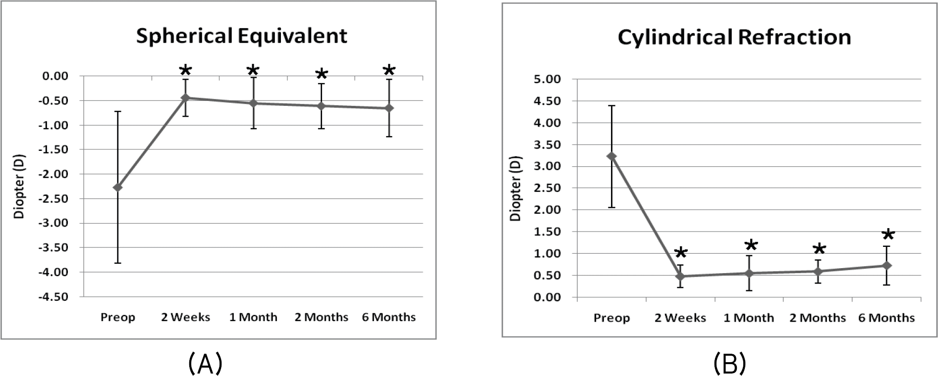 | Figure 1.Spherical equivalent refraction (A) and cylindrical refraction (B) over 6 months after excimer laser surgery to correct residual refractive error following cataract surgery in 11 eyes. Spherical equivalent and cylindrical refraction decreased significantly after excimer laser surgery (* p<0.05). |
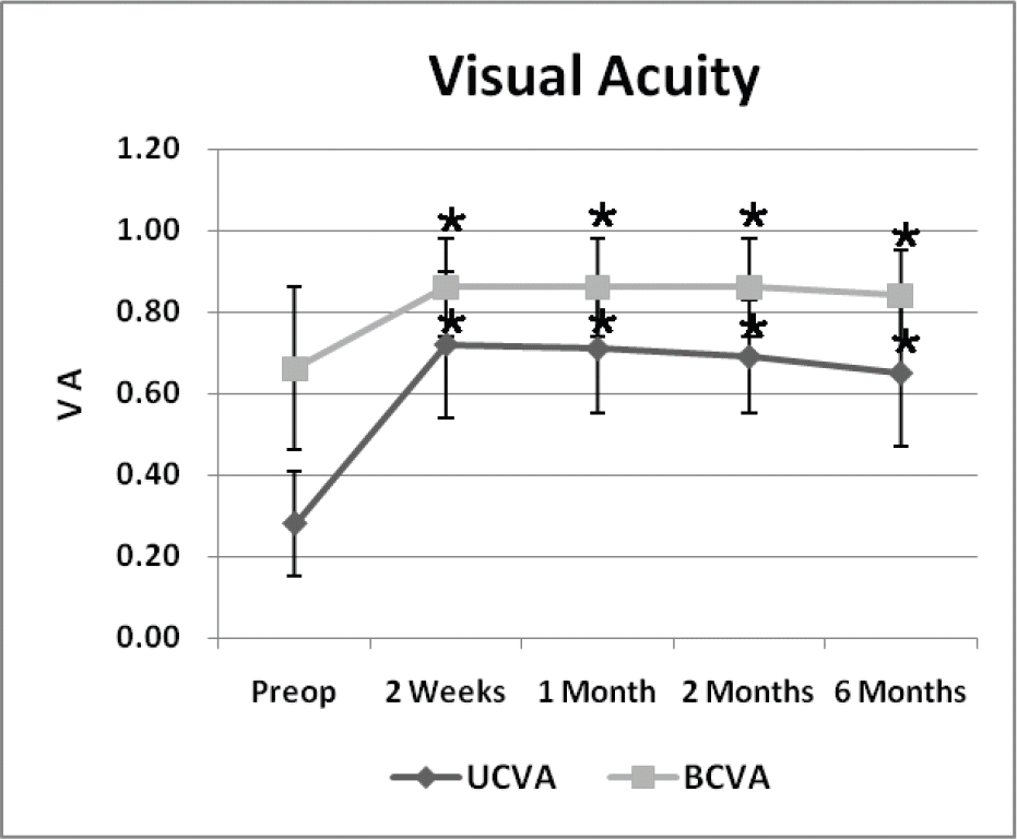 | Figure 2.Time course of uncorrected visual acuity (UCVA) and best corrected visual acuity (BCVA) for 11 eyes with excimer laser surgery for residual refractive error after cataract surgery. UCVA and BCVA improved at 2 weeks after surgery and were stable until 6 months after surgery. (* p<0.05) |
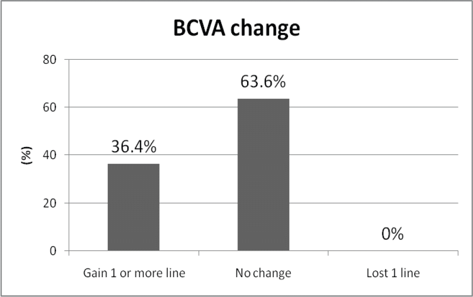 | Figure 3.Postoperative change in best corrected visual acuity (BCVA) in 11 eyes after excimer laser surgery for induced astigmatism following cataract surgery. The final BCVA improved by 1 line in 4 eyes (36.4%) and was unchanged in 7 eyes (63.6%) at 6 months. |
Table 1.
Patient characteristics and results before and after cataract surgery but before eximer laser surgery
| Exam | Preoperative state | Postoperative state |
|---|---|---|
| Age | 65.73±10.05 | |
| Sex (Male:Female) | 6:5 | |
| Axial Length (mm) | 24.52±1.59 | |
| * UCVA | 0.18±0.134 | 0.27±0.13 |
| † BCVA | 0.38±0.20 | 0.76±0.24 |
| Cylinderical refraction (D) | 2.80±1.01 | 3.23±1.02 |
| Spherical equivalent (D) | -3.63±4.99 | -2.27±1.43 |
| Pachymetry (um) | 544.71±43.64 | 551.45±52.57 |
| Keratometry | 43.10±2.24 | 44.99±1.97 |
| ‡ Sim K’s Astigmatism | 2.61±1.27 | 3.02±1.07 |
Table 2.
Refractive and visual acuity data before and after excimer laser surgery following cataract surgery in 11 eyes
| Preoperative state | Postoperative 6 months | |||||||
|---|---|---|---|---|---|---|---|---|
| NO | Age/Sex | Procedure | * UCVA | Manifest Refraction | † BCVA | UCVA | Manifest Refraction | BCVA |
| 1 | 86/M | PRK | 0.1 | +1.00 -5.5×180˚ | 0.4 | 0.63 | +0.50 -1.25×180˚ | 0.8 |
| 2 | 59/F | PRK | 0.25 | +0.75 -3.5×150˚ | 1.0 | 1.0 | +0.50 -0.25×150˚ | 1.0 |
| 3 | 66/M | LASIK | 0.2 | -2.50 -2.00×85˚ | 0.8 | 0.63 | -0.50 -0.75×90˚ | 0.8 |
| 4 | 52/F | LASIK | 0.2 | -1.75 -3.00×175˚ | 1 | 0.63 | -0.50 -0.75×170˚ | 1.0 |
| 5 | 78/F | LASIK | 0.1 | -3.00 -4.50×180˚ | 0.32 | 0.4 | -0.25 -1.00×180˚ | 0.63 |
| 6 | 60/M | LASIK | 0.5 | +1.00 -2.50×25˚ | 1.0 | 0.8 | +0.50 -0.50×30˚ | 1.0 |
| 7 | 67/M | LASIK | 0.3 | +0.50 -3.50×155˚ | 0.8 | 0.63 | -0.25 -1.25×155˚ | 0.8 |
| 8 | 68/M | LASIK | 0.4 | -1.25 -2.50×175˚ | 0.8 | 0.8 | -0.75 sphere | 0.8 |
| 9 | 72/F | LASIK | 0.2 | -1.5 -3.25×95˚ | 0.5 | 0.4 | -1.00 -1.00×90˚ | 0.63 |
| 10 | 57/M | LASIK | 0.4 | +0.25 -2.75×30˚ | 1.0 | 0.8 | -0.50 -0.50×30˚ | 1 |
| 11 | 58/F | LASIK | 0.4 | -0.75 -2.50×165˚ | 0.8 | 0.63 | -0.75 -0.75×170˚ | 0.8 |
Table 3.
Postoperative change in refractive astigmatism in 11 eyes after excimer laser surgery following cataract surgery
Table 4.
Predictability of excimer laser surgery for spherical equivalent refraction (SE) and cylinder (C) following cataract surgery in 11 eyes
Table 5.
Stability of eximer laser surgery for spherical equivalent refraction (SE) and cylinder (C) following cataract surgery in 11 eyes
 XML Download