

 PDF
PDF ePub
ePub Citation
 Print
Print


Abstract
Purpose
To describe the number of cases and cause of Avellino corneal dystrophy (ACD) exacerbated after LASIK (laser in situ keratomileusis), to study the possible mechanism of exacerbation, and to suggest possible preventive measures and the most suitable treatment modality.
Methods
Twenty-eight (56 eyes) ACD patients who underwent LASIK were enrolled, and 24 parameters including pre-and post-operative best corrected visual acuity, contrast sensitivity, and corneal opacity were analyzed. To evaluate the effect of treatment modality for exacerbated cases after LASIK, the progression of corneal opacity, visual acuity, and contrast sensitivity were compared between the two surgical methods (mechanical removal from LASIK interface with removal of surgical flap and deep lamellar keratoplasty (DLKP)).
Results
All patients showed progression of corneal opacities after LASIK regardless of the type of laser instruments, microkeratomes, or the size or thickness of the LASIK flap. As for treatment of exacerbated ACD patients after LASIK, DLKP showed better results than mechanical removal from the LASIK interface after removal of the surgical flap.
Go to : 
References
1. Holland EJ, Daya SM, Stone EM. . Avellino corneal dystrophy. Clinical manifestations and natural history. Ophthalmology. 1992; 99:1564–8.
2. Kennedy SM, McNamara M, Hillery M. . Combined granular lattice dystrophy (Avellino corneal dystrophy). Br J Ophthalmol. 1996; 80:489–90.

3. Dolmetsch AM, Stockl FA, Folberg R. . Combined granular-lattice corneal dystrophy (Avellino) in a patient with no known Italian ancestry. Can J Ophthalmol. 1996; 31:29–31.
4. Kocak-Atlintas AG, Kocak-Midillioglu I, Akarsu AN, Duman S. BIGH3 gene analysis in the different diagnosis of corneal dystrophies. Cornea. 2001; 20:64–8.
5. Klintworth GK. Advances in the molecular genetics of corneal dystrophies. Am J Ophthalmol. 1999; 128:747–54.
6. Konishi M, Mashima Y, Nakamura Y. . Granular-lattice (Avellino) corneal dystrophy in Japanese patients. Cornea. 1997; 16:635–8.
7. Bas AM, Nano HD. In situ myopic keratomileusis results in 30 eyes at 15 months. J Refract Corneal Surg. 1991; 7:223–31.
8. Pallikaris IG, Siganos DS. Excimer laser in situ keratomileusis and photorefractive keratectomy for correction of high myopia. J Refract Corneal Surg. 1994; 10:498–510.
9. Fiander DC, Tayfour F. Excimer lase in situ keratomileusis in 124 myopic eyes. J Refract Surg. 1995; 11:S234–8.
10. Wan XH, Lee HC, Stulting RD. . Exacerbation of Avellino corneal dystrophy after laser in situ keratomileusis. Cornea. 2002; 21:223–6.
11. Jun RM, Tchah H, Kim TI. . Avellino corneal dystrophy after LASIK. Ophthalmology. 2004; 111:463–8.
12. Chung SH, Kim CY, Kim EK. The Classification and Clinical Characteristics in Korean Patients with Avellino Corneal Dystrophy. J Korean Ophthalmol Soc. 2005; 46:938–44.
13. Song QH, Singh RP, Richardson TP. . Transforming growth factorbeta1 expression in cultured corneal fibroblasts in response to injury. J Cell Biochem. 2000; 77:186–99.
14. Brown CT, Applebaum E, Banwatt R. . Synthesis of stromal glycosaminoglycans in response to injury. J Cell Biochem. 1995; 59:57–68.
15. Kay EP, Lee MS, Seong GJ. . TGF-betas stimulate cell proliferation via an autocrine production of FGF-2 in corneal stromal fibroblasts. Curr Eye Res. 1998; 17:286–93.
16. Chen C, Michelini-Norris B, Stevens S. . Measurement of mRNAs for TGFs and extracellular matrix proteins in corneas of rats after PRK. Invest Ophthalmol Vis Sci. 2000; 41:4108–16.
17. Jun RM, Tchah H, Kim TI. . Avellino corneal dystrophy after LASIK. Ophthalmology. 2004; 111:463–8.
18. Rho MI, Grossniklaus HE., Chung SH. . Avellino Corneal Dystrophy Exacerbated After LASIK: Scanning Electron Microscopic Findings. Cornea. 2006; 25:306–11.
19. Moon JW, Kim SW, Kim TI. . Homozygous granular corneal dystrophy type II (Avellino corneal dystrophy): natural history and progression after treatment. Cornea. 2007; 26:1095–100.
20. Escribano J, Hernando N, Ghosh S. . cDNA from human ocular ciliary epithelium homologous to beta ig-h3 is preferentially expressed as an extracellular protein in the corneal epithelium. J Cell Physiol. 1994; 160:511–21.
21. Akiya S, Takahashi H, Nakano N. . Granular-lattice (Avellino) corneal dystrophy. Ophthalmologica. 1999; 213:58–62.
22. Korvatska E, Munier FL, Chaubert P. . On the role of kerato-epithelin in the pathogenesis of 5q31-linked corneal dystrophies. Invest Ophthalmol Vis Sci. 1999; 40:2213–9.
23. Kim TI, Pak JH, Chae JB. . Mitomycin C inhibits recurrent Avellino dystrophy after phototherapeutic keratectomy. Cornea. 2006; 25:220–3.
24. Kanai A, Yamaguchi T, Nakajima A. The histochemical and analytical electron microscopy studies of the corneal granular dystrophy. Nippon Ganka Gakkai Zasshi. 1977; 81:145–54.
25. Wittebol-Post D, van der Want JJ, van Bijsterveld OP. Granular dystrophy of the cornea (Groenouw’s type I). Is the keratocyte the primary source after all? Ophthalmologica. 1987; 195:169–77.
26. Menasche M, Savoldelli M, Pouliquen Y. The keratocyte or fibroblast of the cornea: morphological and biochemical characteristics in normal stroma and a few cases of corneal dystrophies. Pathol Biol. 1992; 40:871–8.
27. Shimazaki J, Shimmura S, Ishioka M, Tsubota K. Randomized clinical trial of deep lamellar keratoplasty vs penetrating keratoplasty. Am J Ophthalmol. 2002; 134:159–65.
Go to :

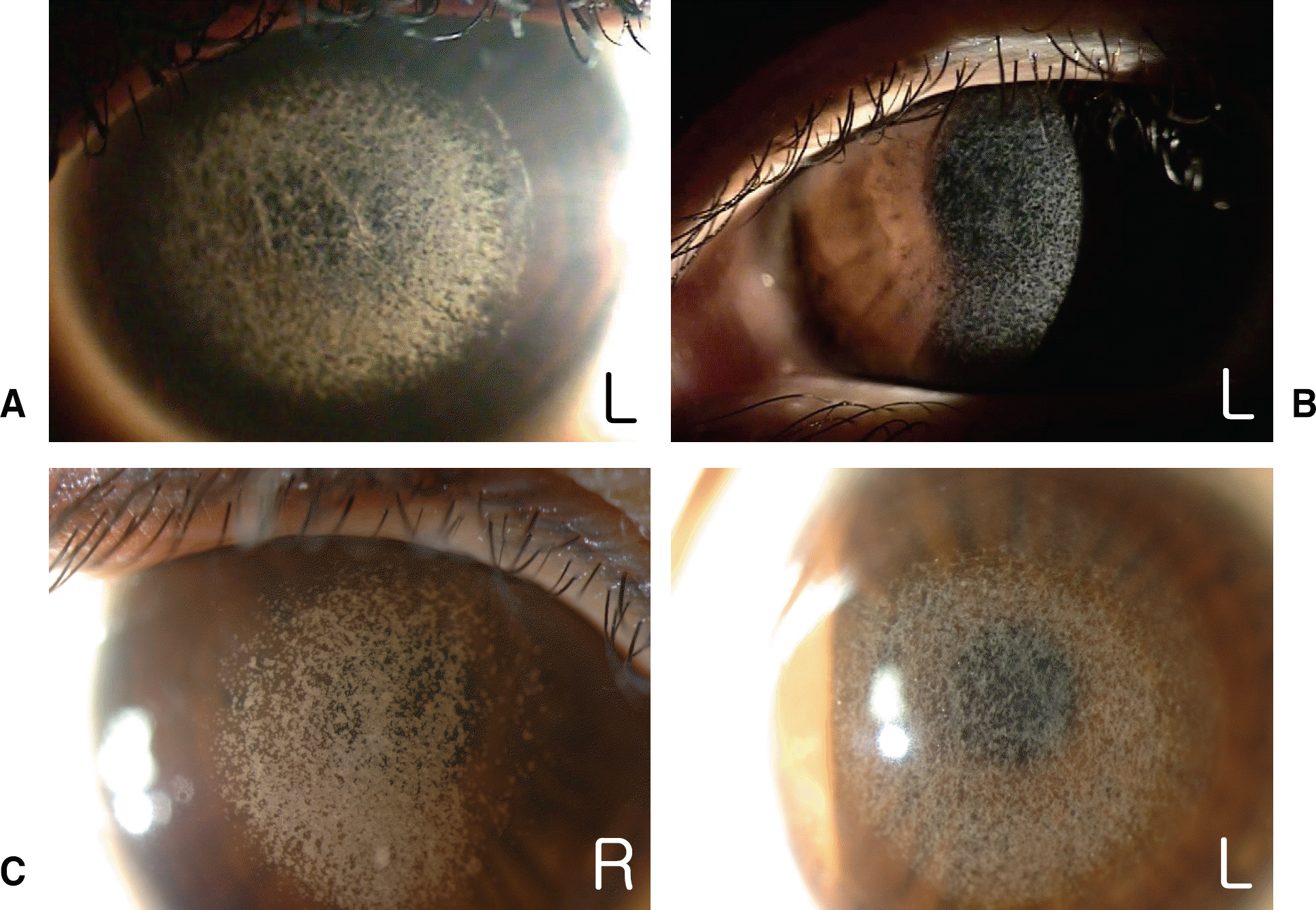
 | Figure 2.Slit-lamp photographs showing exacerbated Avellino corneal dystrophy after LASIK. Corneal images from case 1: (A) Right eye (B) Left eye |
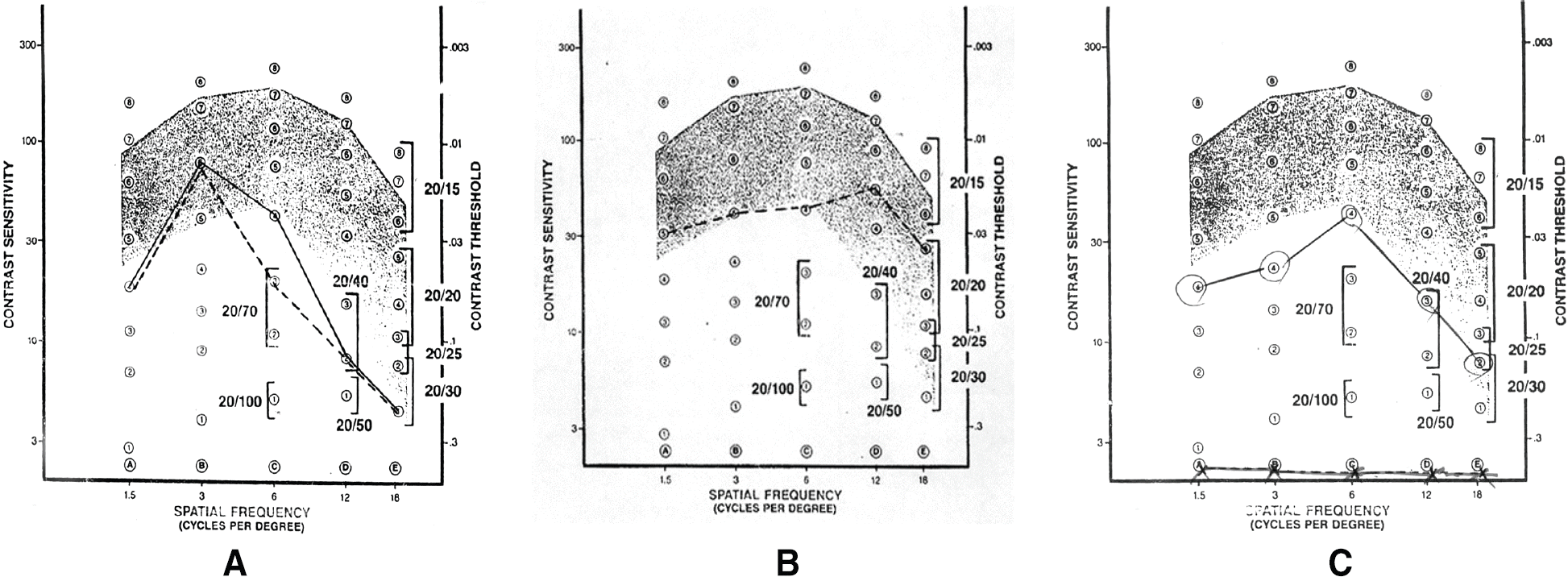 | Figure 3.Contrast sensitivity test: (A) before mechanical removal of corneal deposits from LASIK interface, (B) at 2 weeks after surgery, (C) at three and a half years after surgery (R=right eye; L=left eye; solid line=right; dotted line=left). |
 | Figure 4.(a) Corneal image from case 1: (A) before removal of LASIK flap (at three and a half years after mechanical removal of corneal deposits from LASIK interface). (B) 2 weeks and (C) 1 year and 1 month after LASIK flap removal (R=right eye; L=left eye). |
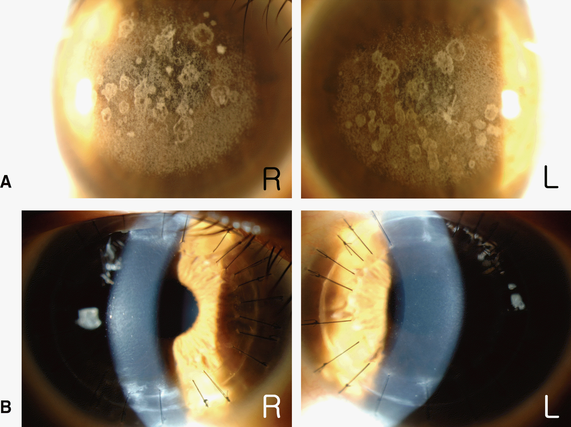 | Figure 5.Corneal images from case 2 (A) before deep lamellar keratoplasty (DLKP). (B) after 6 months of DLKP (R) and after 4 months of DLKP (L). (R=right eye; L=left eye) |
Table 1.
Clinical Characteristics of Avellino Corneal Dystrophy Patients Exacerbated after LASIK∗
| Characteristics | No. of eyes | ||
|---|---|---|---|
| Male | Female | ||
| Sex | 9 (18 eyes) | 19 (38 eyes) | 56 |
| Age (years) | 30.32±7.20 (22~48) | 56 | |
| Time to exacerbation after surgery (months) | 47.80±26.67 (7~108) | 56 | |
| Schirmer test (mm/5min) | 7.17±5.75 (2~24) | 18 | |
| preoperative | postoperative | ||
| UCVA† | 0.09±0.09 (HM‡~0.4) | 0.56±0.27 (HM~1.25) | 30/56 |
| BCVA§ | 0.99±0.14 (0.7~1.25) | 0.73±0.23 (0.4~1.25) | 44/54 |
| Refractive error (Diopter) | -5.89±3.23 (-14.50~+1.50) | -0.56±1.09 (-3.63~+1.00) | 56/54 |
| Corneal Refractive Power (Diopter) | 44.13±1.38 (41.13~47.25) | 40.54±2.72 (36.00~44.50) | 42/18 |
| Corneal thickness (μ m) | 536.39±36.02 (460~626) | 338.25±105.64 (225~474) | 36/8 |
| Amounts of correction (Diopter) | -5.45±3.14 (-10.25~+1.38) | 36 | |
| Thickness (μ m) | Diameter (mm) | ||
| LASIK flap | 135±18.67 (100~160) | 8.44±1.03 (5.5~10) | 32/32 |
Table 2.
Comparison of BCVA* between Pre- and Post- LASIK†
| Eyes (%) | ||
|---|---|---|
| BCVA∗ | Worsened | 34 (60.7) |
| No change | 6 (10.7) | |
| Improved | 0 (0) | |
| Unknown | 16 (28.6) |
Table 3.
Comparison of CST* between Pre- and Post-LASIK†
| Eyes (%) | ||
|---|---|---|
| CST∗ | Normal range | 9 (16.0) |
| Subnormal | 31 (55.4) | |
| N/A‡ | 16 (28.6) |
Table 4.
Status of Corneal Deposits at Pre- and Post-LASIK∗
| Eyes (%) | |||
|---|---|---|---|
| preoperative | postoperative | ||
| Presence | 34 (60.71) | 56 (100) | |
| Absence | 5 (8.93) | 0 (0) | |
| N/A | 17 (30.36) | 0 (0) | |
| Amounts | 1-5 | 3 (5.36) | 0 (0) |
| 6-10 | 17 (30.36) | 0 (0) | |
| 11-19 | 4 (7.14) | 0 (0) | |
| 20≤ | 2 (3.57) | 56 (100) | |
| N/A† | 30 (53.57) | 0 (0) | |
Table 5.
Presence of Diffuse Fine Deposits and Exacerbation after LASIK∗
| Eyes (%) | ||
|---|---|---|
| Diffuse fine deposits | Exacerbation | |
| Presence | 56 (100) | 56 (100) |
| Absence | 0 (0) | 0 (0) |
Table 6.
Location of Corneal Deposits after LASIK∗
| Eyes (%) | |
|---|---|
| Superior | 0 (0) |
| Superior, center, inferior | 4 (7.1) |
| Center | 8 (14.3) |
| Center, inferior | 43 (76.8) |
| Inferior | 1 (1.8) |
Table 7.
Types of LASER instruments Used
 XML Download