Abstract
Purpose
We compared the results of implanting an implantable contact lens (ICL) and an iris claw lens (Artisan® lens) in patients’ wtih myopia and sought to determine a basis for choosing between the two lenses.
Methods
ICLs were implanted in 32 eyes of 18 patients, and Artisan® lenses were implanted in 40 eyes of 23 patients. Uncorrected visual acuity, refraction, endothelial cell density, intraocular pressure, and slit lamp measurements were taken for 12 months.
Results
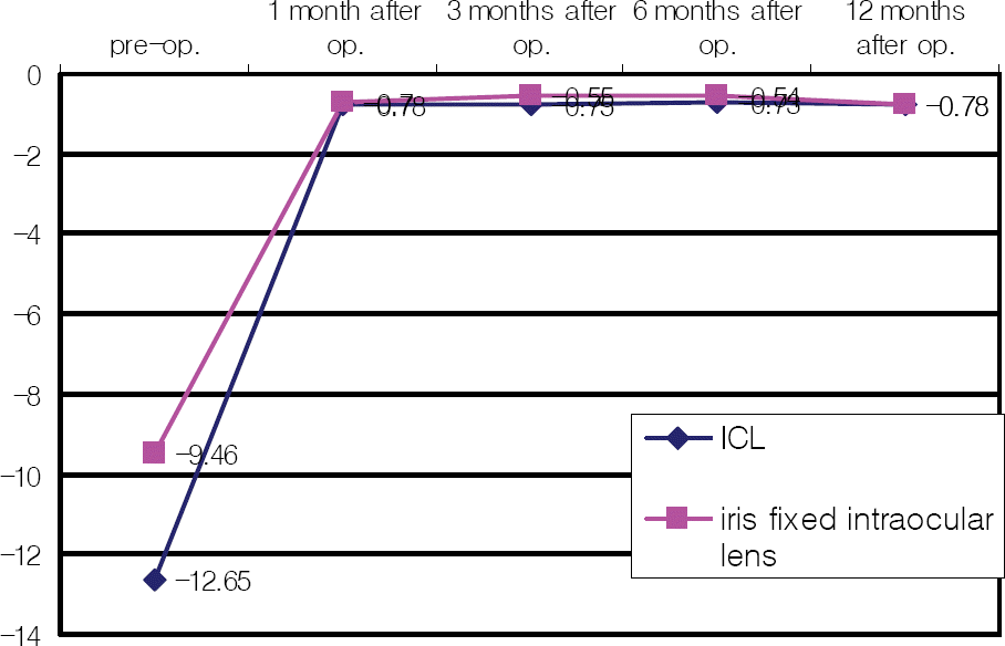
All the patients had improved UCVA from the day after the operation until the 12th month. The mean spherical equivalent refraction at postoperative month 12 was -0.78±0.54D in the ICL group and -0.78 ±0.59D in the Artisan® lens group. In the same period, endothelial cell density loss was 5.34% in the ICL group but was not significant in the Artisan® lens group. There were no significant complications in either group.
Conclusions
Both ICL and Artisan® lens implantation resulted in immediate visual improvement and stability of vision during the follow-up period. There was no significant difference in post-operative results. Further study will be needed to decide which lens is the better choice for delicate conditions in myopic patients.
Go to : 
References
1. Barraquer J. Anterior chamber plastic lenses. Results of conclusions from five years experience. Trans Ophthal Soc U K. 1959; 79:393–424.
2. Zaldivar R, Rocha G. The current status of phakic intraocular lenses. Int Ophthalmol Clin. 1996; 36:107–11.

3. Assetto V, Benedetti S, Pesando P. Collamer intraocular contact lens to correct high myopia. J Cataract Refract Surg. 1996; 22:551–6.

4. Fechner PU, Worst JFG. A new concave intraocular lens for the correction of high myopia. European journal of Implant and Refractive Surgery. 1989; 1:41–3.
5. Fechner PU, Strobel J, Wichmann W. Correction of myopia by implantation of a concave Worst-iris claw lens into phakic eyes. Refract Corneal Surg. 1991; 7:286–98.

6. Landesz M, Worst JG, Siertsema JV, van Rij G. Correction of high myopia with the Worst myopia claw intraocular lens. J Refract Surg. 1995; 11:16–25.

7. Izak MGJ, Izak A. Inflammatory reaction associated to Artisan phakic refractive IOL implantation. Budo CJ, editor. The Artisan lens. 1st. Panama: Highlights of Ophthalmology;2004. v. 1. chap. 14.
8. Heitzmann J, Binder PS, Kassar BS, Nordan LT. The correction of high myopia using excimer laser. Arch Ophthalmol. 1993; 111:1627–34.
9. Holladay JT, Dudeja KR, Chang J. Functional vision and corneal changes after laser in situ keratomileusis determined by contrast sensitivity, glare test and corneal topography. J Cataract Refract Surg. 1991; 25:663–9.
10. Fyodorov SN, Zuev VK, Tumanyan ER. . Analysis of long-term clinilcal and functional results of intraocular correction of high myopia. Ophthalmosurg (Moscow). 1990; 2:3–6.
11. Maloney RK, Nguyen LH, John ME. Artisan phakic intraocular lens for myopia. Short-term results of a prospective, multicenter study. Ophthalmology. 2002; 109:1631–41.

12. Benedetti S, Casamenti V, Marcaccio L. . Correction of myopia of 7 to 24 diopters with the Artisan phakic intraocular lens: two-year follow-up. J Refract Surg. 2005; 21:116–26.

13. Maloney RK, Nguyen LH, John ME. Artisan phakic intraocular lens for myopia. Short-term results of a prospective, multicenter study. Ophthalmology. 2002; 109:1631–41.

14. Dejaco-Ruhswurm I, Scholz U, Pieh S. . Long-term endothelial changes in phakic eyes with posterior chamber intraocular lenses. J Cataract Refract Surg. 2002; 28:1589–93.

15. Pe´rez-Santonja JJ, Iradier MT, Benı´tez del Castillo JM. . Chronic subclinical inflammation in phakic eyes with intraocular lenses to correct myopia. J Cataract Refract Surg. 1996; 22:183–7.
16. Budo C, Hessloehl JC, Izak M. . Multicenter study of the Artisan phakic inraocular lens. J Cataract Refract Surg. 2000; 26:1163–71.
17. Menezo JL, Cisneros AL, Salvador VR. Endothelial study of iris-claw phakic lens: Four year follow-up. J Cataract Refract Surg. 1998; 24:1039–49.

18. Maroccos R, Vaz F, Marinho A. . Glare and halos after Phakic IOL. Surgery for the correction of high myopia. Ophthalmology. 2001; 98:1055–9.
Go to : 
 | Figure 1.Stability of spherical equivalent (SE) refraction. Spherical equivalent at post-operative 1 month remained until 3, 6 and 12 months postoperatively. There was no significant difference between the two groups at 1, 3, 6, and 12 months after operation. |
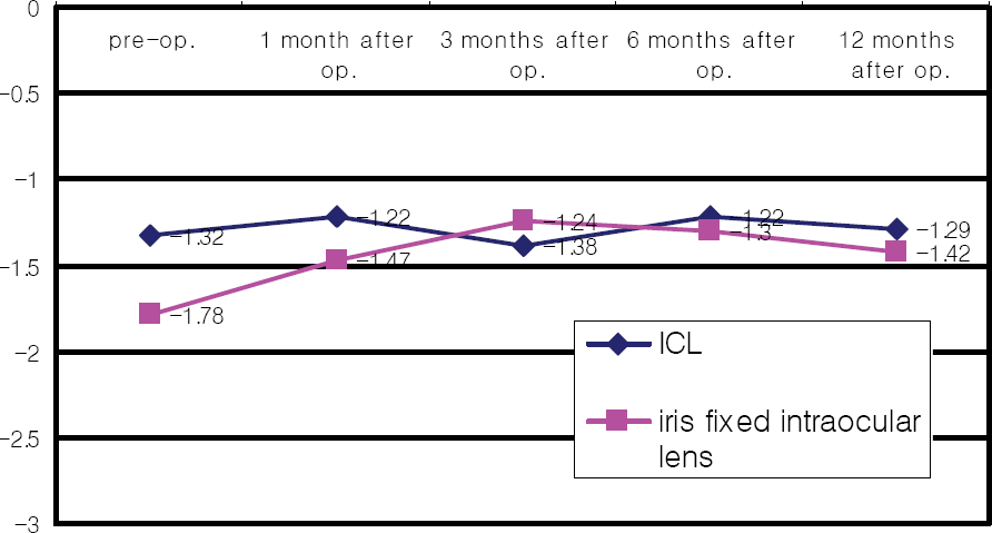
 | Figure 2.Stability of refractive astigmatism. There was no significant difference between the two groups at 1, 3, 6, and 12 months after operation. |
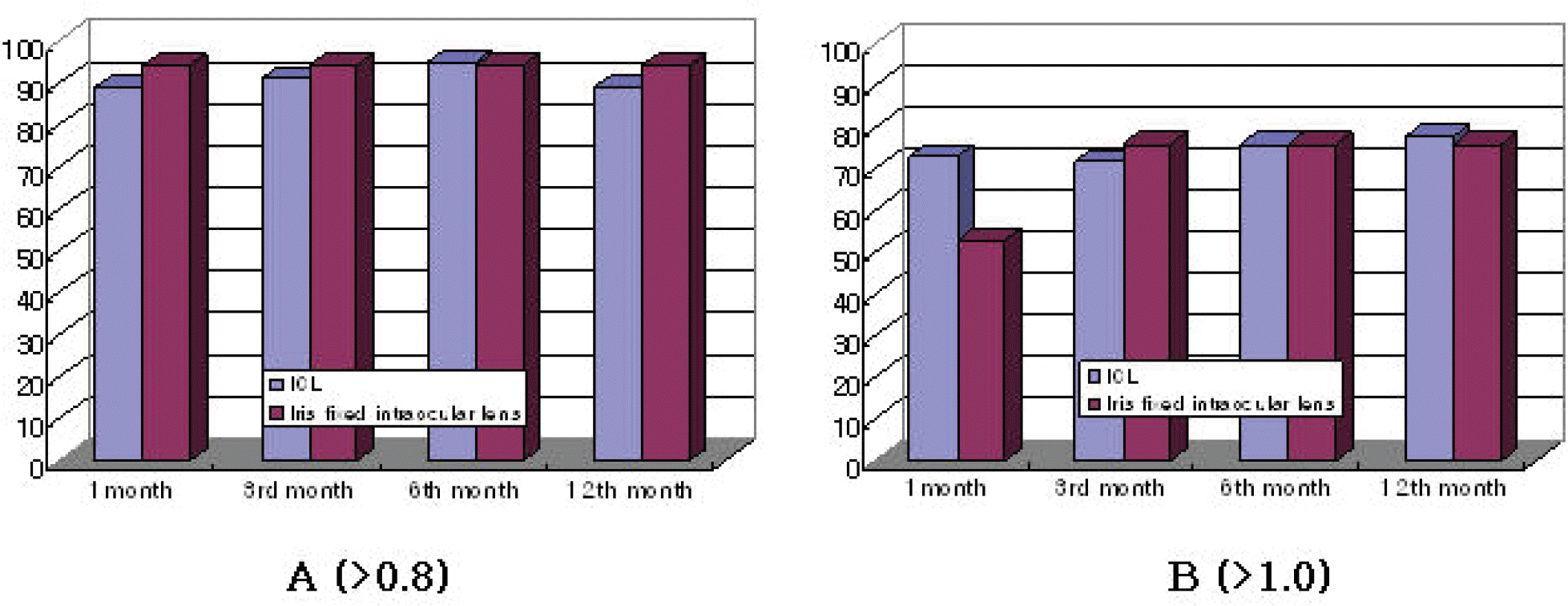
 | Figure 3.Percentage of one-year postoperative uncorrected visual acuity over 0.8 (A) and 1.0 (B) in ICL and iris fixed intraocular lens groups at 1, 3, 6, and 12 months. There was no significant change in difference between the two groups. |
Table 1.
Demographics of patients
Table 2.
Comparison of spherical equivalent over time between ICL group and iris fixed intraocular lens group
Table 3.
Comparison of refractive astigmatism over time between ICL group and iris fixed intraocular lens group
Table 4.
Uncorrected visual acuity over time in ICL group and iris-fixed intraocular lens group (%)
| Uncorrected visual acuity | > 0.8 | >1.0 | ||
|---|---|---|---|---|
| ICL | Iris fixed intraocular lens | ICL group | Iris fixed intraocular lens | |
| 1 st month | 89 | 94 | 73 | 53 |
| 3 rd month | 91 | 94 | 72 | 76 |
| 6 th month | 95 | 94 | 76 | 76 |
| 12 th month | 89 | 94 | 68 | 76 |
Table 5.
Endothelial cell density over time in ICL group and iris-fixed intraocular lens group
Table 6.
Postoperative Complications in ICL group and iris-fixed intraocular lens group


 PDF
PDF ePub
ePub Citation
 Print
Print


 XML Download