

 PDF
PDF ePub
ePub Citation
 Print
Print


Abstract
Purpose
To evaluate the effect of Optibiol®, which is composed of multiple anti-oxidative and anti-inflammatory dietary elements, on the symptoms, clinical manifestations, and expression of various inflammatory cytokines in dry eye patients.
Methods
Patients suffering from dry eye were given Optibiol® for 3 months. They completed questionnaires regarding dry eye symptom and underwent slit lamp biomicroscopic examinations, tear film breakup times, Shirmer tests, and conjunctival fluorescein staining examinations on a monthly basis during the intake of Optibiol®. Sampling of serum and tears was conducted at baseline and 3 months after taking Optibiol®, and various inflammatory cytokines in the serum and tears were measured with multiplex bead immunoassay.
Result
Sixty-three patients were included in this study. After taking Optibiol®, the dry eye symptoms, Schirmer scores, tear film breakup times, and conjunctival staining scores (Oxford scale) showed significant improvement, and the expression of most inflammatory cytokines had decreased: in particular, IL-1? and MIP-1? in serum, and IL-17 and MIP-1? in tears were significantly lower.
Go to : 
References
1. Calder PC. Polyunsaturated fatty acids, inflammation and immunity. Lipids. 2001; 36:1007–24.

2. Calder PC. n-3 polyunsaturated fatty acids, inflammation and immunity: pouring oil on troubled waters or another fishy tale? Nutr Res. 2001; 21:309–41.
3. Calder PC. n-3 fatty acids and cardiovascular disease: evidence explained and mechanisms explored. Clin Sci. 2004; 107:1–11.
4. Paiva SA, Russell RM. Beta carotene and other carotenoids as antioxidants. J Am Coll Nutr. 1999; 18:426–33.
5. Snodderly DM. Evidence for protection against age-related macular degeneration by carotenoids and antioxidant vitamins. Am J Clin Nutr. 1995; 62:S1448–61.
6. Seddon JM, Ajani UA, Sperduto RD. . Dietary carotenoids, vitamins A, C, and E, and advanced age-related macular degeneration. JAMA. 1994; 272:1413–20.
7. Age-Related Eye Disease Study Research Group A randomized, placebo-controlled, clinical trial of high-dose supplementation with vitamins C and E, beta carotene, and zinc for age-related macular degeneration and vision loss: AREDS report no. 8. Arch Ophthalmol. 2001; 119:1417–36.
8. Age-Related Eye Disease Study Research Group A randomized, placebo controlled, clinical trial of high-dose supplementation with vitamins C and E and beta carotene for age-related cataract and vision loss: AREDS report no. 9. Arch Ophthalmol. 2001; 119:1439–52.
9. Bron AJ. The Doyne Lecture. Reflections on the tears. Eye. 1997; 11:583–602.
10. Lemp MA. The definition and classification of dry eye disease: report of the Definition and Classification Subcom mittee of the International Dry Eye WorkShop (2007). Ocul Surf. 2007; 5:75–92.
11. Her J, Yu SI, Seo SG. Clinical effects of various antiinflammatory therapies in dry eye syndrome. J Korean Ophthalmol Soc. 2006; 47:1901–10.
12. Stephen C, Pflugfelder SC. Antiinflammatory therapy for dry eye. Am J Ophthalmol. 2004; 137:337–42.
13. Tatlipinar S, Akpek EK. Topical cyiclosporin in the treatment of ocular surface disorder. Br J Ophthalmol. 2005; 89:1363–7.
14. Mrukwa-Kominek E, Rogowska-Godela A, Gierek-Ciaciura S. Effect of anti-inflammatory therapy on the treatment of dry eye syndrome. Klin Oczna. 2007; 109:79–84.
15. Creuzot C, Passemard M, Viau S. . Improvement of dry eye symptoms with polyunsaturated fatty acids. J Fr Ophtalmol. 2006; 29((8)):868–73.
16. Sherry B, Tekamp-Olson P, Gallegos C. . Resolution of the two components of macrophage inflammatory protein 1, and cloning and characterization of one of those components, macrophage inflammatory protein 1? . J Exp Med. 1988; 168:2251–9.
17. Menten P, Wuyts A, Van Damme J. Macrophage inflammatory protein-1. Cytokine Growth Factor Rev. 2002; 13:455–81.
18. Mrugacz M, Zelazowska B, Bakunowicz-Lazarczyk A. . Elevated Tear Fluid Levels of MIP-1? in Patients with Cystic Fibrosis. J Interferon Cytokine Res. 2007; 27:491–6.
19. Yoon KC, De Paiva Cs, Qi H. . Expression of Th-1 Chemokines and Chemokine Receptors on the Ocular Surface of C57BL/6 Mice: Effects of Desiccating Stress. Invest Ophthalmol Vis Sci. 2007; 48:2561–9.
20. Suzuki Y, Ohgami K, Shiratori K. . Suppressive effects of astaxanthin against rat endotoxin-induced uveitis by inhibiting the NF-kappaB signaling pathway. Exp Eye Res. 2006; 82:275–81.
21. Zhao Y, Yu W, Hu W, Yuan Y. Anti-inflammatory and anticoagulant activities of lycopene in mice. Nutrition Research. 2003; 23:1591–5.
22. Nakamura S, Shibuya M, Nakashima H. . Involvement of Oxidative Stress on Corneal Epithelial Alterations in a Blink-Suppressed Dry Eye. Invest Ophthalmol Vis Sci. 2007; 48:1552–8.
23. Tüzün A, Erdil A, Inal V. . Oxidative stress and antioxidant capacity in patients with inflammatory bowel disease. Clin Biochem. 2002; 35:569–72.
24. Francisco PG, Esther M, Salvador C, Tomas A. Anti-inflamatory action of pluchea sagittalis: involvement of an antioxidant mechanism. Life Sci. 1996; 59:2033–40.
Go to :
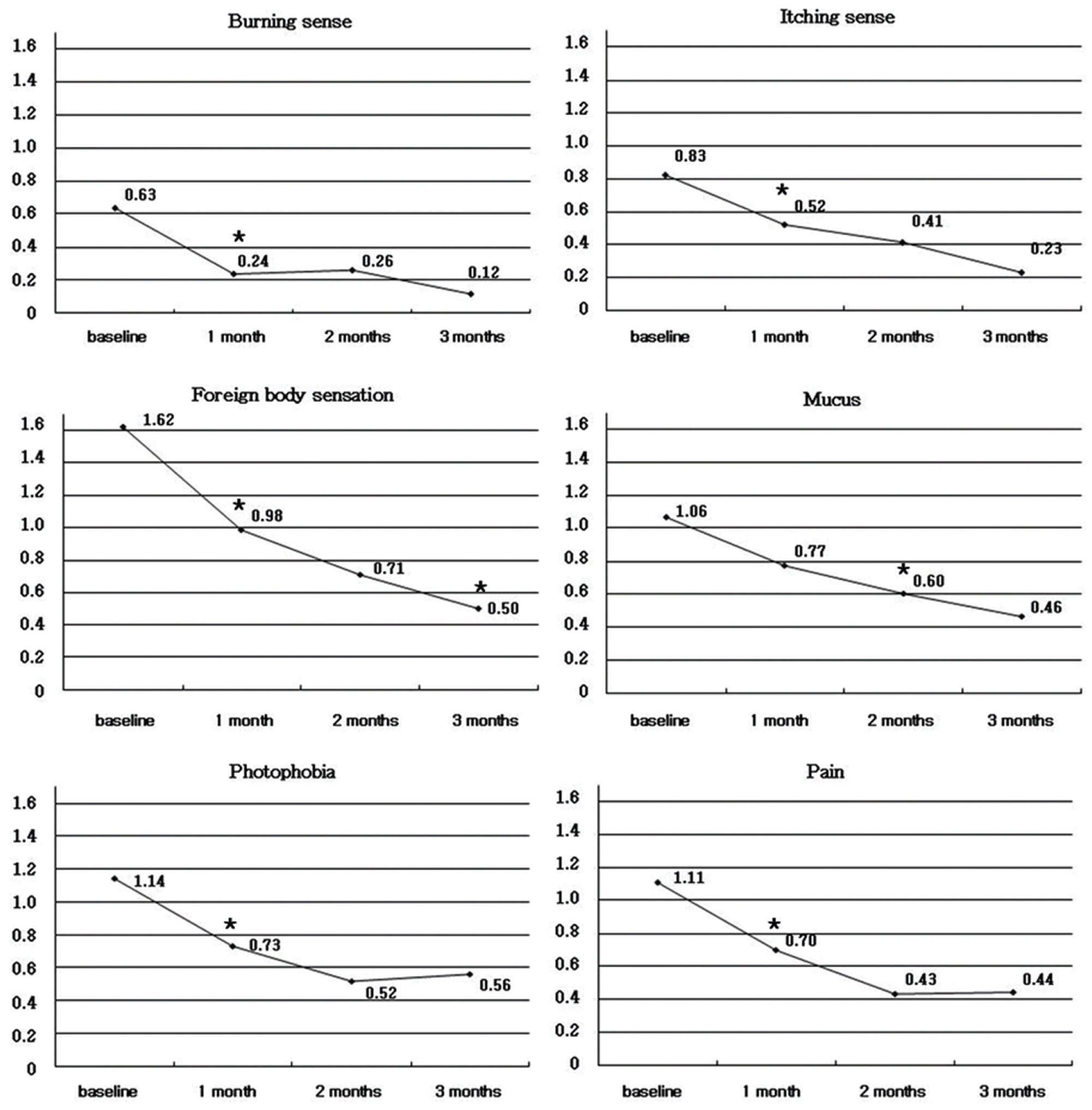 | Figure 1.Changes in scores of 6 ocular symptoms before and after taking Optibiol®.* p<0.05 : One way ANOVA, Duncan multiple range test. |
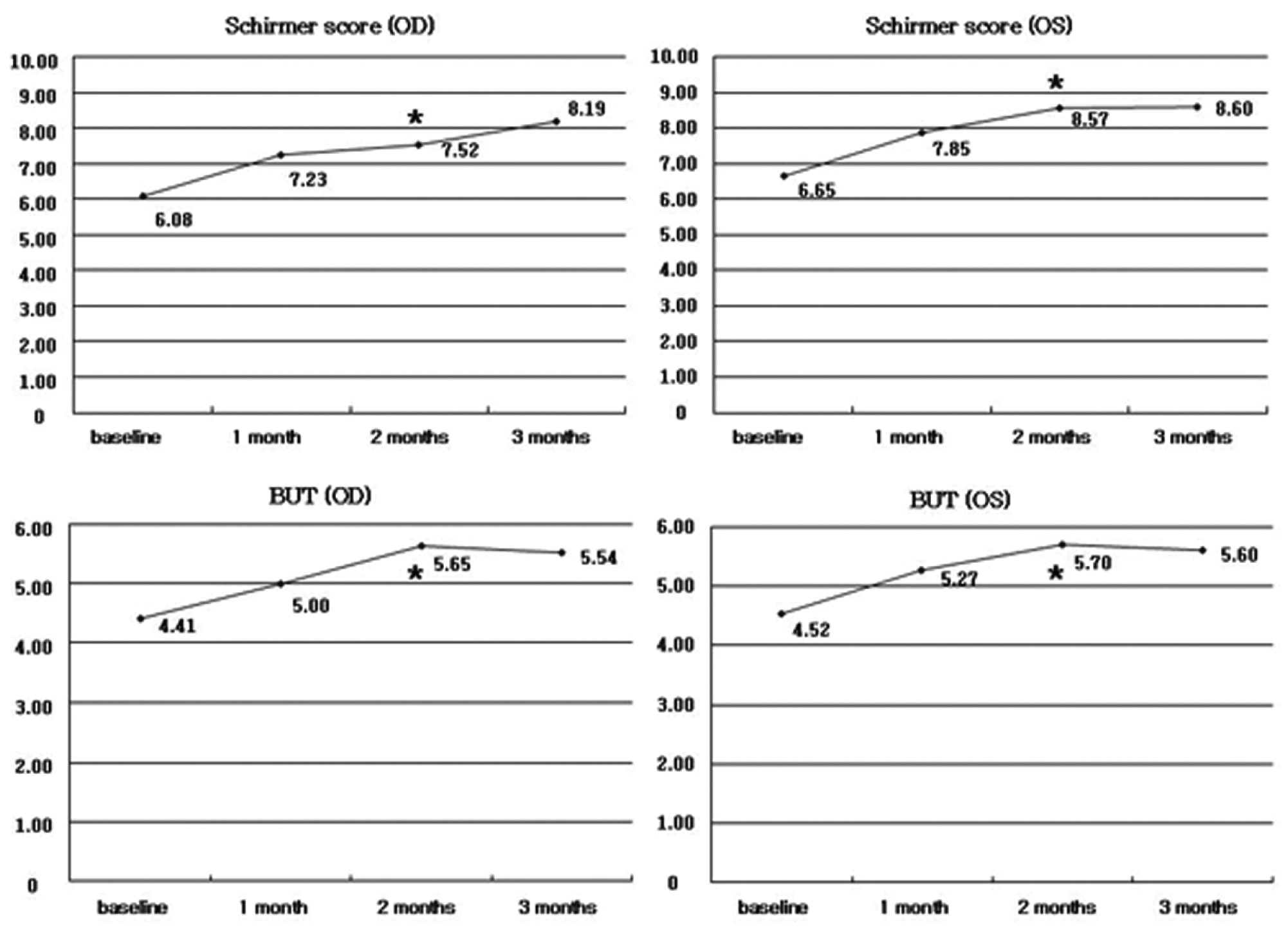 | Figure 2.Changes in Schirmer score and tear break-up time before and after taking Optibiol®. BUT=tear film breakup time,* p<0.05 : One way ANOVA, Duncan multiple range test. |
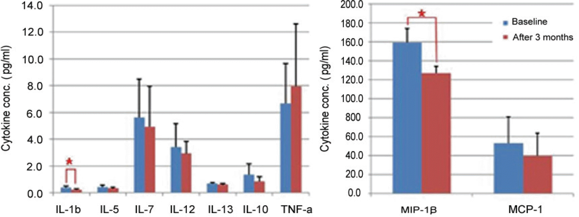 | Figure 3.Concentration of inflammatory cytokines in serum, before and after taking Optibiol®. After taking Optibiol®, the expression of most inflammatory cytokines had decreased in serum, especially IL-1β and MIP-1β were significantly lower; IL=interleukin; TNF-α=tumor necrosis factor-α; MIP-1β=macrophage inflammatory protein-1β; MCP-1=monocyte chemoattractant protein-1; * p<0.05 : Student T-test. |
 | Figure 4.According to age groups, changes of inflammatory cytokines in serum after taking Optibiol®. No significant difference in changes of inflammatory cytokines in serum after taking Optibiol®, according to age groups; IL=interleukin; TNF-α=tumor necrosis factor-α; MIP-1β=macrophage inflammatory protein-1β; MCP-1=monocyte chemoattractant protein-1. |
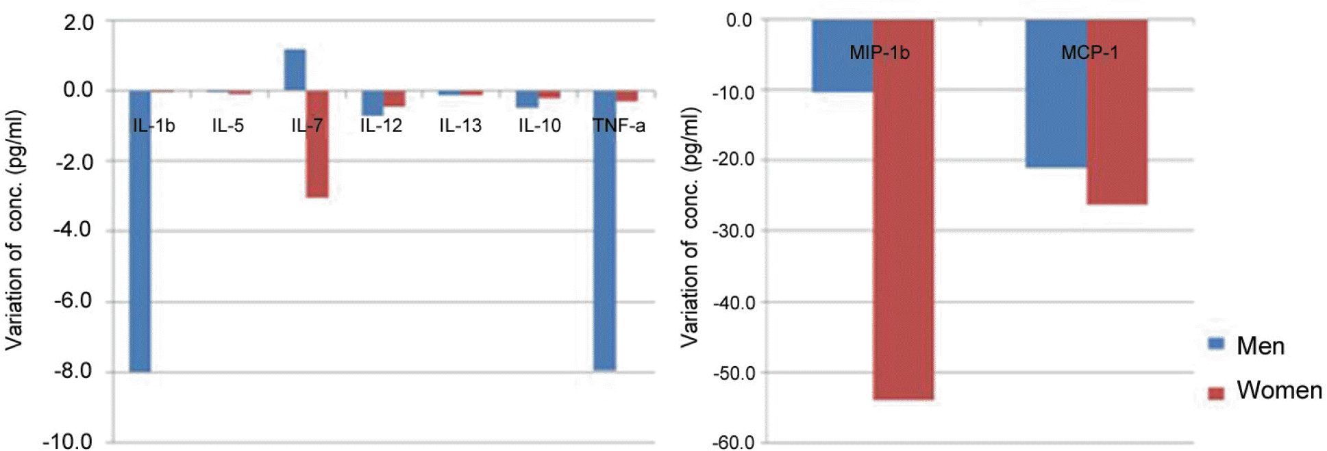 | Figure 5.Changes of inflammatory cytokines in serum according to sex after taking Optibiol®. No significant difference in changes of inflammatory cytokines in serum after taking Optibiol®, according to sex. IL=interleukin; TNF-α=tumor necrosis factor-α; MIP-1β=macrophage inflammatory protein-1β; MCP-1=monocyte chemoattractant protein-1. |
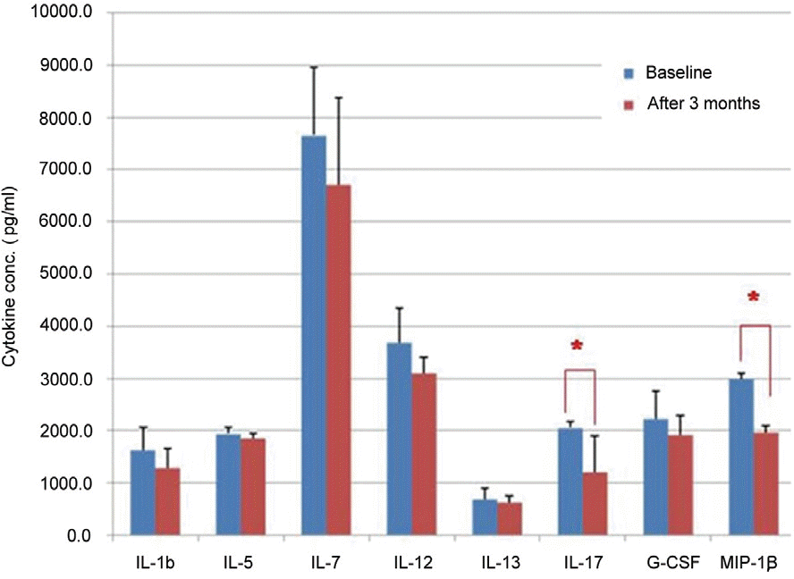 | Figure 6.Concentrations of inflammatory cytokines in tear, before and after taking Optibiol®. After taking Optibiol®, the expression of all inflammatory cytokines had decreased in tear, especially IL-17 and MIP-1β were significantly lower. IL=interleukin; G-CSF=granulocyte colony stimulating factor; MIP-1β=macrophage inflammatory protein-1β. |
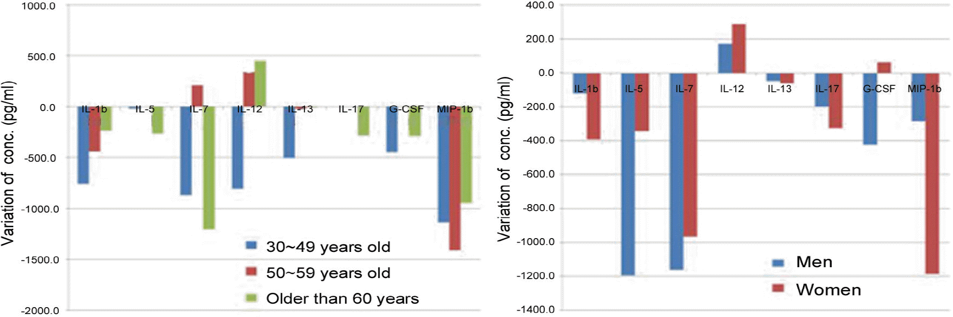 | Figure 7.Changes of concentrations of inflammatory cytokines in tear according to age groups and sex after taking Optibiol®. No significant difference in changes of inflammatory cytokines in serum after taking Optibiol®, according to age groups and sex. IL=interleukin; G-CSF=granulocyte colony stimulating factor; MIP-1β=macrophage inflammatory protein-1β. |
Table 1.
Nutritional facts in 1 capsule of Optibiol®
Table 2.
Changes of conjunctival staining scores and serum lipid level before and after taking Optibiol
| Baseline | After 3 months | p-value∗ | |
|---|---|---|---|
| Conjunctival stain score (OD) | 1.48 | 0.94 | 0.000 |
| Conjunctival stain score (OS) | 1.48 | 0.98 | 0.000 |
| Total cholesterol | 182.77 mg/dl | 182.49 mg/dl | 0.939 |
| Triglyceride | 130.93 mg/dl | 133.02 mg/dl | 0.818 |
| Low density lipoprotein (LDL) | 114.63 mg/dl | 110.46 mg/dl | 0.217 |
| High density lipoprotein (HDL) | 55.76 mg/dl | 59.42 mg/dl | 0.018 |
 XML Download