Abstract
Purpose
To assess the effect of age and gender on intraocular pressure (IOP) in a large Korean population.
Methods
This cross-sectional study included 102,218 healthy Koreans who were aged between 20 and 79 and had no preexisting ocular conditions that could affect the IOP. All the subjects had undergone a physical check up between 1996 and 2005, and their medical records were reviewed retrospectively. Anthropometric measurements, blood tests, noncontact tonometry, and fundus examination were performed on all participants. Subjects were grouped according to decade of age. For all subjects and each age group, age and systemic variables were analyzed by multiple regression analysis on their relationship with IOP.
Results
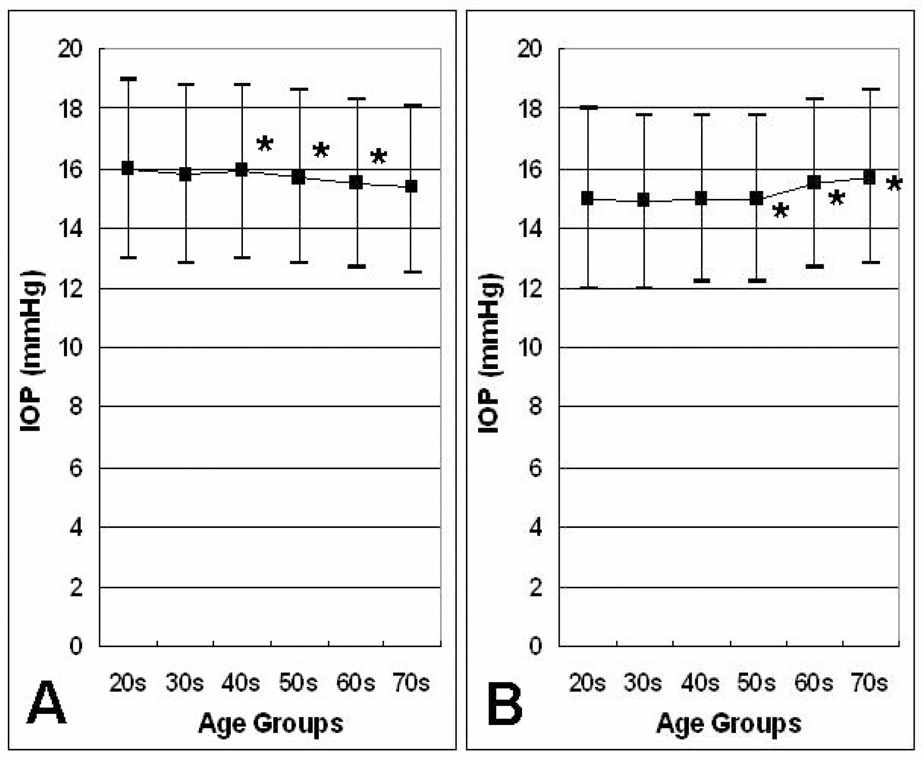
A significant trend of decreasing IOP was observed in the 40s, 50s, and 60s in men, while a significant trend of increasing IOP was found in the 50s, 60s, and 70s in women. Multiple regression analysis revealed different IOP trends with age between age groups in both men and women. In general, the IOP had a significant positive correlation with systolic blood pressure, body-mass index (BMI), hematocrit, and serum cholesterol, especially with BMI in men and hematocrit in women.
Conclusions
In a multiple regression analysis, the IOP trend in each age group was quite different from each other in a large Korean population, and it was suggested that women may have a steeper increasing slope (or less steep decreasing slope) of IOP with age than men. Further investigations with longitudinal study would be required to clarify the age- and gender-related physiologic changes of IOP.
References
1. Morgan RW, Drance SM. Chronic open-angle glaucoma and ocular hypertension. An epidemiological study. Br J Ophthalmol. 1975; 59:211–5.

2. Collaborative Normal-Tension Glaucoma Study Group. The effectiveness of intraocular pressure reduction in the treatment of normal-tension glaucoma. Am J Ophthalmol. 1998; 126:498–505.
3. Mitchell P, Smith W, Attebo K, Healey PR. Prevalence of open-angle glaucoma in Australia. The Blue Mountains Eye Study. Ophthalmology. 1996; 103:1661–9.
4. Sommer A, Tielsch JM, Katz J, et al. Relationship between intraocular pressure and primary open angle glaucoma among white and black Americans. The Baltimore Eye Survey. Arch Ophthalmol. 1991; 109:1090–5.
5. Dielemans I, Vingerling JR, Wolfs RC, et al. The prevalence of primary open-angle glaucoma in a population-based study in The Netherlands. The Rotterdam Study. Ophthalmology. 1994; 101:1851–5.
6. Leske MC, Connell AM, Schachat AP, Hyman L. The Barbados Eye Study. Prevalence of open angle glaucoma. Arch Ophthalmol. 1994; 112:821–9.
7. Rochtchina E, Mitchell P. Projected number of Australians with glaucoma in 2000 and 2030. Clin Experiment Ophthalmol. 2000; 28:146–8.

8. Klein BE, Klein R. Intraocular pressure and cardiovascular risk variables. Arch Ophthalmol. 1981; 99:827–9.

9. Armaly MF. On the distribution of applanation pressure. I. Statistical features and the effect of age, sex, and family history of glaucoma. Arch Ophthalmol. 1965; 73:11–8.
10. David R, Zangwill L, Stone D, Yassur Y. Epidemiology of intraocular pressure in a population screened for glaucoma. Br J Ophthalmol. 1987; 71:766–71.

11. Bonomi L, Marchini G, Marraffa M, et al. Prevalence of glaucoma and intraocular pressure distribution in a defined population. The Egna-Neumarkt Study. Ophthalmology. 1998; 105:209–15.
12. Colton T, Ederer F. The distribution of intraocular pressures in the general population. Surv Ophthalmol. 1980; 25:123–9.

14. Kahn HA, Leibowitz HM, Ganley JP, et al. The Framingham Eye Study. I. Outline and major prevalence findings. Am J Epidemiol. 1977; 106:17–32.
15. Klein BEK, Klein R, Linton KLP. Intraocular pressure in an American community. The Beaver Dam Eye Study. Invest Ophthalmol Vis Sci. 1992; 33:2224–8.
16. Carel RS, Korczyn AD, Rock M, Goya I. Association between ocular pressure and certain health parameters. Ophthalmology. 1984; 91:311–4.

17. Rochtchina E, Mitchell P, Wang JJ. Relationship between age and intraocular pressure: the Blue Mountains Eye Study. Clin Experiment Ophthalmol. 2002; 30:173–5.

18. Weih LM, Mukesh BN, McCarty CA, Taylor HR. Association of demographic, familial, medical, and ocular factors with intraocular pressure. Arch Ophthalmol. 2001; 119:875–80.

19. McLeod SD, West SK, Quigley HA, Fozard JL. A longitudinal study of the relationship between intraocular and blood pressures. Invest Ophthalmol Vis Sci. 1990; 31:2361–6.
20. Wu SY, Leske MC. Associations with intraocular pressure in the Barbados Eye Study. Arch Ophthalmol. 1997; 115:1572–6.

21. Shiose Y. The aging effect on intraocular pressure in an apparently normal population. Arch Ophthalmol. 1984; 102:883–7.

22. Nomura H, Ando F, Niino N, et al. The relationship between age and intraocular pressure in a Japanese population: The influence of central corneal thickness. Curr Eye Res. 2002; 24:81–5.

23. Nomura H, Shimokata H, Ando F, et al. Age-related changes in intraocular pressure in a large Japanese population. Ophthalmology. 1999; 106:2016–22.

24. Mori K, Ando F, Nomura H, et al. Relationship between intraocular pressure and obesity in Japan. Int J Epidemiol. 2000; 29:661–6.

25. Nakano T, Tatemichi M, Miura Y, et al. Long-term physiologic changes of intraocular pressure. Ophthalmology. 2005; 112:609–16.
26. Kashiwagi K, Shibuya T, Tsukahara S. De novo age-related retinal disease and intraocular-pressure changes during a 10-year period in a Japanese adult population. Jpn J Ophthalmol. 2005; 49:36–40.

27. Lee JS, Kim CM, Choi HY, Oum BS. A relationship between intraocular pressure and age and body mass index in a Korean population. J Korean Ophthalmol Soc. 2003; 44:1559–66.
28. Bankes JL, Perkins ES, Tsolakis S, Wright JE. Bedford Glaucoma Survey. Br Med J. 1968; 1:791–796.

29. Leske MC, Connel AM, Wu SY, et al. Distribution of intraocular pressure: the Barbados Eye Study. Arch Ophthalmol. 1997; 115:1051–7.
30. Kupfer C. Clinical significance of pseudofacility. Sanford R. Gifford Memorial Lecture. Am J Ophthalmol. 1973; 75:193–204.
31. Bloom JN, Levene RZ, Thomas G, Kimura R. Fluorophotometry and the rate of aqueous flow in man. I. Instrumentation and normal values. Arch Ophthalmol. 1976; 94:435–43.
32. Brubaker RF, Nagataki S, Townsend DJ, et al. The effect of age on aqueous humor formation in man. Ophthalmology. 1981; 88:283–7.

33. Miyazaki M, Segawa K, Urakawa Y. Age-related changes in the trabecular meshwork of the normal human eye. Jpn J Ophthalmol. 1987; 31:558–69.
34. Altintas O, Caglar Y, Yuksel N, et al. The effects of menopause and hormone replacement therapy on quality and quantity of tear, intraocular pressure and ocular blood flow. Ophthalmologica. 2004; 218:120–9.

35. Qureshi IA. Ocular hypertensive effect of menopause with and without systemic hypertension. Acta Obstet Gynecol Scand. 1996; 75:266–9.

36. Toker E, Yenice O, Temel A. Influence of serum levels of sex hormones on intraocular pressure in menopausal women. J Glaucoma. 2003; 12:436–40.

37. Affinito P, Di Spiezio Sardo A, Di Carlo C, et al. Effects of hormone replacement therapy on ocular function in postmenopause. Menopause. 2003; 10:482–7.

38. Lee AJ, Mitchell P, Rochtchina E, et al. Female reproductive factors and open angle glaucoma: the Blue Mountains Eye Study. Br J Ophthalmol. 2003; 87:1324–8.

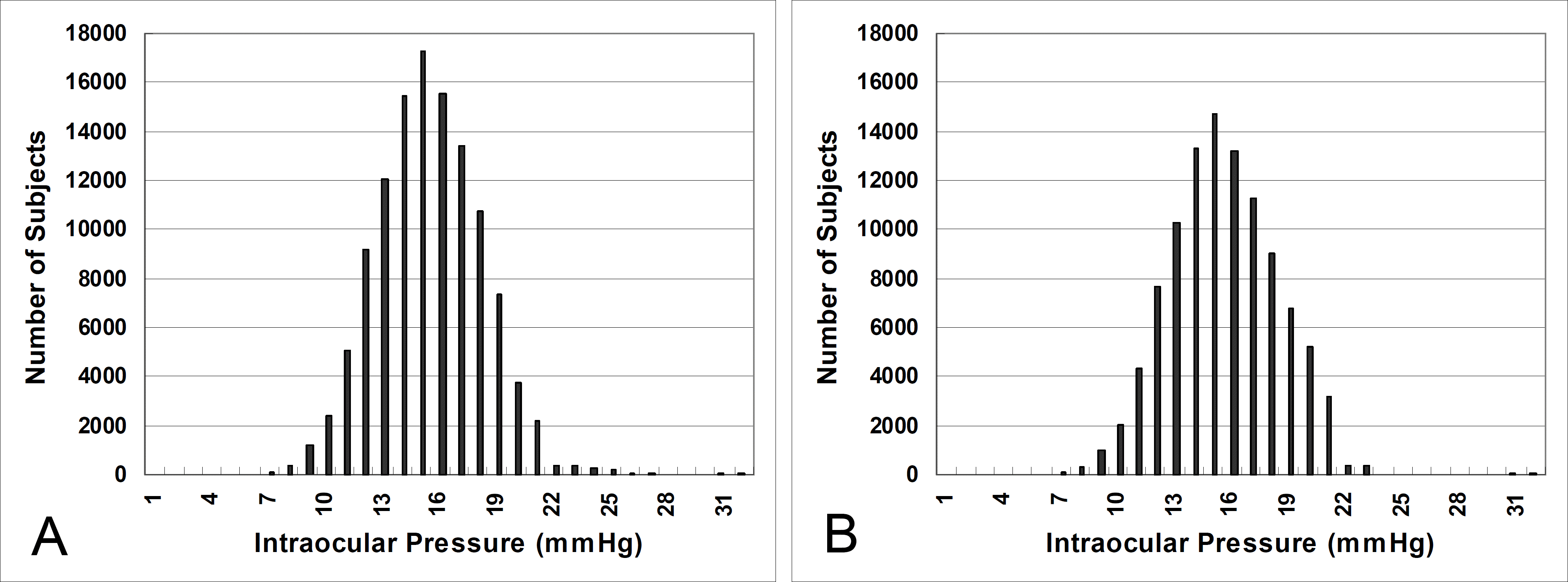
Figure 1.
The distribution of intraocular pressure in total subjects. (A) Before and (B) after excluding subjects with doubtable findings, such as optic disc abnormality and fundus abnormality based on fundus photography, IOP over 21 mm Hg, participant’s own history or family history of medical and/or ocular conditions that could possibly affect the IOP (glaucoma, proliferative diabetic retinopathy, hypertension, hyperlipidemia, etc.), and drug history of antiglaucoma, antihypertensive, or antihyperlipidemic medications.

Figure 2.
The distribution of mean intraocular pressure (IOP) by age group. (A) In men, there was a significant difference in the mean IOP between those in their 40s and 50s and between those in their 50s and 60s (P<0.001, respectively; by one-way ANOVA with multiple comparison)*). (B) In women, there was a significant difference in the (mean IOP between those in their 50s and 60s and between those in their 60s and 70s (P<0.001, respectively; by one-way ANOVA with multiple comparison) (*).

Table 1.
The number of participants by age group
|
Age Groups* |
Total (n=102,218) | ||||||
|---|---|---|---|---|---|---|---|
| 20s (n=3,209) | 30s (n=17,848) | 40s (n=37,479) | 50s (n=29,820) | 60s (n=12,465) | 70s (n=1,397) | ||
| Men | 1,603 | 9,672 | 21,186 | 16,725 | 7,329 | 855 | 57,370 |
| Women | 1,606 | 8,176 | 16,293 | 13,095 | 5,136 | 542 | 44,848 |
Table 2.
Cross-sectional analysis between intraocular pressure (IOP) and other variables (age, body mass index (BMI), systolic blood pressure (SBP), hematocrit, and cholesterol) using a multiple regression model
| Men (OD) |
Age Groups (at the baseline) |
Total | |||||
|---|---|---|---|---|---|---|---|
| 20s | 30s | 40s | 50s | 60s | 70s | ||
| R2 | 0.042* | 0.028* | 0.026* | 0.019* | 0.020* | 0.016* | 0.024* |
| Age (years) | -0.04±0.026 | 0.004±0.010 | -0.006±0.006 | -0.034*±0.006 | -0.016±0.010 | -0.069†±0.028 | -0.019*±0.001 |
| BMI (kg/m2) | 0.076*±0.020 | 0.070*±0.010 | 0.066*±0.006 | 0.063*±0.007 | 0.098*±0.011 | 0.082‡±0.027 | 0.074*±0.004 |
| SBP (mmHg) | 0.027*±0.006 | 0.022*±0.002 | 0.024*±0.001 | 0.017*±0.001 | 0.015*±0.002 | 0.011‡±0.004 | 0.019*±0.001 |
| Hematocrit (%) | 0.062†±0.028 | 0.015±0.010 | 0.013†±0.006 | 0.011±0.006 | -0.020†±0.009 | -0.027±0.022 | 0.009†±0.003 |
| Cholesterol (mg/dL) | 0.002±0.002 | 0.006*±0.001 | 0.004*±0.001 | 0.011*±0.001 | 0.003*±0.001 | 0.002±0.002 | 0.004*±0.001 |
| Women (OD) |
Age Groups (at the baseline) |
Total | |||||
|
20s |
30s |
40s |
50s |
60s |
70s |
||
| R2 | 0.016* | 0.021* | 0.022* | 0.020* | 0.030* | 0.026‡ | 0.027* |
| Age (years) | -0.076‡±0.027 | -0.017±0.011 | 0.020‡±0.007 | 0.004±0.007 | 0.001±0.013 | 0.029±0.045 | 0.006±0.001 |
| BMI (kg/m2) | 0.066†±0.027 | 0.064*±0.011 | 0.028*±0.007 | 0.002±0.007 | -0.025†±0.012 | 0.031±0.034 | 0.020*±0.004 |
| SBP (mmHg) | 0.008±0.006 | 0.022*±0.002 | 0.019*±0.001 | 0.018*±0.001 | 0.020*±0.002 | 0.005±0.005 | 0.019*±0.001 |
| Hematocrit (%) | 0.082†±0.028 | 0.030‡±0.010 | 0.025*±0.006 | 0.036*±0.008 | 0.078*±0.012 | 0.112‡±0.034 | 0.037*±0.004 |
| Cholesterol (mg/dL) | -0.004±0.003 | 0.006*±0.001 | 0.006*±0.001 | 0.004*±0.001 | 0.006*±0.001 | 0.005±0.003 | 0.005*±0.001 |
Table 3.
Previous cross-sectional studies on the physiologic changes in the intraocular pressure (IOP) with age
| Ethnic Group | Previous Study | Range of Age (years) | The No. of Subjects | Method of IOP Measurement |
Association with Age |
|
|---|---|---|---|---|---|---|
| Univariate Analysis | Multivariate Analysis | |||||
| Cross-sectional Studies | ||||||
| Caucasian | The USA Beaver Dam Eye Study15 | 43-86 | 4,926 | Applanation (GAT*) | Positive | Not significant |
| The USA Framingham Eye Survey14 | 52-85 | 2,631 | Applanation (GAT*) | Positive† | Not done | |
| The Italian Egna-Neumarkt Study11 | ≥40 | 4,297 | Applanation (GAT*) | Positive | Not done | |
| The Australia Melbourne Visual Impairment Project18 | ≥40 | 4,576 | Applanation (Tono-Pen) | Negative | Not significant | |
| The Blue Mountains Eye Study17 | ≥49 | 3,260 | Applanation (GAT*) | Positive | Not significant | |
| The Baltimore Longitudinal Study of Aging19 | 19-89 | 572 | Schiotz | Not significant | Not significant | |
| African | The Barbados Eye Study20 | 40-84 | 4,601 | Applanation (GAT*) | Positive | Positive |
| American | ||||||
| Japanese | Shiose Y21 | 10s-70s | 27,969 S | Schiotz or noncontact | Negative | Negative |
| Nomura H et al22 | 40-80 | 1,317 | Noncontact | Negative | Negative | |
| Nomura H et al23 | 20-79 | 69,643 | Noncontact | Negative | Negative | |
| Mori K et al24 | 14-94 | 70,139 | Noncontact | Negative | Negative | |
| Korean | Lee et al27 | 20s-70s | 13,212 | Noncontact | Not | Negative |
| significant | ||||||
| Longitudinal Studies | ||||||
| Caucasian | The Baltimore Longitudinal Study of Aging19 | 21-85 | 413 | Schiotz | Not significant | |
| Japanese | Nakano T et al25 | 21-49 | 2,330 | Applanation (GAT*) | Negative | |
| Kashiwagi K et al26 | ≥40 | 219 | Noncontact | Negative | ||
| Nomura H et al23 | 20-79 | 38,554 | Noncontact | Positive | ||


 PDF
PDF ePub
ePub Citation
 Print
Print


 XML Download