

 PDF
PDF ePub
ePub Citation
 Print
Print


Abstract
Purpose
To investigate the epidemiologic and clinical features of uveal melanomas in Korean patients.
Methods
The medical records of 35 patients (35 eyes) with the diagnosis of uveal melanoma between September 2004 and December 2006 were reviewed retrospectively.
Results
Of the 35 patients, 22 were males (62.9%) and 13 were females (37.1%), with a mean age at diagnosis of 48.2±14.1 years (range, 24-82 years). Decreased visual acuity was the most common symptom in 23 patients (65.7%), while 6 patients (17.1%) were detected without prior symptoms. The shape of the uveal melanoma was dome-shaped in 27 eyes (77.1%) and mushroom-shaped in 8 eyes (22.9%). The mean largest basal diameter of the tumors was 9.0±3.3 mm (2.4-19.0 mm), and the mean apical height was 6.2±2.6 mm (1.1-13.0 mm). The tumors were classified according to their size; a small melanoma was found in 4 eyes (11.4%), a medium melanoma in 29 eyes (82.9%), and a large melanoma in 2 eyes (5.7%).
Go to : 
References
1. Scotto J, Fraumeni JF Jr, Lee JAH. Melanomas of the eye and other noncutaneous sites: Epidemiologic aspects. J Natl Cancer Inst. 1976; 56:489–91.
2. Egan KM, Seddon JM, Glynn RJ. . Epidemiologic aspects of uveal melanoma. Surv Ophthalmol. 1988; 32:239–51.

3. Singh AD, Topham A. Incidence of uveal melanoma in the United States: 1973-1997. Ophthalmology. 2003; 110:956–61.
4. Kaneko A. Incidence of malignant melanoma of the eye in Japan, 1977-1979. Nippon Ganka Gakkai Zasshi. 1982; 86:1042–5.
5. Kuo PK, Puliafito CA, Wang KM. . Uveal melanoma in China. Int Ophthalmol Clin. 1982; 22:57–71.
6. Shuangshoti S, Panyathanya R. Retinoblastoma and uveal melanoma: A study of 206 cases. J Med Asst Thai. 1973; 56:331–6.
7. Mork T. Malignant neoplasms of the eye in Norway: Incidence, treatment, and prognosis. Acta Ophthalmol. 1961; 39:824–31.
8. Gragoudas ES, Seddon JM, Egan KM. . Long-term results of proton beam irradiated uveal melanomas. Ophthalmology. 1987; 94:349–53.
9. Jensen OA. Malignant melanomas of the uvea in Denmark 1943-1952. A clinical, histopathological and prognostic study. Acta Ophthalmol. 1963; 43:S1–220.
10. Shammas HF, Blodi FC. Prognostic factors in choroidal and ciliary body melanomas. Arch Ophthalmol. 1977; 95:63–9.
11. Tucker MA, Shields JA, Hartge P. . Sunlight exposure as risk factor for intraocular malignant melanoma. N Engl J Med. 1985; 313:789–92.
12. Rhee HC, Lee WR. Two cases of choroidal malignant melanoma and a case of malignant melanoma of the ciliary body. J Korean Ophthalmol Soc. 1990; 31:1107–11.
13. Collaborative Ocular Melanoma Study Group. Design and methods of a clinical trial for a rare condition: COMS Report No.3. Control Clin Trials. 1993; 14:362–73.
14. Virgili G, Gatta G, Ciccolallo L. . Incidence of uveal melanoma in Europe. Ophthalmology. 2007; 114:2309–15.
15. Singh AD, Shields CL, Shields JA, Sato T. Uveal melanoma in young patients. Arch Ophthalmol. 2000; 118:918–23.
16. Shields CL, Shields JA, Milite J. . Uveal melanoma in teenagers and children. A report of 40 cases. Ophthalmology. 1991; 98:1662–6.
17. Rodriguez-Sains R. Ocular findings in patients with dysplastic nevus syndrome. Ophthalmology. 1986; 93:661–5.
18. Mukai S, Dryja T. Loss of alleles at polymorphic loci on chromosome 2 in uveal melanoma. Cancer Genet Cytogenet. 1986; 22:45–53.
19. Char DH, Kroll S, Phillips TL. Uveal melanoma: Growth rate and prognosis. Arch Ophthalmol. 1997; 115:1014–8.
20. Ah-Fat FG, Damato BE. Delays in diagnosis of uveal melanoma and effect on treatment. Eye. 1998; 12:781–2.
21. Lin DT, Munk PL, Maberley AL. . Ultrasonography of pathologically proved choroidal melanoma with a high-resolution small-parts scanner. Can J Ophthalmol. 1987; 22:161–4.
22. Collaborative Ocular Melanoma Study Group. Comparison of clinical, echographic, and Histopathological measurements from eyes with medium-sized choroidal melanoma in the Collaborative Ocular Melanoma Study: COMS Report No. 21. Arch Ophthalmol. 2003; 121:1163–71.
23. Sobottka B, Kreissig I. Ultrasonography of metastases and melanomas of the choroid. Curr Opin Ophthalmol. 1999; 10:164–7.
24. Chang AE, Karnell LH, Menck HR. The National Cancer Data Base report on cutaneous and noncutaneous melanoma: a summary of 84836 cases from the past decade. The American College of Surgeons Commission on Cancer and the American Cancer Society. Cancer. 1998; 83:1664–78.
25. Raivio I. Uveal melanoma in Finland. An epidemiological, clinical, histological and prognostic study. Acta Ophthalmol. 1977; 133:S1–64.
26. Jakobiec FA, Silbert G. Are most iris “melanomas” really nevi? A clinicopathologic study of 189 lesions. Arch Ophthalmol. 1981; 99:2117–32.
27. Shields CL, Shields JA, Materin M. . Iris melanoma: risk factors for metastasis in 169 consecutive patients. Ophthalmology. 2001; 108:172–8.
28. Jensen OA. Malignant melanomas of the human uvea: 25-year follow-up of cases in Denmark, 1943-1952. Acta Ophthalmol. 1982; 60:161–82.
29. Collaborative Ocular Melanoma Study Group. Histopathologic characteristics of uveal melanomas in eyes enucleated from the Collaborative Ocular Melanoma Study: COMS Report No.6. Am J Ophthalmol. 1998; 125:745–66.
Go to :
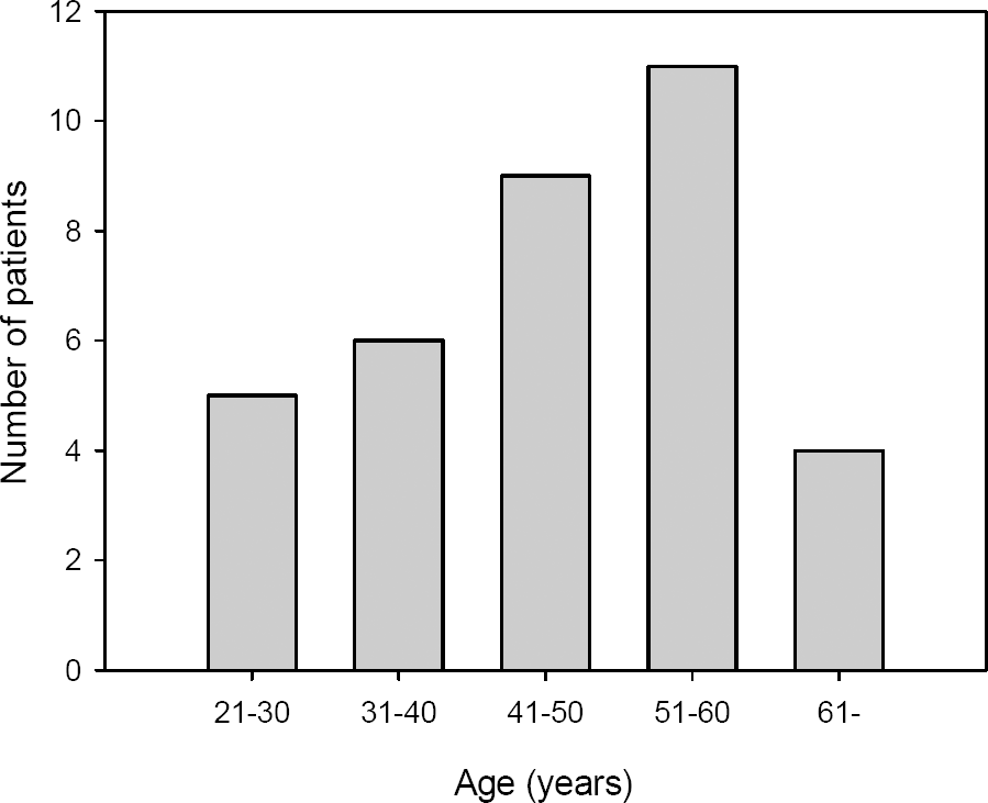 | Figure 1.The number of patients with uveal melanoma increases gradually with age, but it decreases suddenly after 61 years old. |
 | Figure 2.(A) A 79-year-old female. Darkly pigmented, medium-sized juxtapapillary tumor is associated with exudative retinal detachment. (B) On B-scan ultrasonography, the tumor is dome-shaped with regular internal echogenecity. (C) A 49-year-old male. ‘Collar-button’, or mushroom-shaped subretinal tumor is a pathognomonic finding of uveal melanoma. (D) The tumor has low to medium internal reflectivity with typical sound attenuation on A-scan ultrasonography. |
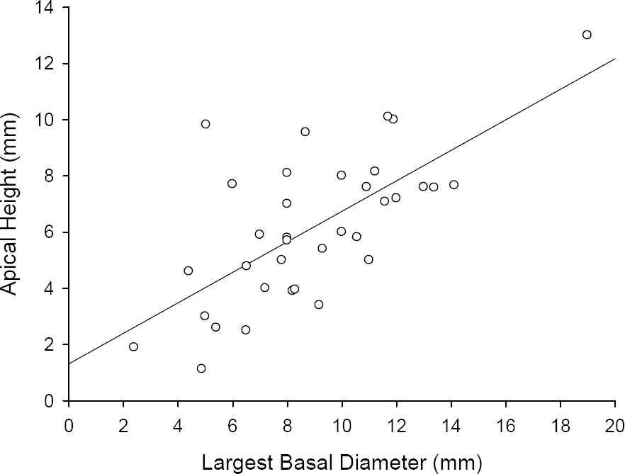 | Figure 3.This graph demonstrates strong linear correlation between the largest basal diameter and apical height of uveal melanoma, with Pearson correlation coefficient of 0.68 (P<0.001). |
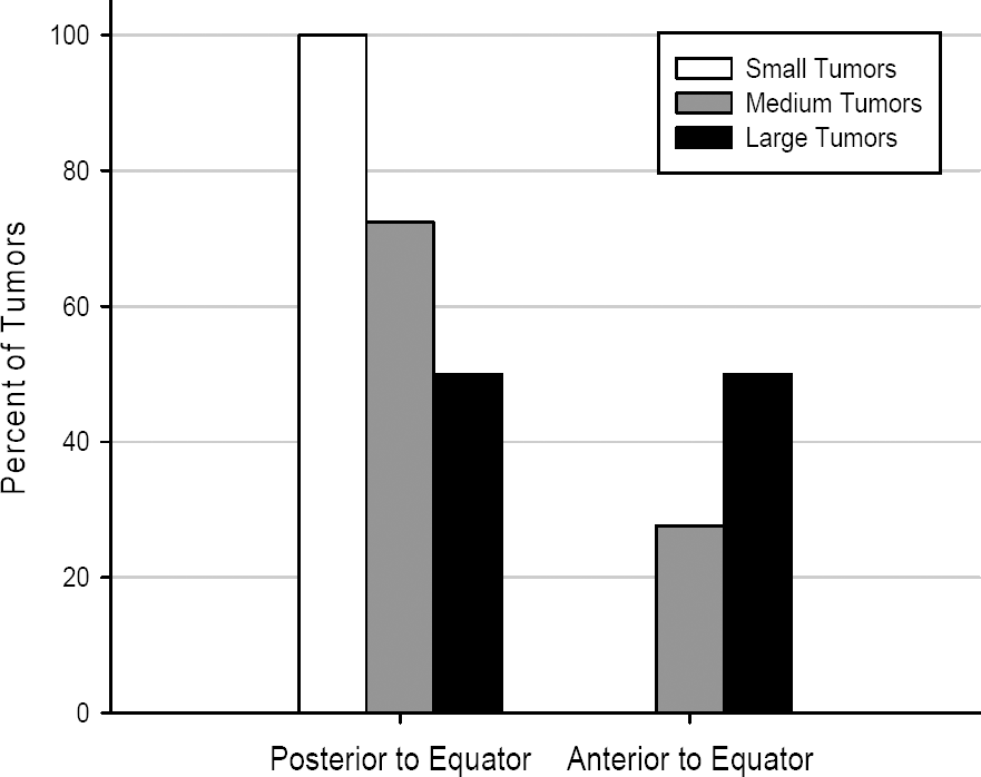 | Figure 4.The tumors were located entirely posterior to the equator for all small tumors and 72.4% of the medium tumors, whereas no small tumor and only 27.6% of the medium tumors were located anterior to the equator. |
Table 1.
Demographic data of uveal melanoma
| Gender (N*) | |
| Male | 22 (62.9%) |
| Female | 13 (37.1%) |
| Age (years) | |
| Range | 24~82 |
| Overall | 48.2±14.1† |
| Male | 48.6±13.7 |
| Female | 47.6±15.5 |
| Eye (N) | |
| Right | 17 (48.6%) |
| Left | 18 (51.4%) |
Table 2.
Distribution of the uveal melanomas by size group
| Tumor size | Number of patients | Apical height (mm) | Largest basal diameter (mm) | H-B ratio* |
|---|---|---|---|---|
| Small | 4 | 2.0±0.7† | 4.8±1.7 | 0.47±0.24 |
| Medium‡ | 29 | 6.4±1.9 | 9.1±2.6 | 0.74±0.31 |
| <5 mm | 9 | 4.2±0.7 | 7.5±2.0 | 0.60±0.20 |
| ≥5 mm | 20 | 7.4±1.4 | 9.8±2.5 | 0.81±0.33 |
| Large | 2 | 11.6±2.1 | 15.4±5.2 | 0.77±0.13 |
|
|
||||
| Total | 35 | 6.2±2.6 | 9.0±3.3 | 0.71±0.31 |
 XML Download