

 PDF
PDF ePub
ePub Citation
 Print
Print


Abstract
Purpose
To compare postoperative wavefront aberration and visual functions between aspherical Tecnis Z9003, a new acrylic aspheric intraocular lens (IOL), and spherical AcrySof SA60AT IOL.
Methods
Fifty patients (56 eyes) who underwent cataract extraction and were implanted with spherical or aspherical IOLs were randomly evaluated by wavefront analysis, including an examination of spherical aberration and higher-order aberrations using two different types of aberrometers (ray tracing and automatic retinoscope), manifested refraction, a contrast sensitivity test, and modulation transfer function (MTF), three months after surgery.
Go to : 
References
1. Owsley C, Sekuler R, Siemsen D. Contrast sensitivity throughout adulthood. Vision Res. 1983; 23:689–99.

2. McLellan JS, Marcos S, Burns SA. Age-related changes in monochromatic wave aberrations of the human eye. Invest Ophthalmol Vis Sci. 2001; 42:1390–5.
3. Guirao A, Redondo M, Artal P. Optical aberrations of the human cornea as a function of age. J Opt Soc Am A Opt Image Sci Vis. 2000; 17:1697–702.
4. Oshika T, Klyce SD, Applegate RA, Howland HC. Changes in corneal wavefront aberrations with aging. Invest Ophthalmol Vis Sci. 1999; 40:1351–55.
5. Artal P, Berrio E, Guirao A, Piers P. Contribution of the cornea and internal surface to the change of ocular aberrations with age. J Opt Soc Am A Opt Image Sci Vis. 2002; 19:137–43.
6. Artal P, Guirao A, Berrio E, Williams DR. Compensation of corneal aberrations by the internal optics in the human eye. J Vis. 2001; 1:1–8.
7. Mester U, Dillinger P, Anterist N. Impact of a modified optic design on visual function: clinical comparative study. J Cataract Refract Surg. 2003; 29:652–60.
8. Rawer R, Stork W, Spraul CW, Lingenfelder C.Imaging quality of intraocular lenses. J Cataract Refract Surg. 2005; 31:1618–31.
9. Guirao A, Redondo M, Geraghty E. . Corneal optical aberrations and retinal image quality in patients in whom monofocal intraocular lenses were implanted. Arch Ophthalmol. 2002; 120:1143–51.
10. Chalita MR, Krueger RR. Correlation of aberrations with visual acuity and symptoms. Ophthalmol Clin North Am. 2004; 17:135–42.
11. Holladay JT, Piers PA, Koranyi G. . A new intraocular lens design to reduce spherical aberration of pseudophakic eyes. J Refract Surg. 2002; 18:683–91.
12. Packer M, Fine IH, Hoffman RS, Piers PA. Prospective randomized trial of an anterior surface modified prolate intraocular lens. J Refract Surg. 2002; 18:692–6.
13. Bellucci R, Scialdone A, Buratto L. . Visual acuity and contrast sensitivity comparison between Tecnis and AcrySof SA60AT intraocular lenses: a multicenter randomized study. J Cataract Refract Surg. 2005; 31:712–7.
14. Denoyer A, Lez ML, Majzoub S, Pisella P. Quality of vision after cataract surgery after Tecnis Z9000 intraocular lens implantation: Effect of contrast sensitivity and wavefront aberration improvements on the quality of daily vision. J Cataract Refract Surg. 2007; 33:210–6.
15. Rozema JJ, Van Dyck D, Tassignon M. Clinical comparison of 6 aberrometers. Part I: Technical specifications. J Cataract Refract Surg. 2005; 31:1114–27.
16. Marcos S, Barbero S, Jimenez-Alfaro I. Optical quality and depth-of-field of eyes implanted with spherical and aspheric intraocular lenses. J Refract Surg. 2005; 21:223–35.
17. Padmanabhan P, Yoon G, Porter J. . Wavefront aberration in eyes with acrysof monofocal intraocular lenses. J Refract Surg. 2006; 22:237–42.
18. Park I, Park C, Ryu K. Corneal Astigmatic Changes by Temporal Incision or Oblique Incision in Sutureless Cataract Surgery. J Korean Ophthalmol Soc. 1995; 36:1467–72.
19. Guirao A, Tejedor J, Artal P. Corneal aberrations before and after small-incision cataract surgery. Invest Ophthalmol Vis Sci. 2004; 45:4312–9.
20. Barbero S, Marcos S, Jime´nez-Alfaro I. Optical aberrations of intraocular lenses measured in vivo and in vitro. J Opt Soc Am A Opt Image Sci Vis. 2003; 20:1841–51.
21. Atchison DA. Design of aspheric intraocular lenses. Ophthalmic Physiol Opt. 1991; 11:137–146.
22. Altmann GE, Nichamin LD, Lane SS, Pepose JS. Optical performance of 3 intraocular lens designs in the presence of decentration. J Cataract Refract Surg. 2005; 31:574–85.
23. Wang L, Koch DD. Effect of decentration of wavefront- corrected intraocular lenses on the higher-order aberrations of the eye. Arch Ophthalmol. 2005; 123:1226–30.
24. Akkin C, Ozler SA, Mentes J. Tilt and decentration of bag fixated lenses: a comparative study between capsulorrhexis and envelope techniques. Doc Ophthalmol. 1994; 87:199–209.
25. Mutlu FM, Bilge AH, Altinsoy HI, Yamusak E. The role of capsulotomy and intraocular lens type on tilt and decentration of polymethylmethacrylate and foldable acrylic lenses. Ophthalmologica. 1998; 212:359–63.
26. Hayashi K, Harada M, Hayashi H. . Decentration and tilt of polymethyl methacrylate, silicone, and acrylic soft intraocular lenses. Ophthalmology. 1997; 104:793–8.
27. Norrby NE, Grossman LW, Geraghty ED. . Determining the imaging quality of intraocular lens. J Cataract Refract Surg. 1998; 24:703–14.
28. Ginsburg AP. Contrast sensitivity: determining the visual quality and function of cataract, intraocular lenses and refractive surgery. Curr Opin Ophthalmol. 2006; 17:19–26.
Go to :
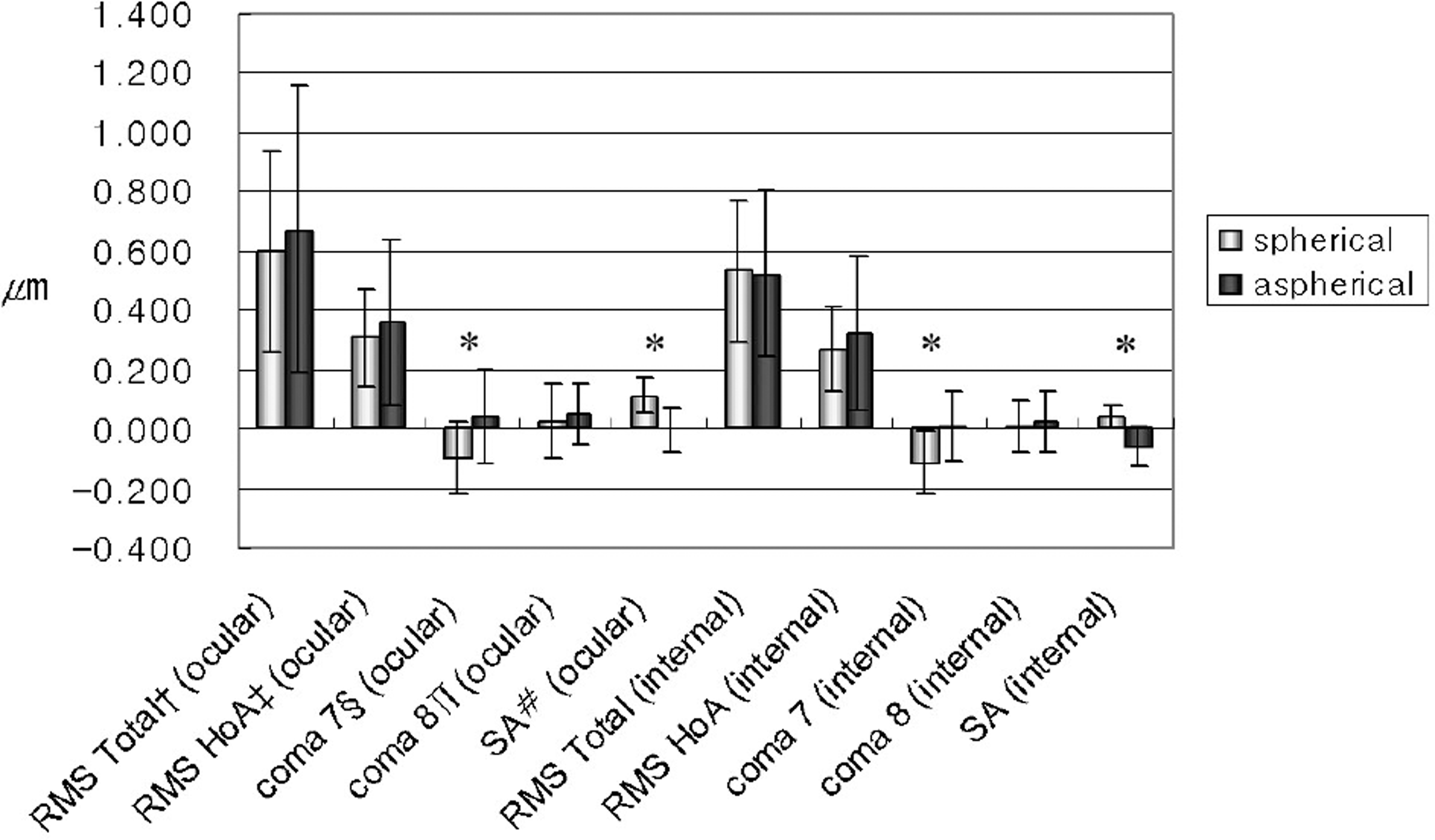 | Figure 1.Ocular and internal aberrations of spherical and aspherical IOL-implanted groups at 4-mm pupil zone † RMS Total=root mean measured by iTrace. (* p<0.05; square of total aberrations; ‡ RMS HoA=root mean square of § 7th total higher-order aberrations from 3rd to 6th order; # SA=spherical ∏8th coma aberration; coma aberration; aberration) |
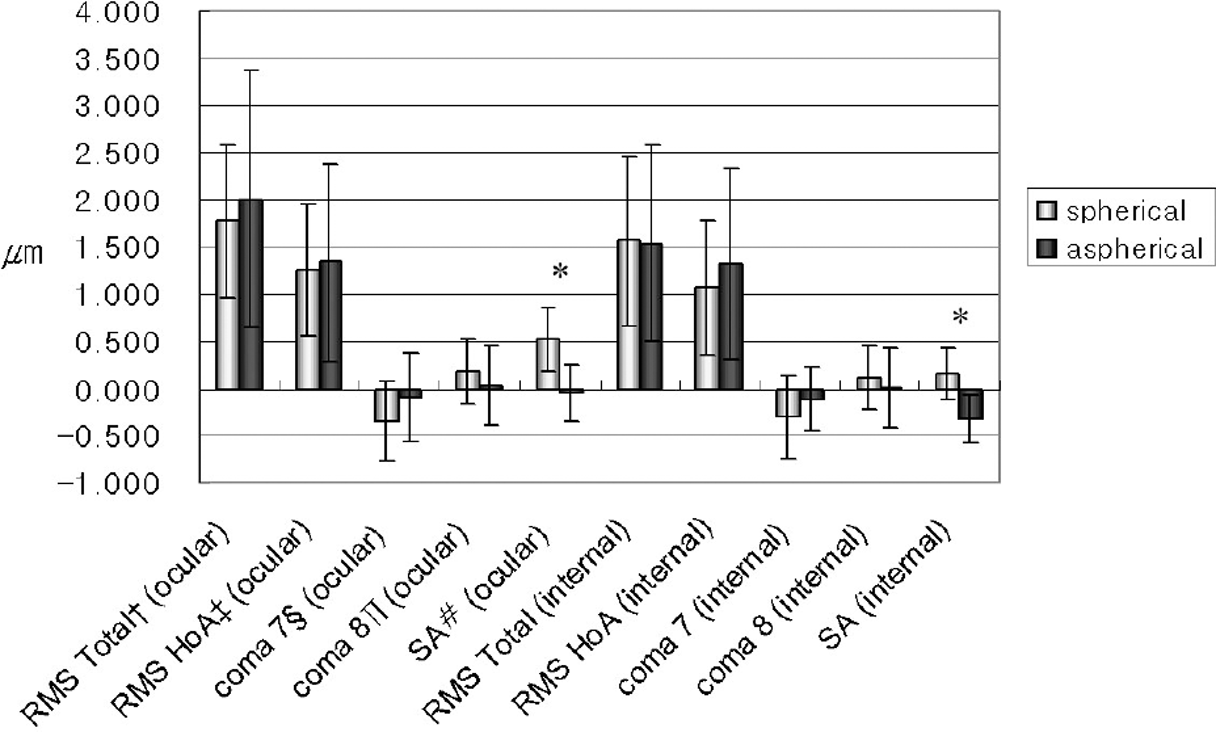 | Figure 2.Ocular and internal aberrations of spherical and aspherical IOL implanted groups at 6-mm pupil zone †: RMS Total=root mean measured by iTrace. (* p<0.05; ‡ RMS HoA=Root mean square square of total aberrations; § 7th of total higher order aberrations from 3rd to 6th order; # SA=spherical ∏8th coma aberration; coma aberration, aberration) |
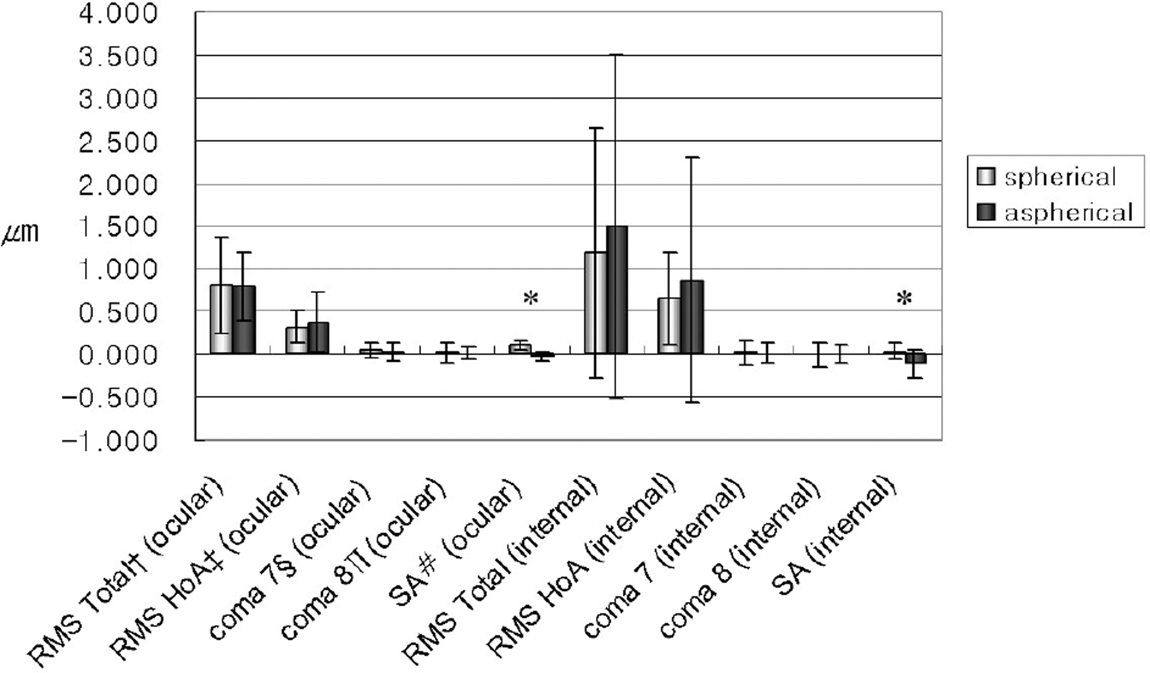 | Figure 3.Ocular and internal aberrations of spherical and aspherical IOL implanted groups at 4 mm pupil zone measured by OPD scan. (* p<0.05; RMS Total=root mean square of total aberrations; ‡ RMS HoA=root mean square of § 7th total higher order aberrations from 3rd to 6th order; # SA=spherical ∏8th coma aberration; coma aberration; aberration). |
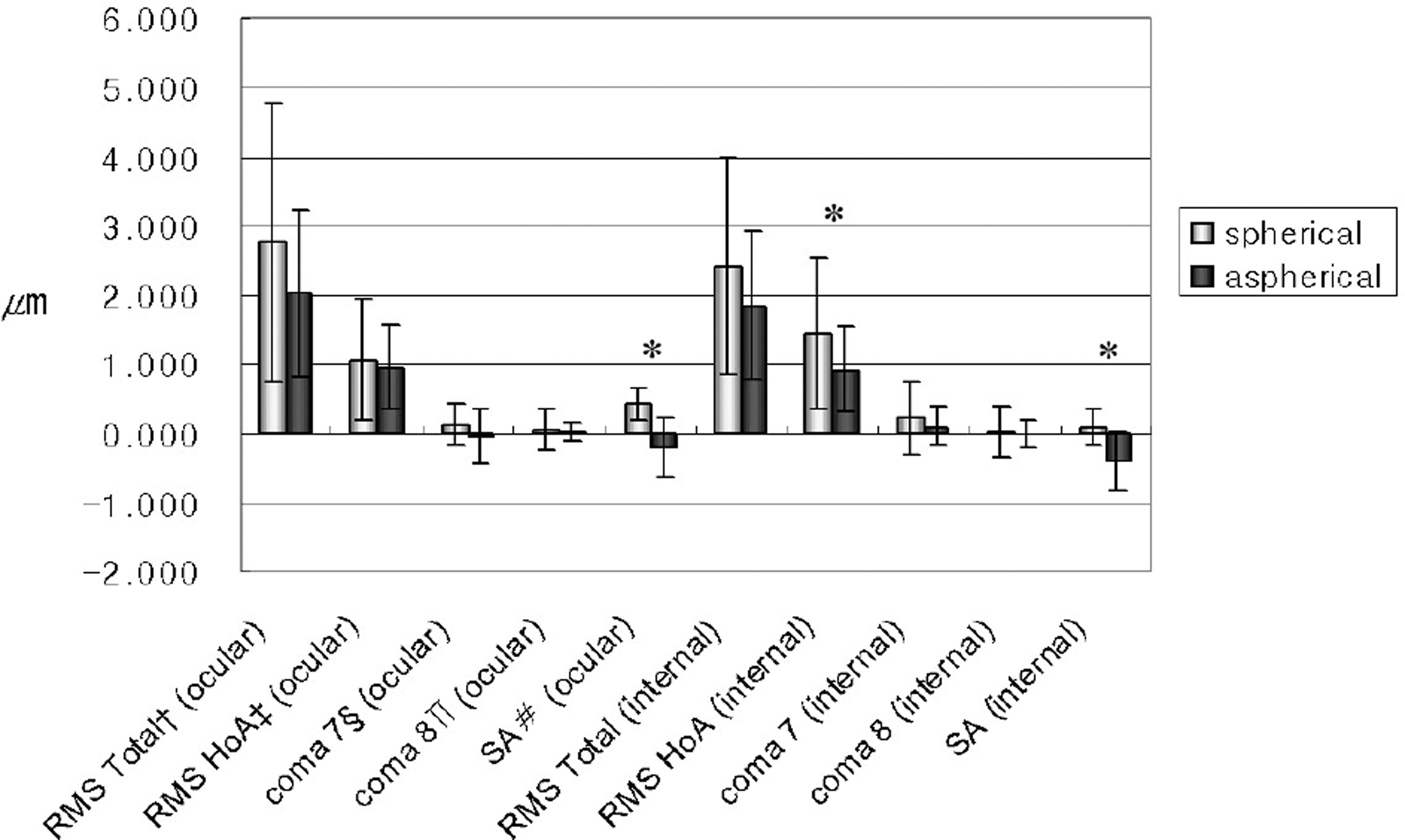 | Figure 4.Ocular and internal aberrations of spherical and aspherical IOL implanted groups at 6-mm pupil zone measured by OPD scan. (* p<0.05; † RMS Total = root mean ‡RMS HoA = root mean square square of total aberrations; § 7th of total higher order aberrations from 3rd to 6th order; # SA=spherical ∏8th coma aberration; coma aberration; aberration). |
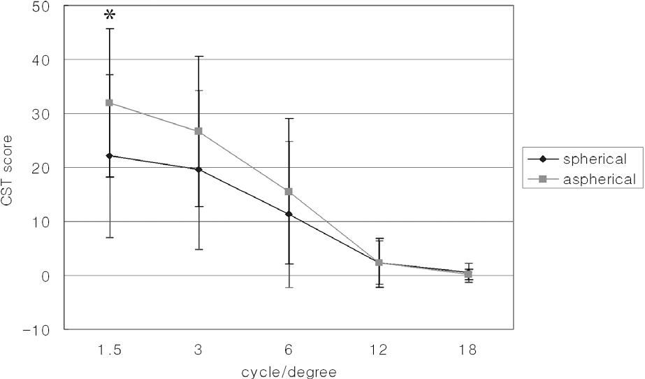 | Figure 5.Contrast sensitivity test result of spherical and aspherical IOL-implanted groups in mesopic condition (* p<0.05). |
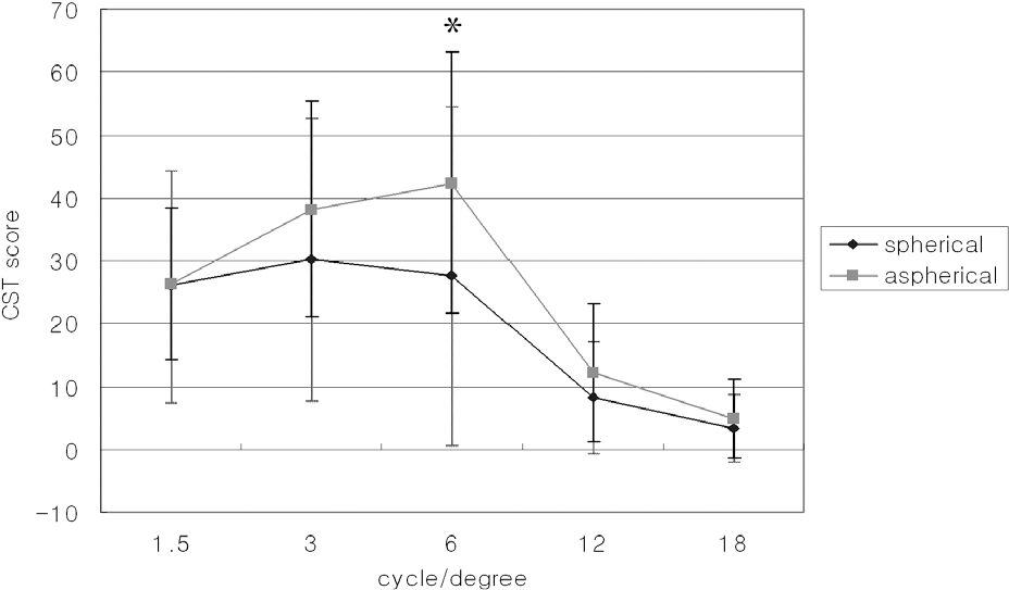 | Figure 6.Contrast sensitivity test result of spherical and aspherical IOL-implanted groups in photopic condition (* p<0.05). |
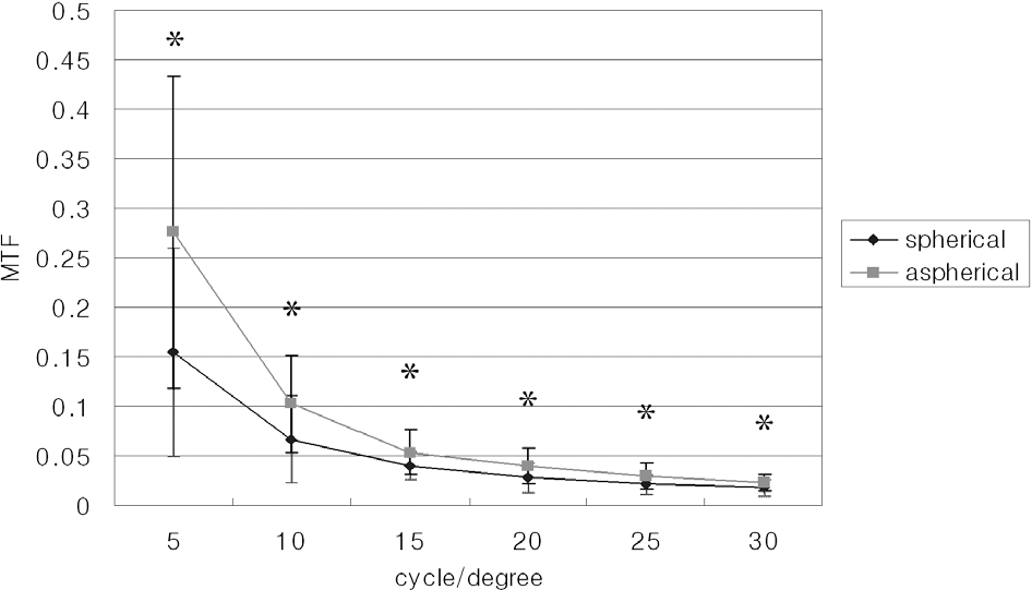 | Figure 7.Modulation transfer function (MTF) of spherical and aspherical IOL implanted groups at 6-mm pupil zone (* p<0.05). |
Table 1.
Comparison of sex, age, manifest refraction, BCVA between spherical IOL-implanted group and aspherical IOL- implanted group
| Spherical | Aspherical | P | |
|---|---|---|---|
| Sex (M:F) | 12:17 | 10:11 | |
| Age | 61.30±13.69 | 59.43±15.32 | 0.634 |
| MR* (SE†) | -0.39±0.91 | -0.70±0.53 | 0.151 |
| BCVA‡(logMAR) | 0.08±0.17 | 0.04±0.08 | 0.290 |
Table 2.
Comparison of ocular and internal aberrations measured by iTrace aberrometer between spherical IOL-implanted group and aspherical IOL-implanted group
| Optic zone | Zernike’s coefficients | Spherical | Aspherical | p | |
|---|---|---|---|---|---|
| 4 mm | ocular aberration | RMS Total† | 0.601±0.339 | 0.671±0.485 | 0.527 |
| RMS HoA‡ | 0.306±0.163 | 0.360±0.279 | 0.410 | ||
| coma 7 | -0.098±0.122 | 0.041±0.155 | 0.000* | ||
| coma 8 | 0.024±0.124 | 0.049±0.104 | 0.430 | ||
| SA§ | 0.114±0.062 | -0.002±0.073 | 0.000* | ||
| Internal aberration | RMS Total | 0.532±0.243 | 0.522±0.277 | 0.892 | |
| RMS HoA | 0.268±0.142 | 0.325±0.259 | 0.341 | ||
| coma 7 | -0.115±0.106 | 0.008±0.119 | 0.000* | ||
| coma 8 | 0.008±0.085 | 0.023±0.104 | 0.568 | ||
| SA | 0.040±0.039 | -0.058±0.066 | 0.000* | ||
| 6 mm | ocular aberration | RMS Total | 1.783±0.812 | 2.000±1.364 | 0.527 |
| RMS HoA | 1.256±0.687 | 1.334±1.050 | 0.773 | ||
| coma 7 | -0.348±0.429 | -0.098±0.459 | 0.055 | ||
| coma 8 | 0.174±0.338 | 0.041±0.419 | 0.220 | ||
| SA | 0.516±0.339 | -0.048±0.292 | 0.000* | ||
| Internal aberration | RMS Total | 1.566±0.901 | 1.536±1.047 | 0.914 | |
| RMS HoA | 1.065±0.723 | 1.320±1.008 | 0.336 | ||
| coma 7 | -0.303±0.450 | -0.113±0.329 | 0.111 | ||
| coma 8 | 0.126±0.335 | 0.010±0.419 | 0.286 | ||
| SA | 0.169±0.275 | -0.328±0.257 | 0.000* |
Table 3.
Comparison of ocular and internal aberrations measured by OPD scan aberrometer between spherical IOL-implanted group and aspherical IOL-implanted group
| Optic zone | Zernike’s coefficients | Spherical | Aspherical | P | |
|---|---|---|---|---|---|
| 4 mm | ocular aberration | RMS Total† | 0.802±0.553 | 0.792±0.390 | 0.937 |
| RMS HoA‡ | 0.319±0.184 | 0.374±0.345 | 0.440 | ||
| coma 7 | 0.046±0.081 | 0.031±0.109 | 0.541 | ||
| coma 8 | 0.017±0.113 | 0.009±0.073 | 0.783 | ||
| SA§ | 0.103±0.061 | -0.022±0.053 | 0.000* | ||
| Internal aberration | RMS Total | 1.185±1.457 | 1.491±2.009 | 0.511 | |
| RMS HoA | 0.653±0.536 | 0.863±1.433 | 0.443 | ||
| coma 7 | 0.022±0.142 | 0.012±0.124 | 0.772 | ||
| coma 8 | -0.002±0.139 | -0.002±0.105 | 0.991 | ||
| SA | 0.035±0.091 | -0.110±0.163 | 0.000* | ||
| 6 mm | ocular aberration | RMS Total | 2.769±2.014 | 2.025±1.208 | 0.119 |
| RMS HoA | 1.051±0.885 | 0.957±0.626 | 0.664 | ||
| coma 7 | 0.111±0.306 | -0.045±0.392 | 0.101 | ||
| coma 8 | 0.044±0.299 | 0.019±0.130 | 0.709 | ||
| SA | 0.416±0.235 | -0.220±0.429 | 0.000* | ||
| Internal aberration | RMS Total | 2.412±1.564 | 1.851±1.079 | 0.140 | |
| RMS HoA | 1.438±1.076 | 0.925±0.619 | 0.039* | ||
| coma 7 | 0.219±0.537 | 0.099±0.277 | 0.308 | ||
| coma 8 | 0.020±0.362 | -0.019±0.199 | 0.634 | ||
| SA(internal) | 0.074±0.268 | -0.421±0.437 | 0.000* |
Table 4.
Comparison of modulation transfer function measured by iTrace scan at 6 mm optic zone between spherical IOL-implanted group and aspherical IOL-implanted group
| Spatial frequency | Spherical | Aspherical | P |
|---|---|---|---|
| 5 | 0.156±0.105 | 0.276±0.158 | 0.005* |
| 10 | 0.067±0.045 | 0.103±0.487 | 0.011* |
| 15 | 0.040±0.014 | 0.054±0.022 | 0.020* |
| 20 | 0.028±0.014 | 0.040±0.018 | 0.017* |
| 25 | 0.022±0.010 | 0.030±0.013 | 0.017* |
| 30 | 0.018±0.009 | 0.023±0.009 | 0.048* |
 XML Download