Abstract
Purpose
To evaluate the outcomes of posterior chamber lens (PCL) exchange in patients with opacified foldable PCLs.
Methods
This study consisted of 31 patients (35 eyes) who had received phacoemulsification and implantation of foldable intraocular lenses in the bag or sulcus and developed late opacification of the PCL. All patients reported a reduction of visual acuity and deterioration in vision. The PCLs were explanted and replaced with new PMMA lenses. The perioperative complications and the best corrected visual acuities (BCVAs) before and after surgery were evaluated.
Results
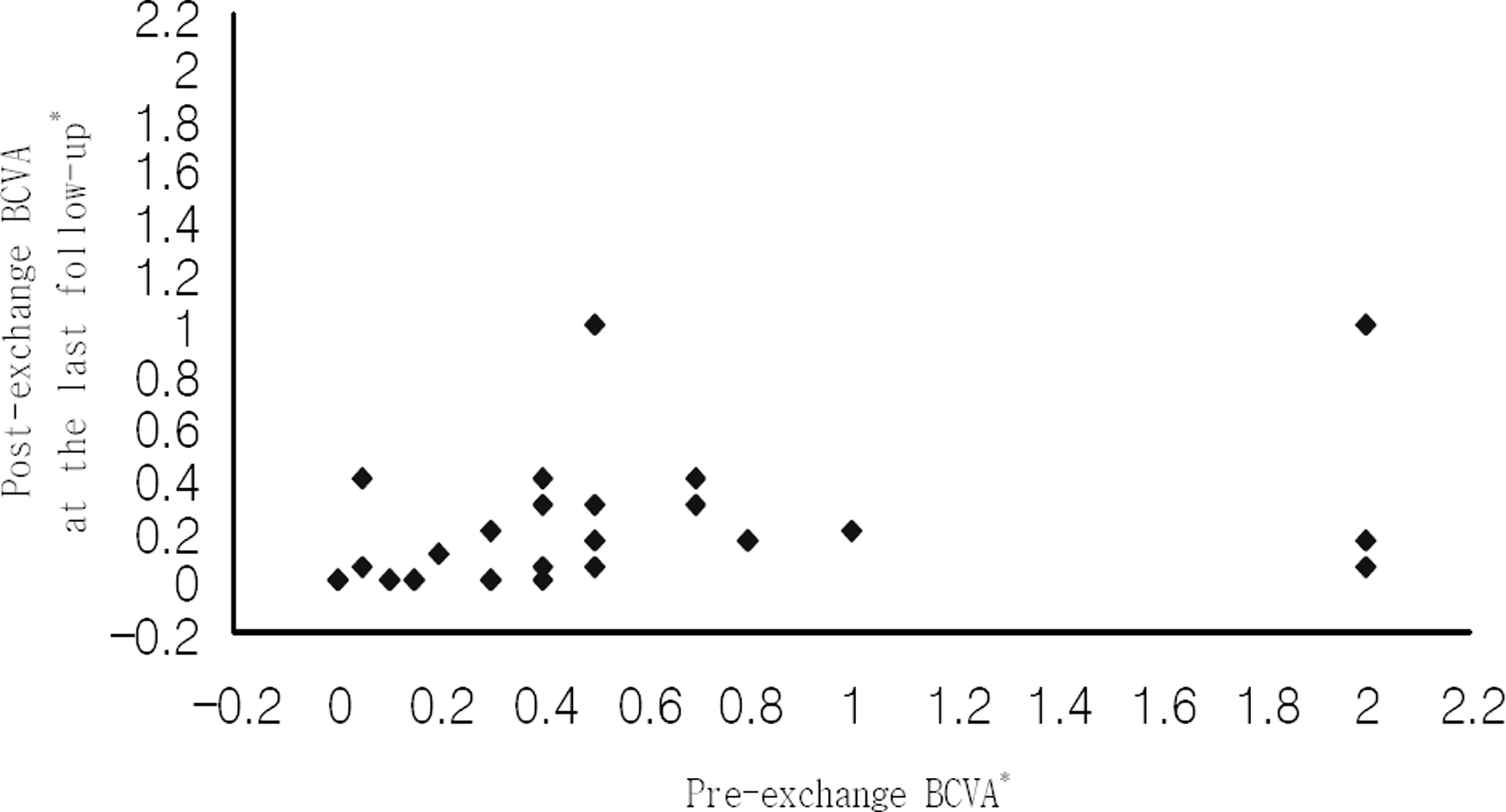
The mean visual acuities (logMAR value) before and after IOL exchange were 0.59±0.80 and 0.21±0.27, respectively. The difference was statistically significant (p=0.005, paired t-test). Intraoperative complications included posterior capsule rupture in six patients, zonular dehiscence in three patients, and both in one patient. Postoperative complications included intraocular pressure elevation in five patients and cystoid macular edema in two patients. One patient showed hypopyon at 6 days postoperatively, which lasted for 3 months, but she showed good visual acuity.
Go to : 
References
1. Kohnen T. The variety of foldable intraocular lens materials. J Cataract Refract Surg. 1996; 22:1255–8.

2. Apple DJ, Mamalis N, Loftfield K. . Complications of intraocular lenses. A historical and histopathological review. Surv Ophthalmol. 1984; 29:1–54.

3. Yu AK, Shek TW. Hydroxyapatite formation on implanted hydrogel intraocular lenses. Arch Ophthalmol. 2001; 119:611–4.

4. Ursell PG, Spalton DJ, Pande MV. . Relationship between intraocular lens biomaterials and posterior capsule opacification. J Cataract Refract Surg. 1998; 24:352–60.

6. Mamalis N, Crandall AS, Pulsipher MW. . Intraocular lens explantation and exchange; a review of lens styles, clinical indications, clinical results, and visual outcome. J Cataract Refract Surg. 1991; 17:811–8.
7. Doren GS, Stern GA, Driebe WT. Indications for and results of intraocular lens explantation. J Cataract Refract Surg. 1992; 18:79–85.

8. Sinskey RM, Amin P, Stoppel JO. Indications for and results a large series of intraocular lens exchanges. J Cataract Refract Surg. 1993; 19:68–71.
9. Tappin MJ, Larkin DK. Factors leading to lens implant decentration and exchange. Eye. 2000; 14:773–6.
10. Farbowitz MA, Zabriskie NA, Crandall AS. . Visual complaints associated with the acrysof acrylic intraocular lens. J Cataract Refract Surg. 2000; 26:1339–45.

11. Saika S, Miyamoto T, Ohnishi Y. Histology of anterior capsule opacification with a poly HEMA/HOHEXMA hydrophilic hydrogel intraocular lens compared to poly(methyl methacrylate), silicone, and acrylic lenses. J Cataract Refract Surg. 2003; 29:1198–203.
12. Apple DJ, Werner L, Escobar-Gomez M, Pandey SK. Deposits on the optical surfaces of hydroview intraocular lenses. J Cataract Refract Surg. 2000; 26:796–7.

13. Murray RI. Two cases of late opacification of the hydroview hydrogel intraocular lens. J Cataract Refract Surg. 2000; 26:1272–3.

14. Apple DJ, Werner L, Pandey SK. Newly recognized complications of posterior chamber intraocular lenses. Arch Ophthalmol. 2001; 119:581–2.

15. Chang BY, Davey KG, Gupta M, Hutchinson C. Late clouding of an acrylic intraocular lens following routine phacoemul- sification. Eye. 1999; 13:807–8.
16. Lee JY, Joo KM, Kim SH. Late opacification of hydrophilic acrylic intraocular lenses. J Korean Ophthalmol Soc. 2002; 43:2419–29.
17. Kim HG, Lee SH, Choi YJ. Late postoperative opacification of the foldable hydrophilic acrylic intraocular lens, ACRL-160. J Korean Ophthalmol Soc. 2003; 44:315–20.
18. Kim JC, Kim CS, Choi SH. . Clinical characteristics of patients with opacification of hydrophilic acrylic intraocular lens after cataract surgery. J Korean Ophthalmol. 2005; 46:1281–90.
19. Yu AK, Nq AS. Complications and clinical outcomes of intraocular lens exchange in patients with calcified hydrogel lenses. J Cataract Refract Surg. 2002; 28:1217–22.

20. Altaie RW, Costigan T, Donegan S. . Investigation and management of an epidemic of hydroview intraocular lens opacification. Graefes Arch Clin Exp Ophthalmol. 2005; 243:1124–33.

21. Dagres E, Khan MA, Kyle GM, Clark D. Perioperative complications of intraocular lens exchange in patients with opacified Aqua-Sense lenses. J Cataract Refract Surg. 2004; 30:2569–73.

22. Moesen I, Maudgal PC, Foets B. Late opacification of hydroview intraocular lenses: report of 11 cases. Bull Soc Belge Ophthalmol. 2006; 299:13–8.
23. van Looveren J, Tassignon MJ. Intraocular lens exchange for late-onset opacification. Bull Soc Belge Ophthalmol. 2004; 293:61–8.
24. Werner L, Apple DJ, Kaskalglu M, Pandey SK. Dense opacification of the optical component of a hydrophilic acrylic intraocular lens. J Cataract Refract Surg. 2001; 27:1485–92.

25. Dorey MW, Brownstein S, Hill VE. . Proposed pathogenesis for the delayed postoperative opacification of the hydroview hydrogel intraocular lens. Am J Ophthalmol. 2003; 135:591–8.

26. Trivedi RH, Werner L, Apple DJ. . Post cataract intraocular lens(IOL) surgery opacification. Eye. 2002; 16:217–41.
27. Werner L, Apple DJ, Escobar-Gometz M. . Postoperative deposition of calcium on the surfaces of a hydrogel intraocular lens. Ophthalmology. 2000; 107:2179–85.
28. Lee DH, Seo Y, Joo CK. Progressive opacification of hydrophilic acrylic intraocular lenses in diabetic patients. J Cataract Refract Surg. 2002; 28:1271–5.

29. Frohn A, Dick HB, Augustin AJ, Grus FH. Late opacification of the foldable hydrophilic acrylic lens SC60B-OUV. Ophthalmology. 2001; 108:1999–2004.

30. Kim CY, Kang SJ, Lee SJ. . Opacification of a hydrophilic acrylic lens with exacerbation of Behcet's uveitis. J Cataract Refract Surg. 2002; 28:1276–8.
31. Oner HE, Durak I, Saatci OA. Late postoperative opacification of hydrophilic acrylic intraocular lenses. Ophthalmic Surg Lasers. 2002; 33:304–8.

32. Fernado GT, Crayford BB. Visually significant calcification of hydrogel intraocular lenses necessitating explantation. Clin Experiment Ophthalmol. 2000; 28:280–6.
33. Olson RJ, Caldwell KD, Crandall AS. . Intraoperative crystallization on the intraocular lens surface. Am J Ophthalmol. 1998; 126:177–84.

34. Farbowitz MA, Zabriskie NA, Crandall AS. . Visual complaints associated with the AcrySof acrylic intraocular lens. J Cataract Refract Surg. 2000; 26:1339–45.

35. Dhaliwal DK, Mamalis N, Olson RJ. . Visual significance of glistenings seen in the AcrySof intraocular lens. J Cataract Refract Surg. 1996; 22:452–7.

36. Busin M, Arffa RC, McDonald MB, Kaufman HE. Intraocular lens removal during penetrating keratoplasty for pseudophakic bullous keratopathy. Ophthalmology. 1987; 94:505–9.

37. Brown DC, Snead JW. Intraocular lens implant exchanges. J Am Intraocul Implant Soc. 1985; 11:376–9.

38. Poort-van Nouhuijs HM, Handrickx KH, van Marle WF. . Corneal astigmatism after clear corneal and corneoscleral incisions for cataract surgery. J Cataract Refract Surg. 1997; 23:758–60.

39. Mendívil A. Frequency of induced astigmatism following phacoemulsification with suturing versuswithout suturing. Ophthalmic Surg Lasers. 1997; 28:377–81.
40. Müller-Jensen K, Barlinn B, Zimmerman H. Astigmatism reduction: no stitch 4.0 mm versus sutured 12.0 mm clear corneal incisions. J Cataract Refract Surg. 1997; 22:1108–12.
41. Grabow HB. Early results of 500 cases of no stitch cataract surgery. J Cataract Refract Surg. 1991; 17:726–30.
42. Ahn DG, Hyung SM, Kim SK. The change of corneal astigmatism after extracapsular cataract extraction with 7 mm scleral tunnel incision. J Korean Ophthalmol Soc. 2000; 41:1344–52.
Go to : 
 | Figure 1.Scattergram depiciting visual acuity before and after exchange of an opaque posterior chamber lens. At the last follow-up, BCVA improved in all eyes except 3 patients (2 with cystoid macular edema, 1 with posterior capsule opacity).* BCVA=best-corrected visual acuity=(logMAR). |
Table 1.
Basal characteristics I
Table 2.
Basal characteristics II
| Ocular comorbidity | No. of patients | Systemic illness | No. of patients |
|---|---|---|---|
| Diabetic retinopathy | 3 | DM‡ | 2 |
| AMD* | 1 | HT§ | 8 |
| Glaucoma | 5 | DM & HT | 4 |
| RVO† | 0 | Ischemic heart disease | 1 |
| HT retinopathy | 0 | Intracerebral hemorrhage | 1 |
| Total | 10 | Total | 16 |
Table 3.
Intraoperative complications
| Complication | No. of eyes (%) |
|---|---|
| Posterior capsular defect | 6 (17.0%) |
| Zonulysis | 3 (8.6%) |
| Posterior capsular defect & zonulysis | 1 (2.9%) |
|
|
|
| Total | 10 (28.5%) |
Table 4.
Post-operative complications
| Complications | No of eyes (%) |
|---|---|
| IOP elevation | 5 (14.0%) |
| Cystoid macular edema | 2 (5.7%) |
| Hypopyon | 1 (2.9%) |
| PC opacity | 1 (2.9%) |
| Hyphema | 0 (0.0%) |
|
|
|
| Total | 10 (28.5%) |
Table 5.
Patient data
F/U=follow-up; Vit. Loss=vitreous loss; Cx=complication; HM=hand movement; AMD=age-related macular edema; DMR=diabetic mellitus retinopathy; POAG=primary open-angle glaucoma; PC=posterior capsule rupture; IIOP=increased intra-ocular pressure; CME=cystoid macular edema; Hypop=hypopyon; Zon=zonulysis; NTG=normal tension glaucoma.


 PDF
PDF ePub
ePub Citation
 Print
Print


 XML Download