

 PDF
PDF ePub
ePub Citation
 Print
Print


Abstract
Purpose
To report a case of fungal endophthalmitis developed early after penetrating keratoplasty (PKP).
Case summary
A 67-year-old man was diagnosed with cataract, bullous keratopathy, and Fuchs' dystrophy. He underwent phacoemulsification, posterior chamber intraocular lens implantation, and PKP. The preoperative visual acuity was counting fingers. One day after surgery, his visual acuity was 20/200, and there was no abnormal finding. In the afternoon, a slit lamp examination showed endothelial plaque and white materials in the anterior chamber, and ultrasonography revealed vitritis. A Gram stain and a KOH smear of donor corneoscleral rim, which was conducted during the operation, revealed yeast-like organisms. An intravitreal amphotericin B injection was performed, and topical and systemic amphotericin B were administered. The donor corneoscleral rim scraping grew Candida albicans on culture. Amphotericin B injection into the anterior chamber and anterior chamber irrigation were performed. The patient’s vitritis worsened, so we performed pars plana total vitrectomy. Two months after treatment, the cornea showed complete clearing, and the endothelial cell density was 1779 cells/mm2 on noncontact specular microscopy. Five months after treatment, the final best corrected visual acuity was 20/20.
Go to : 
References
1. Kloess PM, Stulting RD, Waring GO. Bacterial and fungal endophthalmitis after penetrating keratoplasty. Am J Ophthalmol. 1993; 115:309–16.

2. Pardos GJ, Gallagher MA. Microbial contamination of donor eyes. Arch Ophthalmol. 1982; 100:1611–3.
3. Leveille A, McMullin F, Cavanaugh H. Endophthalmiits following penetrating keratoplasty. Ophthalmology. 1983; 90:38–9.
4. Everts RJ, Fowler WC, Chang DH. . Corneoscleral rim cultures: lack of utility and implications for clinical decision- making and infection prevention in the care of patients undergoing corneal transplantation. Cornea. 2001; 20:586–9.
5. Wiffen SJ, Weston BC, Maguire LJ, Boume Wm. The value of routine corneal rim cultures in penetrating keratoplasty. Arch Ophthalmol. 1997; 115:719–24.
6. Seedor JA, Stulting RD, Epstein RJ. . Survival of corneal grafts from donors supported by mechanical ventilation. Ophthalmology. 1987; 94:101–8.
7. Gomes JA, Dana MR, Dua HS. . Positive donor rim culture in penetrating keratoplasty. Cornea. 1995; 14:457–62.
8. Antonios SR, Cameron JA, Badr IA. . Contamination of donor cornea:postpenetrating keratoplasty endophthalmitis. Cornea. 1991; 10:217–20.
9. Sutphin JE, Pfaller MA, Hollis RJ, Wagoner MD. Donor-to-host transmission of Candida albicans after corneal transplantation. Am J Ophthalmol. 2002; 134:120–1.
10. Harris DJ, Stulting RD, Waring GO. . Late bacterial and fungal keratitis after corneal transplantation. Ophthalmology. 1988; 95:1450–7.
11. Keyhani K, Seedor JA, Shah MK. . The incidence of fungal keratitis and endophthalmitis following penetrating keratoplasty. Cornea. 2005; 24:288–91.
12. Merchant A, Zacks CM, Wilhemus K. . Candidal endophthalmitis after keratoplasty. Cornea. 2001; 20:226–9.
13. Garcia Serrano JL, Dominguez I, Serrano Laborda D. Endoftalmitis recurrente en queratoplastia penetrante, por flora mixta. Arch Soc Esp Oftalmol. 2004; 79:89–92.
14. Cameron JA, Badr IA, Miguel Risco J. . Endophthalmitis cluster from contaminated donor corneas following penetrating keratoplasty. Can J Ophthalmol. 1998; 33:8–13.
15. Hassan SS, Wilhelmus KR. Medical Review Subcommittee of the Eye Bank Association of America. Eye-banking risk factors for fungal endophthalmitis compared with bacterial endophthalmitis after corneal transplantation. Am J Ophthalmol. 2005; 139:685–90.
16. Lee SB, Han JW, Chung SK, Baek NH. Factors associated with visual outcomes of postoperative endophthalmitis following cataract surgery. J Korean Ophthalmol Soc. 2005; 46:1618–23.
17. Johnson MW, Doft BH, Kelsey SF. . The Endophthalmitis Vitrectomy Study. Relationship between clinical presentation and microbiologic spectrum. Ophthalmology. 1997; 104:261–72.
18. Wilhelmus KR, Hassan SS. The prognostic role of donor corneoscleral rim cultures in corneal transplantation. Ophthalmology. 2007; 114:440–5.
19. Al-Assiri A, Al-Jastaneiah S, Al-Khalaf A. . Late-onset donor-to-host transmission of Candida glabrata following corneal transplantation. Cornea. 2006; 25:123–5.
20. Manger TF. Mauger TF, Craig EL, editors. Antimicrobials. Havener’s ocular pharmacology. 1994. 6th ed. St. Louis: Mosby;v. 1:p. chap. 6.
21. Souri EN, Green WR. Intravitreal amphotericin-B toxicity. Am J Ophthalmol. 1974; 78:77–81.
22. Kuriakose T, Kothari M, Paul P. . Intracameral Amphotericin B Injection in the Management of Deep Keratomycosis. Cornea. 2002; 21:653–6.
Go to :
 | Figure 1.(A) Preoperative anterior segment photograph shows severe corneal opacity and diffuse stromal edema. (B) Anterior segment photograph shows mild corneal edema and epithelial defect at one day after penetrating keratoplasty (PKP). |
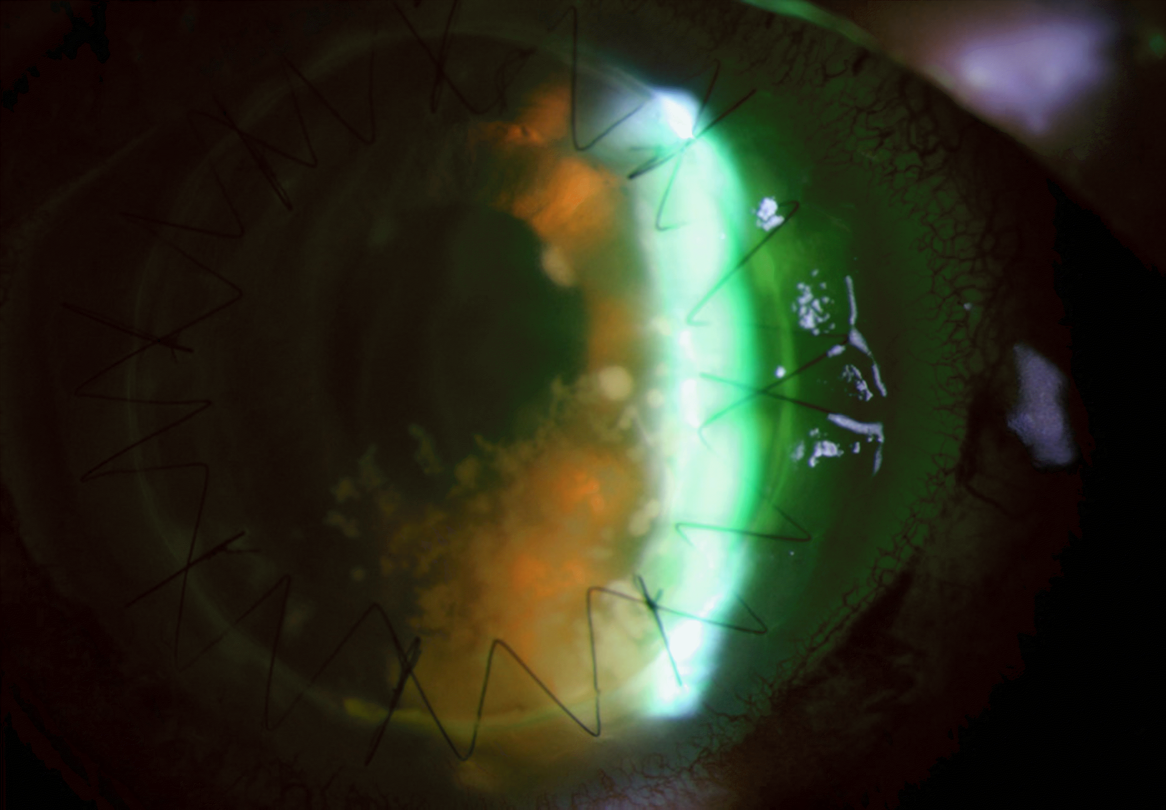 | Figure 1.Anterior segment photograph shows round or fluffy white materials of various size in the anterior chamber at 2 days after PKP. |


 XML Download