Abstract
Purpose
To make an anatomical evaluation of extraocular muscles, macular and optic nerve in enucleated eyes of live subjects and to compare the results with a previous autopsy study.
Methods
Twenty-four surgically enucleated eyes were studied. The distance from the limbus to the muscle insertion site, muscle width, and the distance between muscle insertion sites were measured and compared with an Apt study. After an incision through the eyeball, a 26-gauge needle was passed perpendicularly through the macula to the sclera. We measured the distances between the oblique muscles and the macula and optic nerves from an external site of the eyeball.
Results
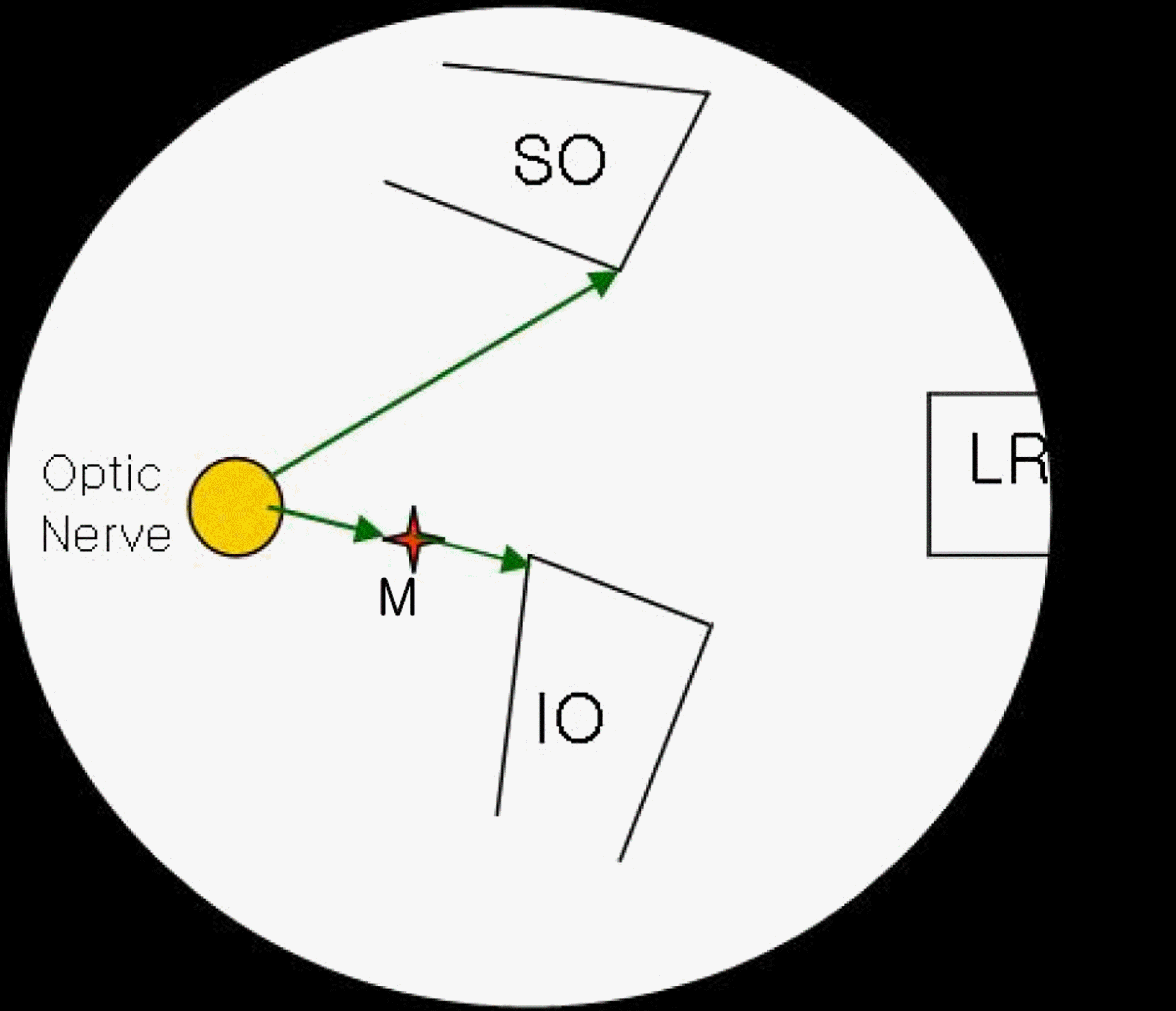
The distance from the limbus to the muscle insertion site showed a significant difference in the superior and inferior rectus muscle. All measurements of muscle widths were significantly narrower than those of autopsy studies. The distances between the muscles were significantly closer between the lateral and superior muscles and between the superior and medial rectus muscles. From the optic nerve to the muscle was 3.99±0.62 mm, to the superior oblique muscle was 7.89±0.88 mm, to the inferior oblique muscle was 5.95±0.83 mm, and the macula to the inferior oblique muscle was 1.35±0.42 mm.
Go to : 
References
1. Fuchs E. Beiträge zur normalen Anatomie des Augapfels. Graefes Arch Clin Exp Ophthalmol. 1884; 30:1–60.

2. Weiss L. Über das Wachstum des menschlichen Auges und ű ber die Veränderung der Muskelinsertionen am wachsenden Auge. Anat Embryol. 1897; 8:191–248.
3. Howe L. On the primary insertions of the ocular muscles. Trans Am Ophthalmol Soc. 1902; 9:668–78.
4. Gát L. Ein Beitrag zur Topographie des Ansatzes der vier geraden Augenmuskeln. Ophthalmologica. 1947; 114:43–51.

5. Apt L. An anatomical reevaluation of rectus muscle insertions. Trans Am Ophthalmol Soc. 1980; 78:365–75.

6. Paik HJ, Cho YA. Insertion of horizontal rectus muscles in strabismus. J Korean Ophthalmol Soc. 1989; 30:761–6.
7. Lee YC, Yang SW. Anatomical evaluations of the location and insertion shape of horizontal rectus muscle. J Korean Ophthalmol Soc. 1995; 36:1357–62.
8. Shin HM, Lew H, Yun YS. Muscle width and distance from limbus to muscle insertion site in strabismus patient. J Korean Ophthalmol Soc. 2005; 46:1387–92.
9. Yamamoto Y, Namiki R, Baba M, Kato M. A study of the measurement of ocular axial length by ultrasound echography. Acta Soc Ophthalmol Jpn. 1960; 64:1333–41.
10. Sorsby A, Leary GA, Richards MJ, Chaston J. Ultrasonographic measurement of the components of ocular refraction in life. Vision Res. 1963; 3:499–505.

11. Lee RH, Kim KS, Cho YA. The axial length of normal emmetropic eyes by ultrasonic biometry. J Korean Ophthalmol Soc. 1983; 24:27–31.
12. O’Sullivan E, Mitchell BS. An improved composition for embalming fluid to preserve cadavers for anatomy teaching in the United Kingdom. J Anat. 1993; 182:295–7.
13. Ward SR, Lieber RL. Density and hydration of fresh and fixed human skeletal muscle. J Biomech. 2005; 38:2317–20.

14. Duke-Elder S, Wyber KC. System of ophthalmology, Vol. 2. 1961. St. Louis: Mosby;p. 426–7.
15. Fink WH. Oblique muscle surgery from the anatomical viewpoint. Trans Am Ophthalmol Soc. 1949; 47:215–53.
16. Feng X, Pilon K, Yaacobi Y, Olsen TW. Extraocular muscle insertions relative to the fovea and optic nerve: humans and rhesus macaque. Invest Ophthalmol Vis Sci. 2005; 46:3493–6.

17. Whitnall SE. Anatomy of the human orbit. 1932. 2nd ed. London: Oxford Medical Publication;p. 257–79.
Go to : 
 | Figure 1.Measurements from an enucleated eyeball. (A) Anterior aspect of the globe showing muscle insertions and the limbus. Arrow indicates distance from the limbus to the anterior midpoint of the muscle insertion. (B) Posterior aspect of the globe showing the optic nerve head and the inferior oblique muscle insertion site. Arrow indicates the distance from the optic nerve to the posterior border of the inferior oblique muscle insertion site. |
 | Figure 2.Macula site (arrow) was externally indicated by passing through a 26-gauge needle from inside the macula to outside. |
 | Figure 3.Relationships among the optic nerve, superior oblique muscle (SO), inferior oblique muscle (IO), and macula (M). Right eye. |
Table 1.
Axial length, age and sex of enucleated patients
| Measurement | ||
|---|---|---|
| Mean±SD | Range | |
| Axial length (mm) | 23.09±0.92 | 22-25 |
| Age (years) | 51.96±13.60 | 25-70 |
| Sex | Male (n=14), F | emale (n=10) |
Table 2.
Distance from the limbus to muscle insertions
| Muscle |
Anterior limbus to anterior muscle insertion (mm) |
P-value | |
|---|---|---|---|
| Present study | Apt† | ||
| Medial rectus | 5.40±0.43 | 5.30±0.70 | 0.1883 |
| Inferior rectus | 6.23±0.42 | 6.80±0.80 | 0.0001* |
| Lateral rectus | 7.02±0.60 | 6.90±0.70 | 0.2202 |
| Superior rectus | 7.56±0.70 | 7.90±0.60 | 0.0087* |
| Superior oblique | 15.87±0.84 | ||
| Inferior oblique | 17.32±0.99 | ||
Table 3.
Width of muscle insertions
| muscle |
Width (mm) |
P-value | |
|---|---|---|---|
| Present study | Apt† | ||
| Medial rectus | 10.22±0.84 | 11.30±0.80 | 0.0001* |
| Inferior rectus | 9.40±1.10 | 10.50±0.80 | 0.0001* |
| Lateral rectus | 9.35±1.14 | 10.10±0.80 | 0.0001* |
| Superior rectus | 10.37±1.41 | 11.50±0.80 | 0.0001* |
| Superior oblique | 10.93±1.30 | 10.80 | |
| Inferior oblique | 10.14±1.49 | 9.60 | |
Table 4.
Distance between extraocular muscle insertions
| Muscle to muscle |
Distance between muscles (mm) |
p-value | |
|---|---|---|---|
| Present study | Apt† | ||
| MR-IR | 6.20±1.08 | 5.90±0.80 | 0.0636 |
| IR-LR | 7.81±1.23 | 8.00±0.08 | 0.1766 |
| LR-SR | 6.65±1.10 | 7.10±0.08 | 0.0119* |
| SR-MR | 6.97±1.71 | 7.50±0.08 | 0.0130* |
| SO-SR | 10.93±1.09 | ||
| IO-LR | 11.99±1.10 | ||


 PDF
PDF ePub
ePub Citation
 Print
Print


 XML Download