Abstract
Purpose
The objective visual acuity test is mandatory in certain cases, such as infants, nonverbal subjects, and subjects who need legal assistance. We designed an objective system for visual acuity test (SOVAT) consisting of three components: stimuli applied via a suppression method, display and evaluation and made a suppression method as stimuli component for SOVAT. Usefulness of the SOVAT was evaluated.
Methods
The visual stimuli were presented on a high-resolution head-mounted display (HMD). An eye movement tracking program and gaze monitoring device allowed us to monitor the patient's fixation status during the test. The suppression method, in addition to a conventional induction method, was developed to use with the SOVAT and its accuracy and confidence level were evaluated.
Results
On the basis of clinical data, we present the reference values for the SOVAT as below. For the induction method, objective visual acuity (smallest pixel size), the presumed subjective visual acuity was 3: 0.35-0.6, 7: 0.05-0.25, 12: below 0.05 and for the suppression method it was 1: 0.6-1.0, 1.5: 0.4-0.7, 3: 0.15-0.4, 5: 0.1-0.2.
Go to : 
References
1. Kim M, Choi YS, Lu WN. . The developement of an objective test for visual acuity assessment using optokinetic nystagmus stimuli presented head-mounted display: Seohan objective visual acuity test. J Korean Ophthalmol Soc. 2000; 41:871–8.
2. Kim MS, Lee IB, Hwang JM. . Method for objective visual acuity test. Annual meeting of ASCRS, U S A. 2000; 20–4.
3. Shin TY. Objective determination of visual acuity using optokinetic nystagmus. J Korean Ophthalmol Soc. 1964; 5:19–25.
4. von Noorden GK.Binocular vision & ocular motility. 1995. 5th ed. St Louis: Mosby;p. 155–8.
5. Goldmann H. Observations on objective determination of distance vision. Klin Monatsblatter Augenheilkd Augenarztl Fortbild. 1950; 117:570–3.
6. Schwarting BH. Testing infants' vision; an apparatus for estimating the visual acuity of infants and young children. Am J Ophthalmol. 1954; 38:714–5.
7. Gunther G, Noteboom E, Plotz C. Objective determination of visual acuity. Albrecht Von Graefes Arch Ophthalmol. 1957; 159:180–90.
8. Voipio H. The objective measurement of visual acuity. Acta Ophthalmol. 1961; 66:S1–70.
9. Dayton GO Jr, Jones MH, Aiu P. . Developmental study of coordinated eye movememts in the human infant. Arch Ophthalmol. 1964; 71:865–70.
11. Weder W, Wiegand H. Determination of visual acuity using optokinetic nystagmus. A newly developed instrument based on Günther's principle. Klin Monatsbl Ausenheilkd. 1987; 191:149–55.
12. Nakamura T, Kato I, Kanayama R, Koike Y. Computer analysis of optokinetic nystagmus for clinical usefulness. Auris Nasus Larynx. 1986; 13:S97–103.

13. Assaf AA. Optokinetic nystagmus testing using personal computers. Optom Vis Sci. 1991; 68:972–5.

14. Pfirsching HP, Frisch N, Wiegand W. Computer-assisted provocation and detection of optokinetic nystagmus. Ophthalmologe. 1994; 91:91–4.
15. Gräf M, Kaufmann H. Clinical application of a new method for the objective estimation of minimal visual acuity. Klin Monatsbl Augenheilkd. 1999; 214:395–400.
16. Ter Braak, Van Vlieta. Subcortical optokinetic nystagmus in the monkey. Psychiatr Neurol Neurochir. 1963; 66:277–83.
17. Aylward GP, Lazzara A, Meyer J. Behavioral and neurologic characteristics of a hydrancephalic infant. Dev Med Child Neurol. 1978; 20:211–7.
Go to : 
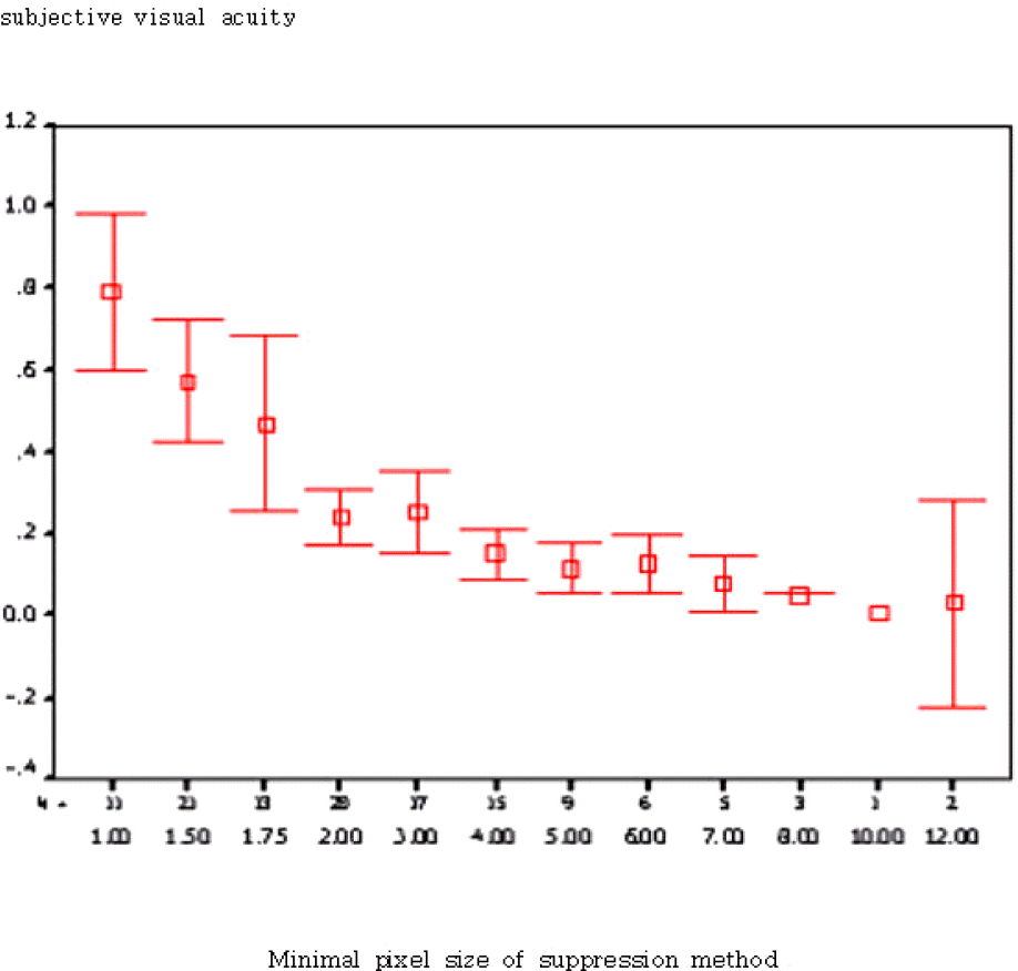 | Figure 6.The objective visual acuity (horizontal) versus subjective visual acuity (vertical) in suppression method. |
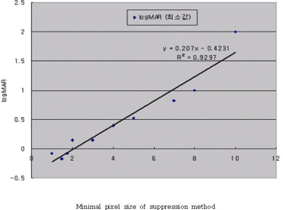 | Figure 7.The correlation of objective visual acuity and subjective visual acuity in suppression method. |
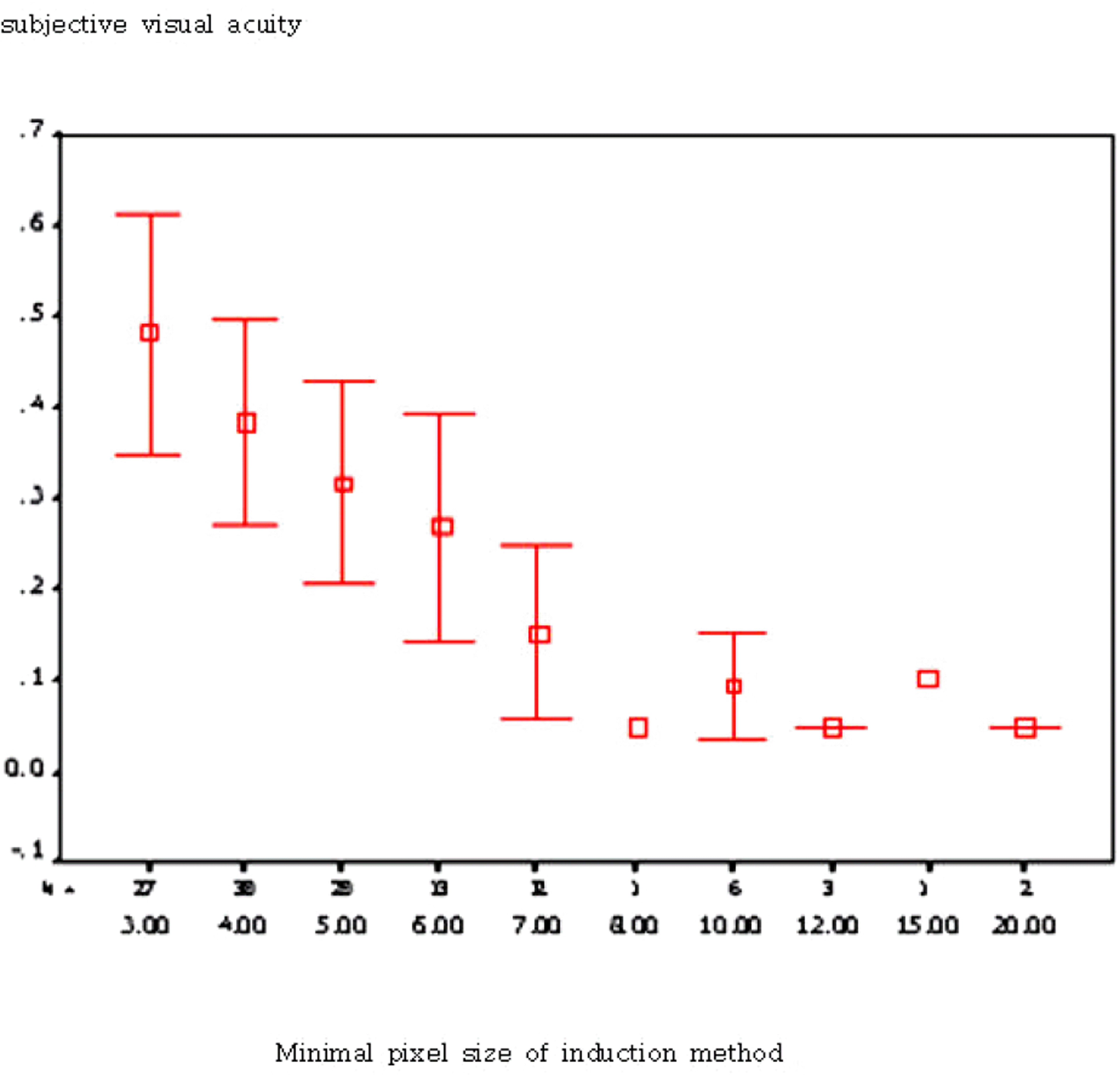 | Figure 8.The objective visual acuity (horizontal) versus subjective visual acuity (vertical) in induction method. |
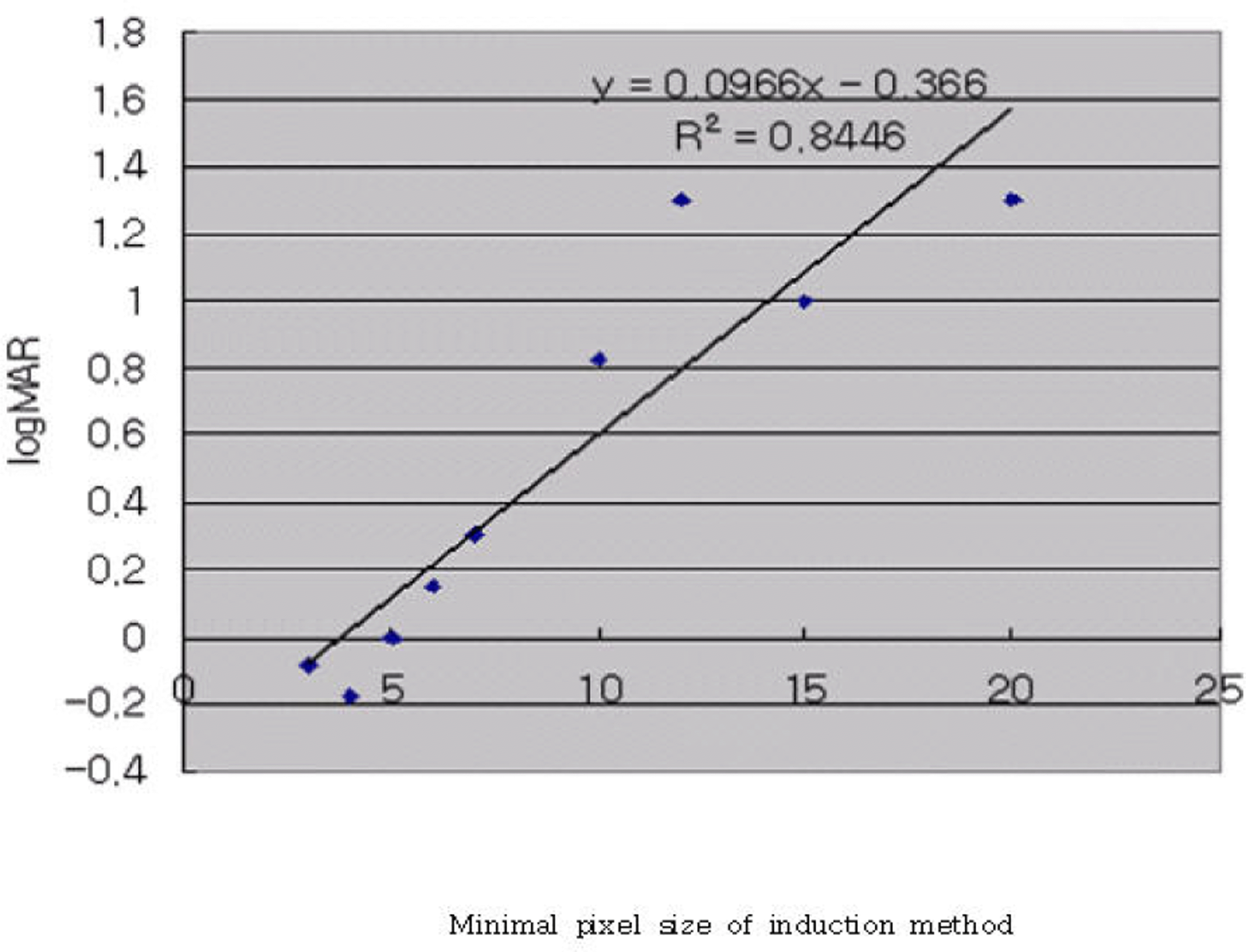 | Figure 9.The correlation of objective visual acuity and subjective visual acuity in induction method. |


 PDF
PDF ePub
ePub Citation
 Print
Print


 XML Download