

 PDF
PDF ePub
ePub Citation
 Print
Print


Abstract
Purpose
Abducens nerve palsy complicating pre-eclampsia during pregnancy occurs very rarely. The authors describe right abducens nerve palsy, disc swelling, and hypertensive retinopathy found in both eyes of a pre-eclampsia patient.
Case summary
A 29-year-old woman, who was in her 31st week of gestation, was admitted to the hospital complaining chiefly of diplopia and abduction limitation in her right eye, which started suddenly 3 weeks prior to admission. Her condition was carefully followed up by a local clinic because this symptom of hypertension manifested one month before her admission to the local clinic. Brain MRI and laboratory tests were performed in order to exclude diseases of the central nervous system and/or other systemic diseases. An alternative prism cover test showed approximately 25PD (prism diopter) right esotropia, with −2 degrees abduction limitation observed in the patient's right eye. Both hypertensive retinopathy and papilledema were observed. Her blood pressure was 155/110 mmHg, and she had moderate proteinuria when she was admitted to the hospital. She was referred to an obstetrician and diagnosed as having pre-eclampsia. Other than pre-eclampsia with proteinuria, the brain imaging examination failed to reveal any particular findings, such as CNS disorders, including brain edema, cerebral infarction, or a brain tumor. In addition, systemic diseases which might have caused papilledema and abducens nerve palsy, including diabetes mellitus, were not evident in this case. Therefore, the patient was diagnosed with ischemic abducens nerve palsy and hypertensive retinopathy complicating moderate pre-eclampsia. The patient's diplopia and right eye abduction limitation were gradually relieved after parturition.
References
1. Patel SV, Mutyala S, Leske DA, et al. Incidence, associations, and evaluation of sixth nerve palsy using a population-based method. Ophthalmology. 2004; 111:369–75.

2. Royburt M, Seidman DS, Serr DM, Mashiach S. Neurologic involvement in hypertensive disease of pregnancy. Obstet Gynecol Surv. 1991; 46:656–64.
3. Peters GB, Bakri SJ, Krohel GB. Cause and prognosis of nontraumatic sixth nerve palsies in young adults. Ophthalmology. 2002; 109:1925–8.
4. Richards BW, Jones FR Jr, Younge BR. Cause and prognosis in 4,278 cases of paralysis of the oculomotor, trochlear, and abducens cranial nerves. Am J Ophthalmol. 1992; 113:489–96.
5. The Korean strabismsus and pediatric ophthalmological society, Current Concepts in Strabismus. 1st ed.Seoul: Naeoei science;2004. p. 236–7.
6. Blade J, Peborde J, Dareguy P. Paralysis of the VI Cranial nerve in during eclampsia. Bull Soc Ophthalmol Fr. 1968; 68:284–7.
7. Sternberg I, Ronen S, Arnon N. Recurrent, isolated, post-febrile abducens nerve palsy. J Pediatr Ophthalmol Strabismus. 1980; 17:323–4.
8. Barry-Kinsella C, Milner M, McCarthy N, Walshe J. Sixth nerve palsy: an unusual manifestation of pre-eclampsia. Obstet Gynecol. 1994; 83:849–51.
9. Fung TY, Chung TK. Abducens nerve palsy complicating pregnancy : a case report. Eur J Obstet Gynecol Reprod Biol. 1999; 83:223–4.
10. Rucker CW. The causes of paralysis of the third, fourth, and sixth cranial nerves. Am J Ophthalmol. 1966; 61:1293–8.
11. Tiffin PA, MacEwen CJ, Craig EA, Clayton G. Acquired palsy of the oculomotor, trochlear and abducens nerves. Eye. 1996; 10:377–84.
12. Berlit P. Isolated and combined pareses of cranial nerves III, IV, and VI. A retrospective study of 412 patients. J Neurol Sci. 1991; 103:10–5.
13. Kim SS, Jin KH, Kim SM. Neuro-ophthalmologic evaluation of the third, fourth, and sixth cranial nerve palsysis. J Korean Ophthalmol Soc. 1991; 32:283–8.
14. Park KH, Chang BL. The etiology and clinical feature of the third, fourth, and sixth cranial nerve palsy. J Korean Ophthalmol Soc. 1997; 103:10–5.
15. Jeon C, Sa HS, Oh SY. Cause and natural course of the sixth cranial nerve palsy. J Koran Ophthalmol Soc. 2006; 47:1776–80.
16. Park UC, Kim SJ, Yu YS. Clinical features and natural history of the acquired thirc, fourth, and sixth cranial nerve palsy. J Korean Ophthalmol Soc. 2005; 46:1555–62.
17. Moster ML, Savino PJ, Sergott RC, et al. Isolated sixth-nerve palsies in younger adults. Arch Ophthalmol. 1984; 102:1328–30.
18. Jacobson DM, McCana TD, Lay de PM. Risk factors for ischemic ocular motor nerve palsies. Arch Ophthalmol. 1994; 112:961–6.
19. Sanders SK, Aki Kawasaki, Purvin VA. Long-term prognosis in patients with vasculopathic sixth nerve palsy. Am J Ophthalmol. 2002; 134:81–4.
20. Jacobson DM, Broste SK. Early progression of ophthalmoplegia in patients with ischemic oculomotor nerve palsies. Arch Ophthalmol. 1995; 113:1535–7.
21. Cullen JF. The swollen optic disc: Further observations of a europian neuroophtalmologist in southeast asia. Asian Journal of ophthalmology. 2001; 3:10–3.
22. Mabie BC, Sibai BMS. Current obstetrics and gynecology diagnosis and treatment. 6th ed.Norwalk, Connecticut: Appleton and Lange;1987. p. 340–52.
Figure 1.
Nine cardinal photography. Patient shows right esotropia in the primary position and limitation of abduction of the right eye at right gaze.
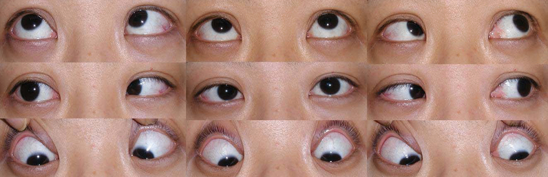
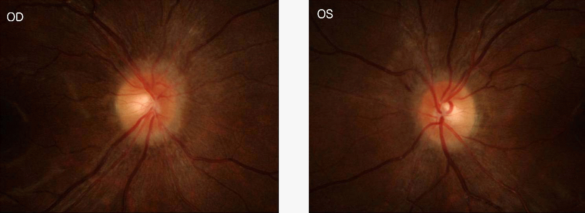
Table 1.
Case summaries of the literature review
 XML Download