

 PDF
PDF ePub
ePub Citation
 Print
Print


Abstract
Purpose
To report a case of acute lens particle glaucoma with an intraocular foreign body that persisted for a long duration.
Case summary
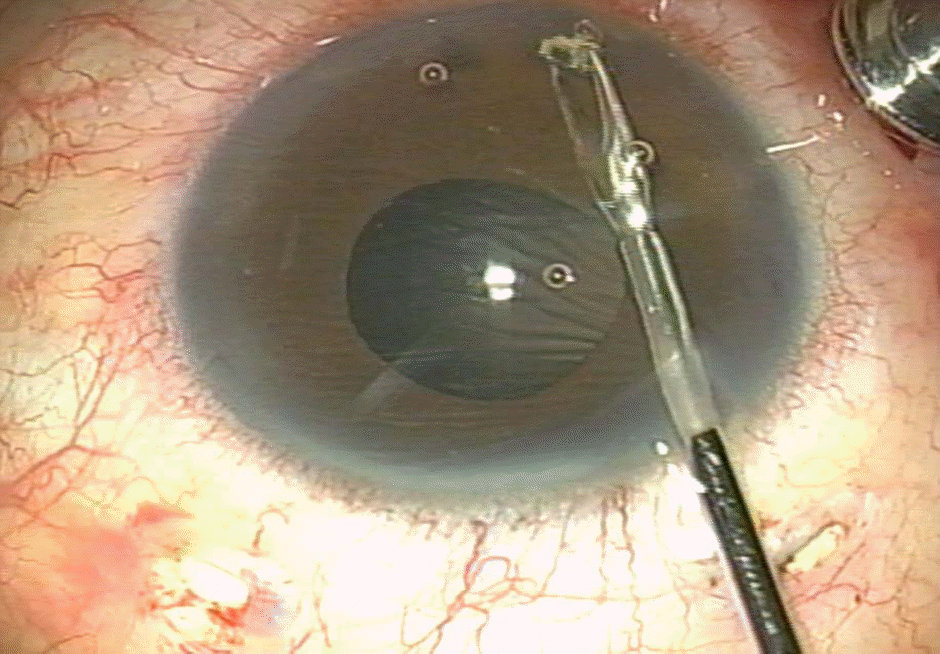
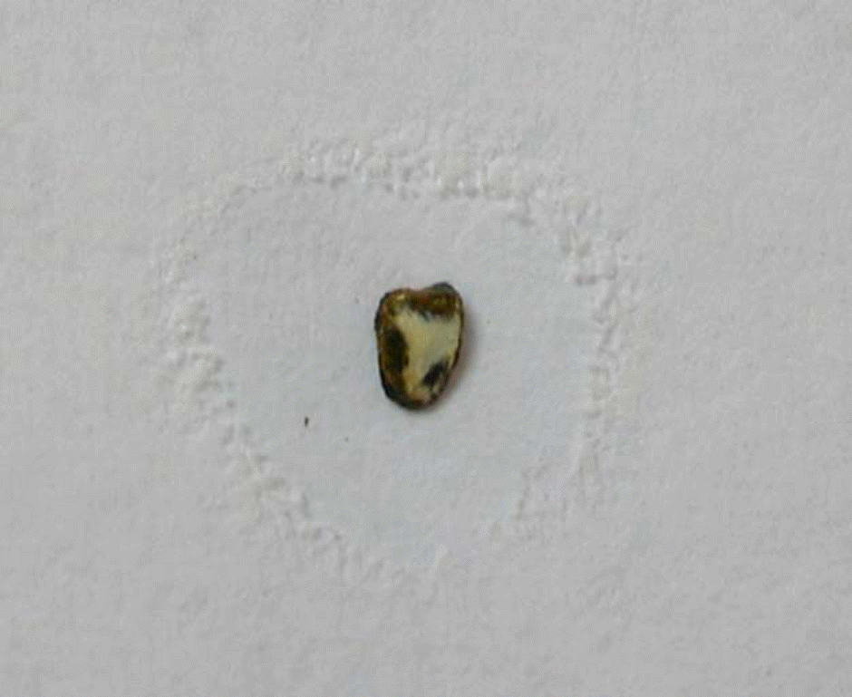
A 47-year-old man visited our clinic due to severe pain in his right eye. His uncorrected visual acuity was hand movement, and intraocular pressure (IOP) measured by a Goldmann applanation tonometer was 76 mmHg in the right eye. Severe corneal edema and floating lens materials in the anterior chamber were revealed by slit-lamp examination. An ultrasonography scan showed that the lens had dislocated into the vitreous cavity and revealed vitreous opacities. To remove lens materials and control IOP, anterior chamber irrigation and trans pars plana vitrectomy with lensectomy were performed under local anesthesia. During the operation, an intraocular foreign body was removed from the anterior chamber. Dislocated lens particles were removed by vitrectomy. After the operation, his best corrected visual acuity was 1.0, and intraocular pressure was 12 mmHg in the right eye.
References
1. Williams DF, Mieler WF, Abrams GW, Lewis H. Results and prognostic factors in penetrating ocular injuries with retained intraocular foreign bodies. Ophthalmology. 1988; 95:911–6.

2. Begle HL. Perforating injuries of the eye by small steel fragments. Am J Ophthalmol. 1929; 12:970–7.
3. Barendtrup P. Two cases of temporary siderosis bulbi with spontaneous resoprtion and without impairment of function. Acta Ophthalmol. 1944; 22:311–6.
4. Ahn M. Noninfectious endophthalmitis caused by an intraocular foreign body retained for 16 years. J Korean Ophthalmol Soc. 2001; 42:793–6.
5. Lin HC, Wang HZ, Lai YH. Occult plastic intravitreal foreign body retained for 30 years: a case report. Kaohsiung J Med Sci. 2006; 22:529–33.
6. Allingham RR. Shields’ textbook of glaucoma. 5th ed.Philadelphia: Lippincott Williams & Wilkins;2005. p. 322–5.
7. Epstein DL. Diagnosis and management of lens-induced glaucoma. Ophthalmology. 1982; 89:227–30.
8. Epstein DL, Jedziniak JA, Grant WM. Obstruction of aqueous outflow by lens particles and by heavy-molecular-weight soluble lens proteins. Invest Ophthlamol Vis Sci. 1978; 17:272–7.
9. Kim JH. Cataract. 1st ed.Seoul: Ilchokak;2002. p. 358–9.
10. Pieramici DJ, Capone A Jr, Rubsamen PE, Roseman RL. Lens preservation after intraocular foreign body injuries. Ophthalmology. 1996; 103:1563–7.
11. Perlman EM, Albert DM. Clinically unsuspected phacoanaphylaxis after ocular trauma. Arch Ophthalmol. 1977; 95:244–6.
12. Rahi AHS, Misra RN, Morgan G. Immunology of the lens. Humoral and cellular immune responses to autologous lens antigens and their roles in ocular inflammation. Br J Ophthalmol. 1977; 61:371–9.
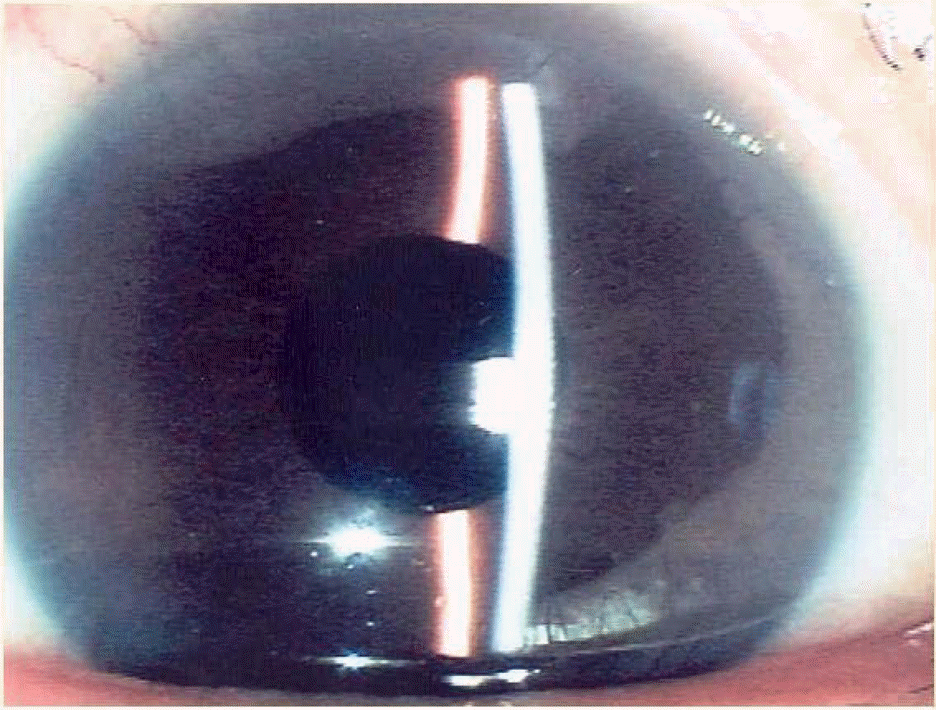
Figure 1.
Photograph of the right eye at first visit. Floating “fluffed-up” lens materials were shown in deep anterior chamber.
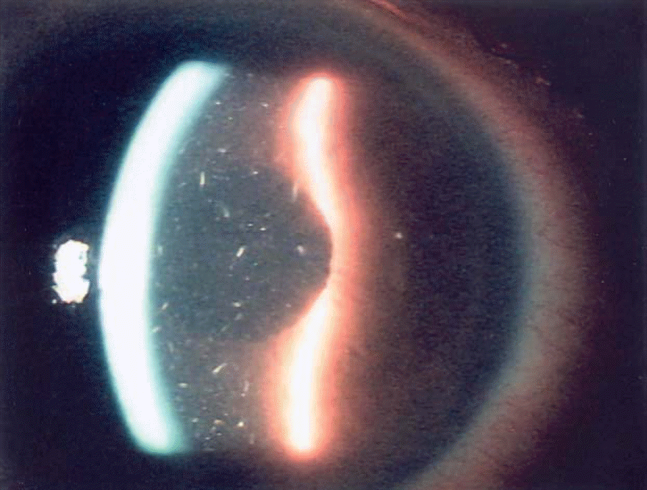
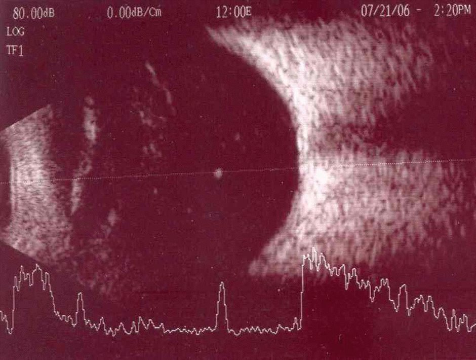
Figure 2.
Ultrasonograph of the right eye at first visit. Dislocated lens and vitreous opacities were revealed.
 XML Download