

 PDF
PDF ePub
ePub Citation
 Print
Print


Abstract
Purpose
To demonstrate the capability of anterior segment optical coherence tomography (OCT), to evaluate central corneal thickness (CCT) and to compare the results with those by Orbscan II and standard ultrasound (US) pachymetry.
Methods
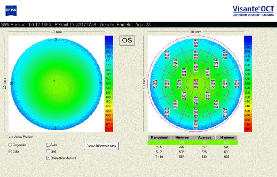
One examiner measured the CCT of 44 normal eyes of 22 subjects using anterior OCT, Orbscan II, and US pachymetry. Non-contact measurements by anterior segment OCT and Orbscan II were performed first, followed by contact measurements using US pachymehy. Three consecutive measurements were taken with each method and the mean values and correlations were analyzed.
Results
The mean value of the CCT was 531.45±32.90 μm with anterior OCT, 537.11±32.21 pm with Orbscan II, and 548.18±34.17 pm with US pachymehy. There was no statistically significant difference among the values obtained by the 3 instruments (P>0.05). CCT measurements by anterior segment OCT were highly correlated with Orbscan II and US pachymetry (P<0.001) measurements.
Conclusion
CCT measurements by anterior segment OCT are highly correlated with Orbscan II or US pachymetry measurements. Using non-contact anterior segment OCT, a closer examination of the anterior segment including the cornea is possible. The measurement of the CCT using anterior segment OCT is applicable because result values are similar to measurements by Orbscan II or US pachymetry.
Go to : 
References
1. Villasenor RA, Santos VR, Cox KC. Comparison of ultrasonic corneal thickness measurements before and during surgery in the Prospective Evaluation of Radial Keratotomy (PERK) study. Ophthalmology. 1988; 93:327–30.
2. Marsich MM, Bullimore MA. The repeatability of corneal thickness measures. Cornea. 2000; 19:792–5.

3. Bechmann M, Thiel MJ, Neubauer AS, et al. Central corneal thickness measurement with a retinal optical coherence tomography device versus standard ultrasonic pachymetry. Cornea. 2001; 20:50–4.
4. Choi SH, Kim JH, Han NS, et al. Comparison of Corneal Thickness Measurements with Optical Low Coherence Reflectometry, Orbscan System and Ultrasound Pachymeter. J Korean Ophthalmol Soc. 2006; 47:19–24.
5. Pierro L, Conforto E, Resti AG, Lattanzio R. High-frequency ultrasound biomicroscopy versus ultrasound and optical pachymetry for the measurement of corneal thickness. Opthalmologica. 1998; 212:1–3.
6. Reader AL III, Salz JJ. Differences among ultrasonic pachymeters in measuring corneal thickness. J Refract Surg. 1987; 3:7–11.
7. Kremer FB, Walton P, Gensheimer G. Determination of corneal thickness using ultrasonic pachometry. Ann Ophthalmol. 1985; 17:506–7.
8. Salz JJ, Azen SP, Berstein J, et al. Evaluation and comparison of sources of variability in the measurement of corneal thickness with ultrasonic and optical pachymeters. Ophthalmic Surg. 1983; 14:750–4.
9. Solomon OD. Corneal indentation during ultrasonic pachymetry. Cornea. 1999; 18:214–5.
10. Kang PS, Kim JD, Yang YS. Comparison of corneal thickness measurements with the orbscan and ultrasonic pachymetry. J Korean Ophthalmol Soc. 2000; 41:1697–703.
11. Kim HS, Kim JH, Kim HM, Song JS. Comparison of thickness measured by specular, US parchymetry, and orbscan in post-PKP eyes. J Korean Ophthalmol Soc. 2007; 48:245–50.
12. Kwon MJ, Seoung YS, Hur DW. Pachymetric measurements using orbscan after excimer refractive surgery. J Korean Ophthalmol Soc. 2004; 45:899–907.
13. Kim SH, Cho JH, Song BJ. Accuracy of Orbscan Pachymetry Measurements and Ultrasonic Pachymetry before and after LASIK with Orbscan II® Topography. J Korean Ophthalmol Soc. 2002; 43:2513–8.
14. Thomas J, Wang J, Rollins AM, Sturm J. Comparison of corneal thickness measured with optical coherence tomography, ultrasonic pachymetry, and a scanning slit method. J Refract Surg. 2006; 22:671–8.
15. Fishman GR, Pons ME, Seedor JA, et al. Assessment of central corneal thickness using optical coherence tomography. J Cataract Refract Surg. 2005; 31:707–11.
16. Wang J, Fonn D, Simpson TL, Jones L. Relation between optical coherence tomography and optical pachymetry measurements of corneal swelling induced by hypoxia. Am J Ophthalmol. 2002; 134:93–8.
17. Thompson RW Jr, Choi DM, Price MO, et al. Noncontact optical coherence tomography for measurement of corneal flap and residual stromal bed thickness after laser in situ keratomileusis. J Refract Surg. 2003; 19:507–15.
18. Yaylali V, Kaufman SC, Thompson HW. Corneal thickness measurements with the Orbscan Topography System and ultrasonic pachymetry. J Cataract Refract Surg. 1997; 23:1345–50.
19. Liu Z, Huang AJ, Pflugfelder SC. Evaluation of corneal thickness and topography in normal eyes using the Orbscan corneal topography system. Br J Ophthalmol. 1999; 83:774–8.
20. Liu Z, Pflugfelder SC. Corneal thickness is reduced in dry eye. Cornea. 1999; 18:403–7.
21. Prydal JI, Artal P, Woon H, Campbell FW. Study of human precorneal tear film thickness and structure using laser interferometry. Invest Ophthalmol Vis Sci. 1992; 33:2006–11.
22. Prisant O, Calderon N, Chastang P, et al. Reliability of pachymetric measurements using orbscan after excimer refractive surgery. Ophthalmology. 2003; 110:511–5.
23. Iskander NG, Peters NT, Penno EA, Gimbel HV. Accuracy of Orbscan pachymetry measurements and DHG ultrasound pachymetry in primary laser in situ keratomileusis and LASIK enhancement procedures. J Cataract Refract Surg. 2001; 27:681–5.
24. Mertz GW. Overnight swelling of the living human cornea. J Am Optom Assoc. 1980; 51:211–3.
25. Wong AC, Wong CC, Yuen NS, Hui SP. Correlational study of central corneal thickness measurements on Hong Kong Chinese using optical coherence tomography, Orbscan and ultrasound pachymetry. Eye. 2002; 16:715–21.
26. Wirbelauer C, Scholz C, Hoerauf H, et al. Corneal optical coherence tomography before and immediately after excimer laser photorefractive keratectomy. Am J Ophthalmol. 2000; 130:693–9.
27. Leung DY, Lam DK, Yeung BY, Lam DS. Comparison between central corneal thickness measurements by ultrasound pachymetry and optical coherence tomography. Clin Experiment Ophthalmol. 2006; 34:751–4.
28. Stucchi CA, Gennari G, Aimino G, et al. Systemic error in computerzied pachymetry. Ophthalmologica. 1993; 207:208–14.
Go to :
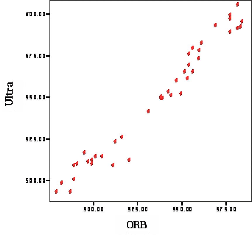
 | Figure 2.Scatterplot showing correlation between ultrasound pachymetry (Ultra) and OCT measurements of central corneal thickness in individuals (Pearson's correlation coefficient r = 0.981, P<0.001). |
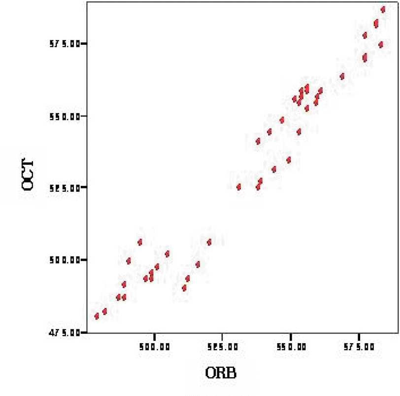
 | Figure 3.Scatterplot showing correlation between Orbscan II (ORB) and OCT measurements of central corneal thickness in individuals (Pearson's correlation coefficient r = 0.976 P<0.001). |
 XML Download