

 PDF
PDF ePub
ePub Citation
 Print
Print


Abstract
Purpose
To determine the incidence and perioperative factors of flap-related complications from Epi-LASIK.
Methods
In this study, 122 eyes of 66 patients who had Epi-LASIK using Centurion SESTM epikeratome (Norwood Eye Care, Australia) were enrolled. Associations of pre-operative corneal curvature, white-to-white distance, central corneal thickness, refractive error, dry eye, punctate corneal erosion, pannus, and history of wearing contact lenses with flap-related complications were investigated. To decrease flap-related complications, surgeons pressed patients’ eyelids with a speculum during epithelial separation, and the effect of this method was verified.
Results
Complete epithelial separation was achieved in 74 eyes (60.6%), incomplete separation in 29 eyes (23.8%), and free epithelial sheet in 19 eyes (15.6%). Thin corneas (P=.041), a history of wearing contact lenses (P=.008), and the duration of contact lens use (P=.003) significantly decreased the incidence of successful epithelial separation. Pressing down the eyelids with a speculum while separating the epithelial sheet increased the incidence of complete separation from 50.6% to 83.8% (P=.003).
Go to : 
References
1. Duffey RJ, Leaming D. US trends in refractive surgery:2003 ISRS/AAO survey. J Refract Surg. 2005; 21:87–91.
2. Lee JB, Seong GJ, Lee JH, et al. Comparison of laser epithelial keratomileusis and photorefractive keratectomy for low to moderate myopia. J Cataract Refract Surg. 2001; 27:565–70.

3. Chen CC, Chang JH, Lee JB, et al. Human corneal epithelial cell viability and morphology after dilute alcohol exposure. Invest Iphthalmol Vis Sci. 2002; 43:2593–602.
4. Pallikaris IG, Katsanevaki VJ, Kalyvianaki MI, Naoumidi II. Advances in subepithelial excimer refractive surgery techniques: Epi-LASIK. Curr Opin Ophthalmol. 2003; 14:207–12.
5. Pallikaris IG, Kalyvianaki MI, Katsanevaki VJ, Ginis HS. Epi-LASIK:preliminary clinical results of an alternative surface ablation procedure. J Cataract Refract Surg. 2005; 31:879–85.
6. Azar DT, Ang RT, Lee JB, et al. Laser subepithelial keratomileusis: electron microscopy and visual outcomes of flap photorefractive keratectomy. Curr Opin Ophthalmol. 2001; 12:323–8.
7. Nichols KK, Nichols JJ, Mitchell L. The relation between tear film tests in patients with dry eye disease. Ophthalmic Physiol Opt. 2003; 23:553–60.
8. Kim JH, Oh CH, Song JS, Kim HM. Inadvertent stromal dissection during mechanical separation of the corneal epithelium using an epikeratome. J Cataract Refract Surg. 2006; 32:1759–63.
9. Matsumoto JC, Chu YS. Epi-LASIK update:overview of techniques and patient management. Int Ophthalmol Clin. 2006; 46:105–15.
10. Shah S, Sebai Sarhan AR, Doyle SJ, et al. The epithelial flap for photorefractive keratectomy. Br J Ophthalmol. 2001; 85:393–6.
11. Vesaluoma M, Perez-Santonja J, Petroll WM, et al. Corneal stromal changes induced by myopic LASIK. Invest Ophthalmol Vis Sci. 2000; 41:369–76.
12. Holden BA, Sweeney DF, Vannas A, et al. Effects of long-term extended contact lens wear on the human cornea. Invest Ophthalmol Vis Sci. 1985; 26:1489–501.
13. Vannas A, Holden BA, Makitie J. The ultrastructure of contact lens induced changes. Acta Ophthalmol. 1984; 62:320–33.
14. Bourne WM, Hodge DO, McLaren JW. Estimation of corneal endothelial pump function in long-term contact lens wearers. Invest Ophthalmol Vis Sci. 1999; 40:603–11.
15. Bruce AS, Brennan NA. Corneal pathophysiology with contact lens wear. Surv Ophthalmol. 1990; 35:25–58.
16. Liesegang TJ. Physiologic changes of the cornea with contact lens wear. CLAO J. 2002; 28:12–27.
17. Liu Z, Pflugfelder SC. The effects of long-term contact lens wear on corneal thickness, curvature, and surface regularity. Ophthalmology. 2000; 107:105–11.
18. Braun DA, Anderson Penno EE. Effect of contact lens wear on central corneal thickness measurements. J Cataract Refract Surg. 2003; 29:1319–22.
19. Madigan MC, Holden BA. Reduced epithelial adhesion after extended contact lens wear correlates with reduced hemidesmosome density in cat cornea. Invest Ophthalmol Vis Sci. 1992; 33:314–23.
Go to :
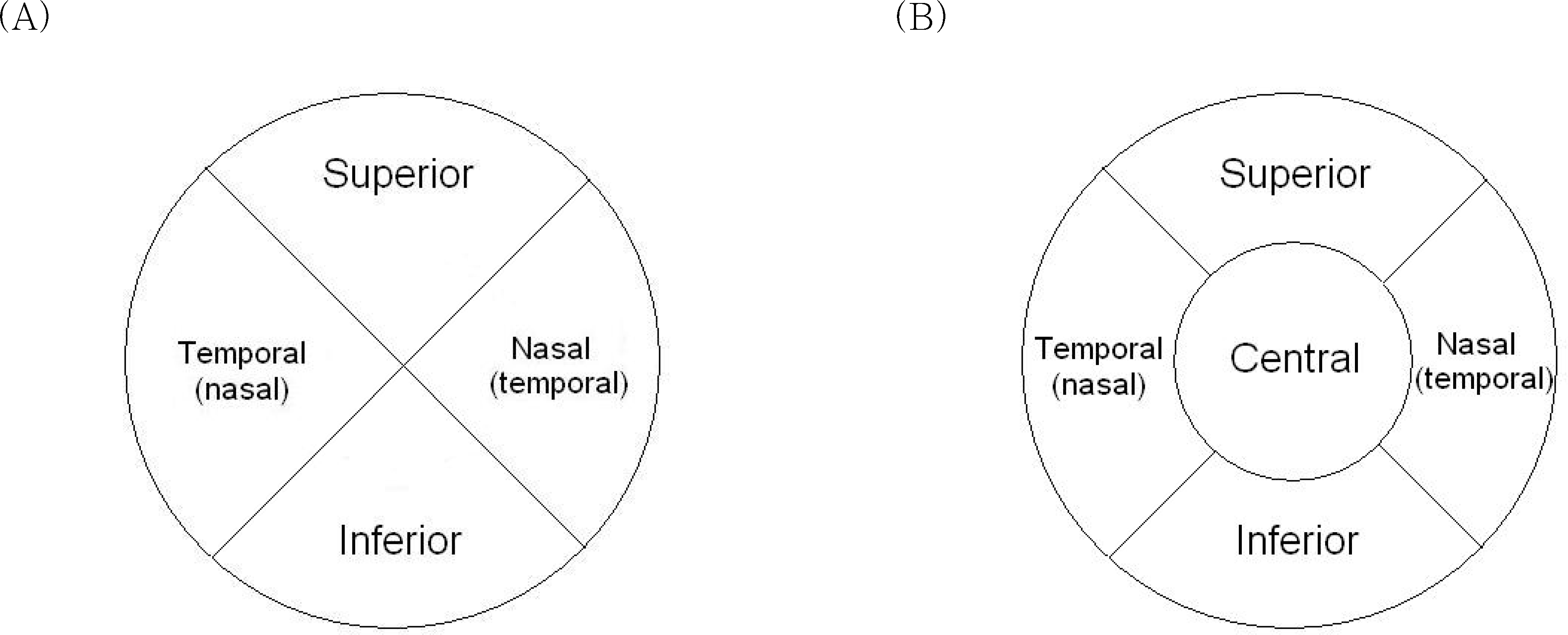 | Figure 1.(A) Diagram of corneal surface to describe punctate erosion. The whole cornea was divided into 5 parts: superior, inferior, nasal, temporal, and central (4 mm in diameter in the center). (B) Diagram of the corneal surface to describe pannus.
The whole cornea was divided into 4 parts: superior, inferior, nasal, and temporal.
|
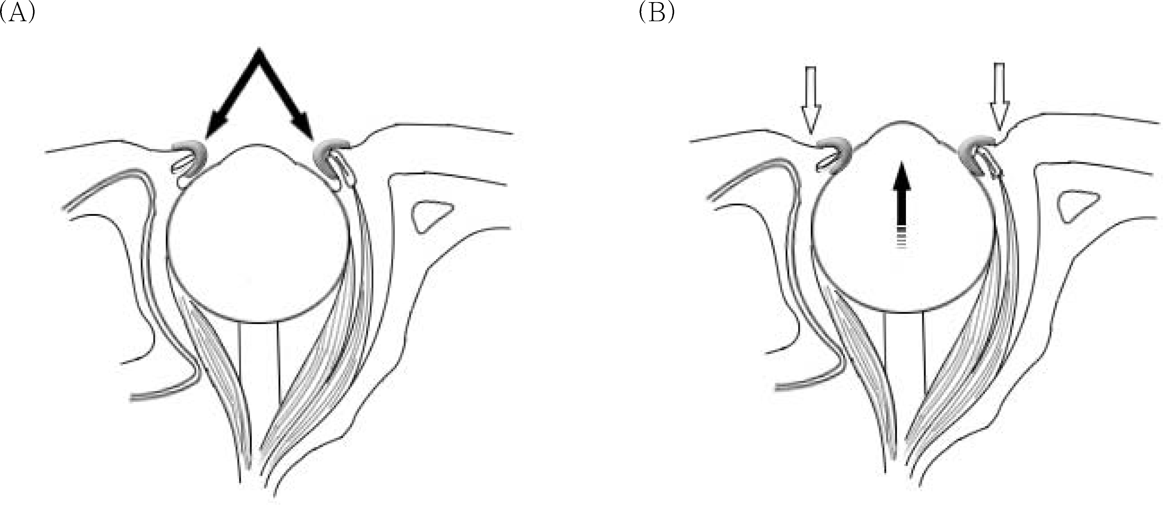 | Figure 2.(A) Normal orbital structure with speculum (arrow). (B) Changes after pressing down the eyelids. Increased mtraorbital pressure makes the eyeball to protrude and corneal curvature steeper. |
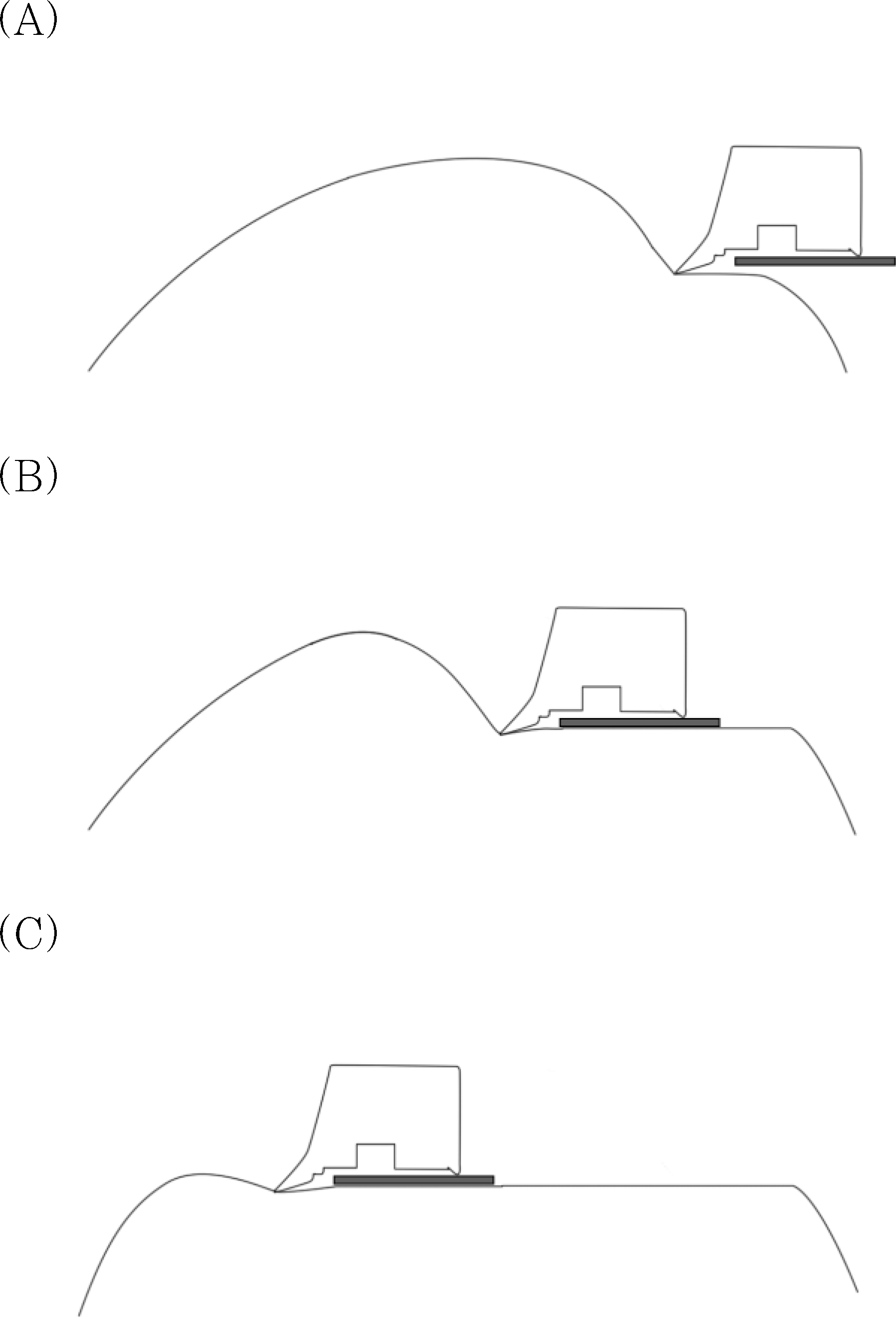 | Figure 3.Hypothesis for incomplete separation. (A) In the early stage, the curvature of the cornea is so steep that it is enough to make counter-force against the separator. (B) In the middle stage, counter-force keeps up. (C) In the late stage, the curvature of the cornea flattens gradually and the decreased counter-force may cause incomplete separation. |
Table 1.
Preoperative patients parameter and flap-related complications
| Parameters | Mean (±SD) | P | ||
|---|---|---|---|---|
| Complete flap | Incomplete flap | Free flap | ||
| No. of cases (%) | 74 (60.6%) | 29 (23.8%) | 19 (15.6%) | |
| Amount of ablation | -4.32±1.8D | -4.31± 1.5 D | -4.17±1.3D | .934 |
| Corneal thickness | 547±27 μm†,‡ | 536±26 μm;† | 534±31 μm;‡ | .041* |
| White-to-white distance | 11.5±0.3 mm | 11.5±0.4 mm | 11.7±0.3 mm | .095 |
| Corneal curvature (5 mm) | 42.8± 1.4 | 43.2±1.5 | 42.2±1.9D | .079 |
Table 2.
Dry eye and flap-related complications
| Tearfilm break-up time (second) | Incidence (%) | Total | ||
|---|---|---|---|---|
| Complete flap | Incomplete flap | Free flap | ||
| < 5 | 5 (62.5) | 1 (12.5) | 2 (25) | 8 |
| 5- 9 | 8 (61.5) | 3 (23.1) | 2 (15.4) | 13 |
| 10 ≤ | 61 (60.4) | 25 (24.8) | 15 (14.8) | 101 |
Table 3.
Punctate erosion and flap-related complications
| Punctate erosion* | Incidence (%) | Total | ||
|---|---|---|---|---|
| Complete flap | Incomplete flap | Free flap | ||
| 0 | 57 (58.2) | 23 (23.5) | 18 (18.3) | 98 |
| 1 | 7 (58.4) | 4 (33.3) | 1 (8.3) | 12 |
| 2 | 5 (71.4) | 2 (28.6) | 0 (0) | 7 |
| 2 < | 3 (60) | 1 (20) | 1 (20) | 5 |
Table 4.
Pannus and flap-related complications
| Pannus* | Incidence (%) | Total | ||
|---|---|---|---|---|
| Complete flap | Incomplete flap | Free flap | ||
| 0 | 71 (60.2) | 28 (23.7) | 19 (16.1) | 118 |
| 1 | 2 (66.7) | 1 (33.3) | 0 (0) | 3 |
| 1 < | 1 (100) | 0 (0) | 0 (0) | 1 |
Table 5.
Contact lens wear and flap-related complications
| Incidence (%) | Total | P | |||
|---|---|---|---|---|---|
| Complete flap | Incomplete flap | Free flap | |||
| Contact lens use | |||||
| Non user | 41 (75.9) | 7 (13) | 6 (11.1) | 54 | .008* |
| user | 33 (48.5) | 22 (32.4) | 13 (19.1) | 68 | |
| Type of contact lens | |||||
| Soft contact lens | 24 (47.1) | 18 (35.3) | 9 (17.6) | 51 | .648 |
| Hard contact lens | 9 (53) | 4 (23.5) | 4 (23.5) | 17 | |
 XML Download