

 PDF
PDF ePub
ePub Citation
 Print
Print


Abstract
Purpose
Based on the questionnaires for dry eyes in 2006, we have investigated the current status and new trends in the recognition and management of the dry eye.
Methods
We ask the 15 questionnaires to 197 ophthalmologists. After reorganizing the data based on each questions, we compared the result with the most current studies in Korea and other countries as well. Then, we report the current trends in the diagnosis and treatment of dry eye.
Results
Dry eye patients account for take up about 20-30% of total outpatients and it is currently accepted etiology that dry eye inflammation is related to the function of the immune system. Patient's symptoms and slit lamp examination are important for diagnosis. An anti-inflammatory therapy is needed for patients with moderate to severe dry eye.
Go to : 
References
1. Lemp MA. Report of the National Eye Institute/Industry workshop on Clinical Trials in Dry Eyes. CL AO J. 1995; 21:221–32.
2. Stern ME, Beuerman RW, Fox RI, et al. The pathology of dry eye: the interaction between the ocular surface and lacrimal glands. Cornea. 1998; 17:584–9.
3. Stern ME, Gao J, Siemasko KF, et al. The role of the lacrimal functional unit in the pathophysiology of dry eye. Exp Eye Res. 2004; 78:409–16.

4. Dana MR, Hamrah P. Role of immunity and inflammation in corneal and ocular surface disease associated with dry eye. Adv Exp Med Biol. 2002; 506:729–38.
5. Behrens A, Doule JJ, Stern L, et al. Dysfunctional tear syndrome: a Delphi approach to treatment recommendations. Cornea. 2006; 25:900–7.
6. Schein OD, Munoz B, Tielsch JM, et al. Prevalence of dry eye among the elderly. Am J Ophthalmol. 1997; 124:723–8.
7. Lin PY, Tsai SY, Cheng CY, et al. Prevalence of dry eye among an elderly Chinese population in Taiwan: the Shihpai Eye Study. Ophthalmology. 2003; 110:1096–101.
8. Brewitt H, Sistani F. Dry eye disease: the scale of the problem. Surv Ophthalmol. 2001; 45:199–202.
9. Moss SE, Klein R, Klein BE. Prevalence of and risk factors for dry eye syndrome. Arch Ophthalmol. 2000; 118:1264–8.
10. Schaumberg DA, Sullivan DA, Buring JE, Dana MR. Prevalence of dry eye syndrome among US women. Am J Ophthalmol. 2003; 136:318–26.
11. Hikichi T, Yoshida A, Fukui Y, et al. Prevalence of dry eye in Japanese eye centers. Graefes Arch Clin Exp Ophthalmol. 1995; 233:555–8.
12. Lin PY, Tsai SY, Cheng CY, et al. Prevalence of dry eye among an elderly Chinese population in Taiwan: the Shihpai Eye Study. Ophthalmology. 2003; 110:1096–101.
13. Stern ME, Beuerman RW, Fox RI, et al. The pathology of dry eye: the interaction between the ocular surface and lacrimal glands. Cornea. 1998; 17:584–9.
14. Kim DK, Song BR. The Concentration of Tear Lysozyme in Normal Subjects and Dry Eye. J Korean Ophthalmol Soc. 1999; 40:2693–700.
15. Puehler W, Rittner HL, Mousa SA, et al. Interleukin-1 beta contributes to the upregulation of kappa opioid receptor mrna in dorsal root ganglia in response to peripheral inflammation. Neuroscience. 2006; (25):141:989–98.
16. Kroemer G, Martinez C. Cytokines and autoimmune disease. Clin Immunol Immunopathol. 1991; 61:275–95.
17. Rowe D, Griffiths M, Stewart J, et al. HLA class I and II, interferon, interleukin 2, and the interleukin 2 receptor expression on labial biopsy specimens from patients with Sjogren's syndrome. Ann Rheum Dis. 1987; 46:580–6.
18. Jones DT, Monroy D, Ji Z, et al. Sjogren's syndrome: cytokine and Epstein-Barr viral gene expression within the conjunctival epithelium. Invest Ophthalmol Vis Sci. 1994; 35:3493–504.
19. Narayanan S, Miller WL, McDermott AM. Conjunctival cytokine expression in symptomatic moderatedry eye subjects. Invest Ophthalmol Vis Sci. 2006; 47:2445–50.
20. Gao J, Gelber-Schwalb TA, Addeo JV, Stern ME. Apoptosis in the lacrimal gland and conjunctiva of dry eye dogs. Adv Exp Med Biol. 1998; 438:453–60.
21. Korb DR. The tear film: its role today and in the future. In : Korb DR, Craig J, Doughty M, editors. The tear film: structure, function and clinical examination. Oxford: Butterworth Heinemann;2002. chap. 7.
22. Lee JK, Ki CW, Roh KG. The Significance of the Tear Film Break-up Time (BUT) in the Diagnosis of the Dry Eye Syndrome. J Korean Ophthalmol Soc. 1985; 26:1311–5.
23. Shyn KH, Kim BH, Kim JC. A Study of the Conjunctival Epithelial Changes in Patients with Dry Eye Syndrome by Impression Cytology and Comparisons of Those Results with BUT, Schirmer Test, Rose Bengal Staining. J Korean Ophthalmol Soc. 1994; 35:362–70.
24. Dogru M, Stern ME, Smith JA, et al. Changing trends in the definition and diagnosis of dry eyes. Am J Ophthalmol. 2005; 140:507–8.
25. Korb DR, Greiner JV, Herman J. Comparison of fluorescein break-up time measurement reproducibility using standard fluorescein strips versus the Dry Eye Test (DET) method. Cornea. 2001; 20:811–5.
26. Dogru M, Ishida K, Matsumoto Y, et al. Strip meniscometry: a new and simple method of tear meniscus evaluation. Invest Ophthalmol Vis Sci. 2006; 47:1895–901.
27. Cho BJ, Lee JH, Shim OJ. The Relation Between Clinical Manifestations of Dry Eye Patients and Their BUTs. J Korean Ophthalmol Soc. 1992; 33:297–302.
28. Calonge M. The treatment of dry eye. Surv Ophthalmol. 2001; 452:S227–39.
29. Nelson JD, Farris RL. Sodium hyaluronate and polyvinyl alcohol artificial tear preparations. A comparison in patients with keratoconjunctivitis sicca. Arch Ophthalmol. 1988; 106:484–7.
30. Kwon DJ, Park DH, Park D, et al. Clinical Investigation of Therapeutic Effect and Extrusion Rate of Punctal Plug for Dry Eye Syndrome. J Korean Ophthalmol Soc. 2006; 47:1204–11.
31. Jeong SH, Lee YJ, Kim HY, et al. The Relationship between the Dacryoscintigraphy and the Satisfaction with Punctal Occlusion in Patients with Dry Eye Syndrome. J Korean Ophthalmol Soc. 2003; 44:1723–7.
32. Lew HM, Jang JW, Lee EH. The Changes of Tear Osmolarity and Protein After Silicone Punctal Plug Insertion In Dry Eye. J Korean Ophthalmol Soc. 2001; 42:1509–14.
33. Her J, Yu SI, Seo SG. Clinical Effects of Various Antiinflammatory Therapies in Dry Eye Syndrome. J Korean Ophthalmol Soc. 2006; 47:1901–10.
Go to :
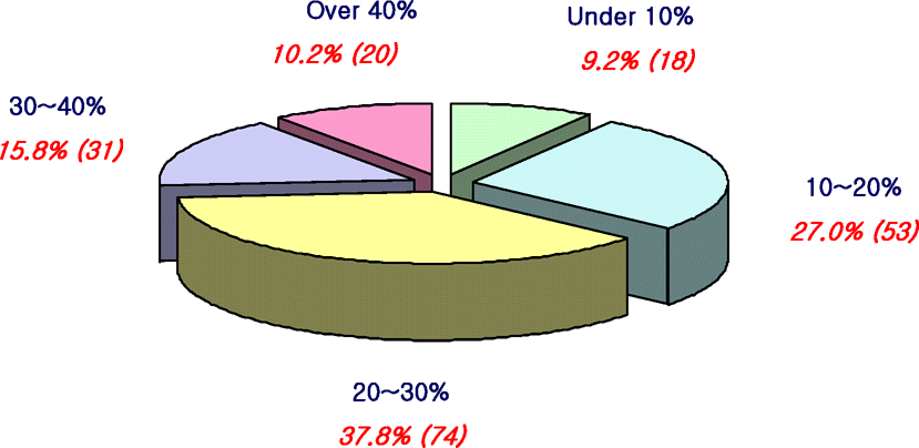 | Figure 1.A proportion of dry eye patients in outpatient clinics.
Red numbers mean the percentage of respondents.
Parenthesized red numbers mean the number of respondents.
|
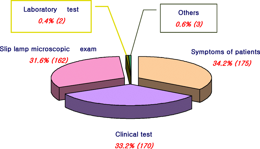 | Figure 2.Most commonly used diagnostic tests for evaluating a patient with probable dry eye.
Red numbers mean the percentage of respondents.
Parenthesized red numbers mean the number of respondents.
|
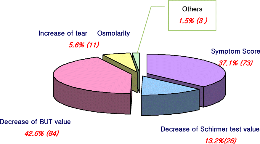 | Figure 3.The gold diagnostic method for dry eye Red numbers mean the percentage of respondents.
Parenthesized red numbers mean the number of respondents.
|
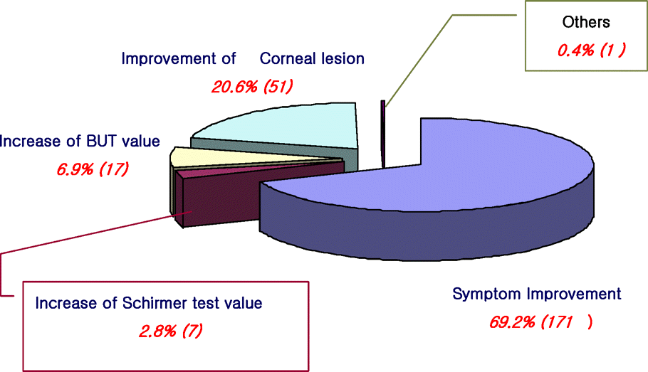 | Figure 4.Most useful indicator of improvement in treatment of dry eye.
Red numbers mean the percentage of respondents.
Parenthesized red numbers mean the number of respondents.
|
Table 1.
Levels of severity of dry eye syndrome without lid margin disease according to symptoms and signs
| Severity* | Patient Profiles |
|---|---|
| Level 1 | Mild to moderate symptoms and no signs |
| Mild to moderate conjunctival staining | |
| Level 2 | Moderate to severe symptoms |
| Tear film signs | |
| Mild corneal punctate staining | |
| Conjunctival staining | |
| Visual signs | |
| Level 3 | Severe symptoms |
| Marked corneal punctate staining | |
| Central corneal staining | |
| Filamentary Keratitis | |
| Level 4 | Severe symptoms |
| Severe corneal staining, erosions | |
| Conjunctival scarring |
Table 2.
Percentage of selected managements of dry eye according to its severity level
Table 3.
Newly suggested treatment strategies of dry eye (N=50)
 XML Download