

 PDF
PDF ePub
ePub Citation
 Print
Print


Abstract
Purpose
We report a case of bilateral tonic pupils and decreased corneal sensitivity in a patient with Vogt-Koyanagi-Harada (VKH) disease during pregnancy.
Case summary
A 33-year-old Korean woman in the sixth month of pregnancy presented with visual impairment in both eyes. Her best corrected visual acuity was 0.2 in the right eye and 0.125 in the left. Slit-lamp examination revealed cells in the anterior chamber and serous retinal detachments in both eyes. The patient was treated with topical corticosteroid in both eyes and posterior subtenon injection of triamcinolone acetonide in the left eye. The serous retinal detachment resolved completely at 2 months in the left eye and at 3 months in the right. As pigmentation of the retina developed, VKH disease was confirmed. The patient complained of photophobia, and both pupils were found to be enlarged to 8 mm without dilation. Light reflex was absent and near reflex was suppressed and slow. After instillation of 0.125% pilocarpine, the pupils were constricted to 3.5 mm and were diagnosed as tonic pupils. Though the patient's corrected visual acuity improved to 0.8 in the right eye and 1.0 in the left at 6 months, her pupils remained unchanged. Corneal sensitivity was decreased.
Go to : 
References
1. Moorthy RS, Inomata H, Rao NA. Vogt-Koyanagi-Harada syndrome. Surv Ophthalmol. 1995; 39:265–92.

2. Sugiura S. Vogt-Koyanagi-Harada disease. Jpn J Ophthalmol. 1978; 22:9–35.
3. Bae HB, Rhee EJ, Ahn BH. A clinical report of 4 cases of Vogt-Koyanagi-Harada syndrome. J Korean Ophthalmol Soc. 1982; 23:1061–6.
4. Chung EH, Oum BS. A clinical analysis of 6 cases of Vogt-Koyanagi-Harada syndrome. J Korean Ophthalmol Soc. 1984; 25:511–23.
5. Doi M, Matsubara H, Uji Y. Vogt-Koyanagi-Harada syndrome in a pregnant patient treated with high-dose systemic corticosteroids. Acta Ophthalmol Scand. 2000; 78:93–6.
6. Miyata N, Sugita M, Nakamura S, et al. Treatment of Vogt-Koyanagi-Harada's disease during pregnancy. Jpn J Ophthalmol. 2001; 45:177–80.
7. Nohara M, Norose K, Segawa K. Vogt-Koyanagi-Harada disease during pregnancy. Br J Ophthalmol. 1995; 19:94–5.
8. Choi DG, Chung H. Clinical analysis of uveitis in Korea. J Korean Ophthalmol Soc. 1989; 30:543–52.
9. Reed L, Henry MK, Shlomo M, Kenneth SP. Williams textbook of endocrinology. 10th ed.Philadelphia: Saunders;2003. p. 811–41.
10. Levy NS, Kramer SG, Barros TD. Pupillary and accommodative abnormalities in the Vogt-Koyanagi-Harada syndrome. Am J Ophthalmol. 1970; 69:582–8.
11. Brouzas D, Chatzoulis D, Galina E, et al. Corneal anesthesia in a case with Vogt-Koyanagi-Harada syndrome. Acta Ophthalmol Scand. 1997; 75:464–5.
12. Kim JS, Yun CH, Moon CS. Bilateral tonic(Adie's) pupils in Vogt-Koyanagi-Harada syndrome. J Neuroophthalmol. 2001; 21:205–6.
Go to :
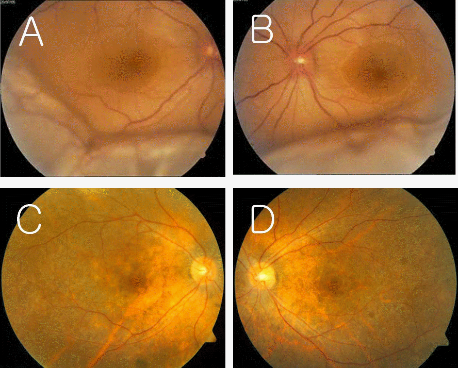 | Figure 1.Fundus Photograph. Serous retinal detachment is seen at presentation (A, B). Serous retinal detachment is resolved at 3 months in the right eye (C), and at 2 months in the left eye (D). Depigmentation is noted around the optic disc of both eyes (C, D). |
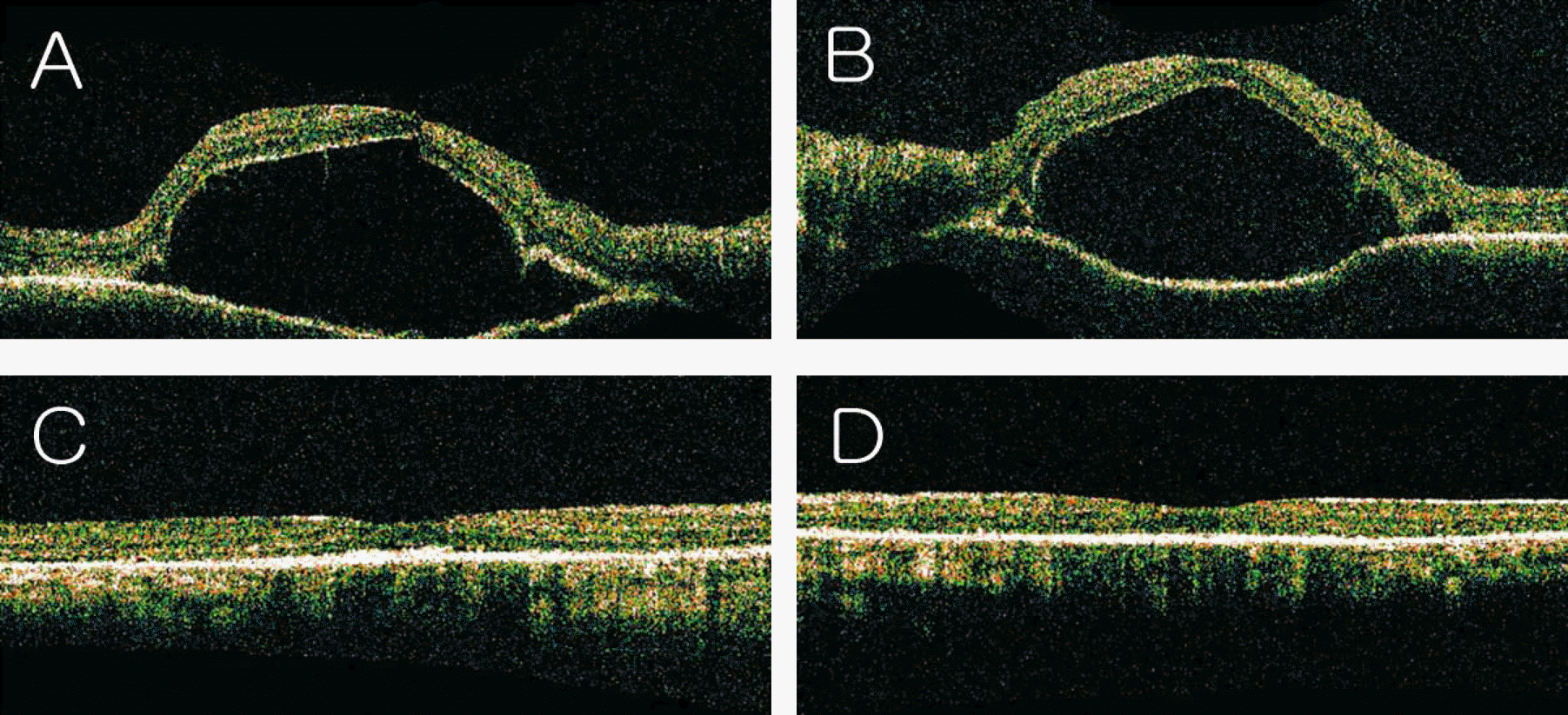 | Figure 2.Optical coherence tomography demonstrates serous retinal detachment at presentation in the right eye(A), and in the left eye (B). Subretinal fluid is resolved at 3 months in the right eye (C), and at 2 months in the left eye (D). |
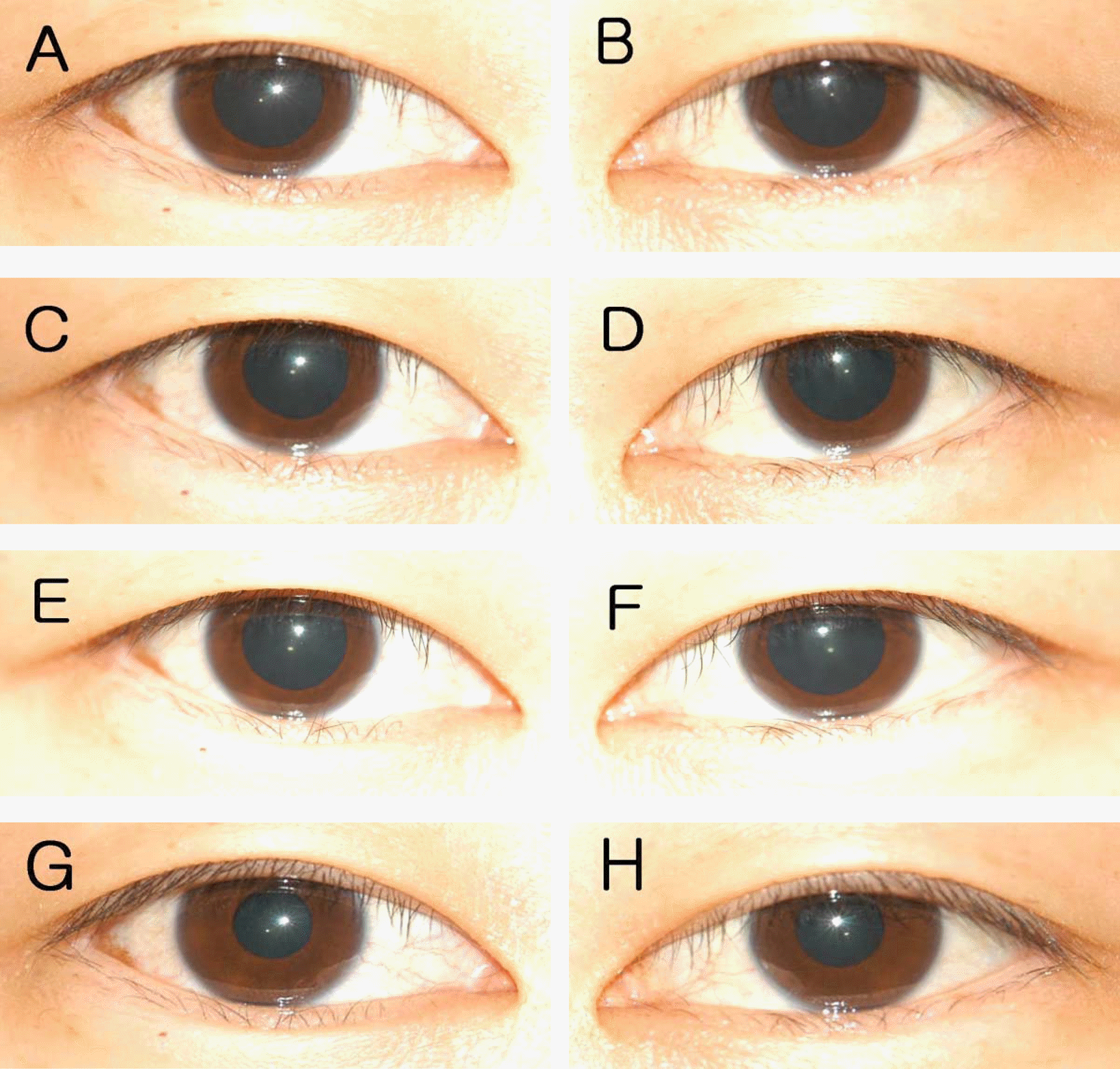 | Figure 3.Photography showing the pupil sizes. (A, B) Looking at the distance in the dark, both pupils are dilated (8 mm). (C, D) During near gazing, near reflex is slow and suppressed (6 mm). (E, F) In bright light, light reflex is suppressed (7 mm). (G, H) After instillation of single drop of 0.125% pilocarpine, both pupils are constricted (3.5 mm). |
 XML Download