

 PDF
PDF ePub
ePub Citation
 Print
Print


Abstract
Case summary
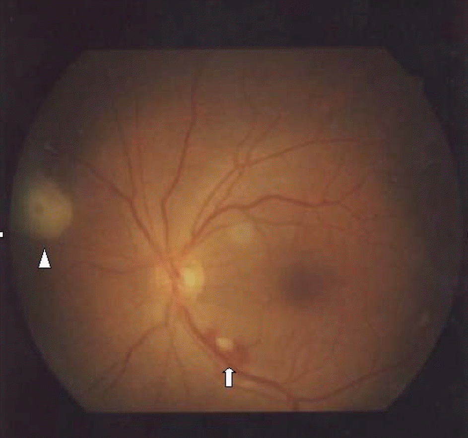
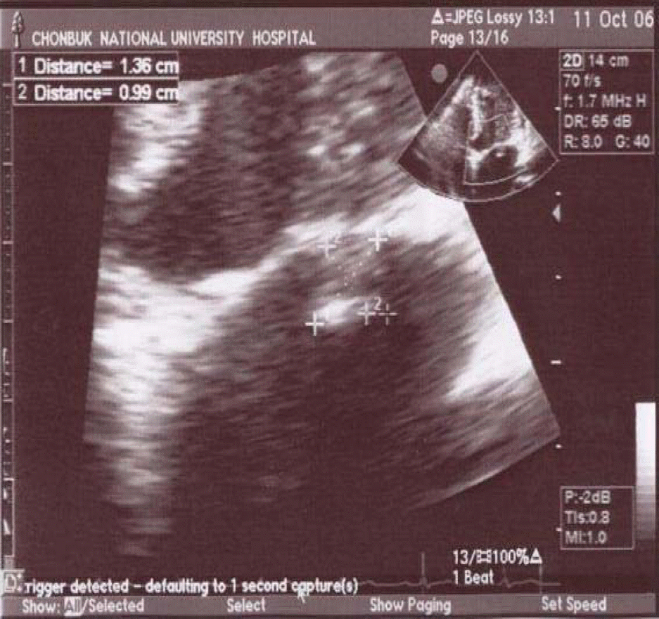
A 45-year-old patient complained of acute visual loss. The patient had a history of epidural anesthesia for the relief of back pain. On the day of admission the patient showed no light perception and had a white-centered retinal hemorrhage and cotton wool spot in the left eye upon fundus examination. Other ocular manifestations were not specific and there were no specific findings on a brain MRI and visual evoked potential. The patient was diagnosed with pyogenic spondylitis and was treated by abscess drainage and systemic antibiotics therapy. The patient's near vision improved up to 0.4/0.5. He was diagnosed with infective endocarditis based on the echocardiogram with epidural and subarachnoid hemorrhage.
Go to : 
References
1. Osler W. Gulstonian lectures on malignant endocarditis. Lancet. 1885; 1:415–505.
2. Roth M. Uber netzhautuffecstionen bei wundfiebrin. Retinal manifestations of wound fever. Deutsch A Chir. 1872; 1:471–84.
3. Roth M. Beitrage zur kenntnis der varicosen hypertrophie der nervenfasern. (Contributions to the knowledge of varicose hypertrophy of nerve fibres.). Virchow's Arch Path Anat. 1872; 55:197–217.
4. Ling R, James B. White-centred retinal haemorrhages (Roth spots). Postgrad Med J. 1998; 74:581–2.

5. Litten M. Ueber acute maligne endocarditis und die dabei vorkommenden retinal veranderungen. Charite-Ann. 1878; 3:135.
6. Byun YC, Lee H, Lee EK, Lee KH. A case of metastatic endophthalmitis originated from bacterial endocarditis. J Korean Ophthalmol Soc. 1993; 35:122–7.
7. Wong VG, Bodey GP. Haemorrhagic retinoschisis due to aplastic anaemia. Arch Ophthalmol. 1968; 80:433–5.
8. Von Barsewisch B. Perinatal retinal haemorrhages. New York: Springer-Verlag,. 1979; 51–2.
9. Duane TD, Osher RH, Green WR. White centered hemorrhages: their significance. Ophthalmology. 1980; 87:66–9.
10. Murtha TJ. Hematologic disorders: leukemia, dysproteinemia, and anemia. In : Albert DM, Jakobiec FA, editors. Principles and practice of ophthalmology: clinical practice. Philadelphia: WB Saunders;1994. v. 5. chap. 243.
11. Green WR. Retina. In : Spencer WH, editor. Ophthalmic pathology an atlas and textbook. Philadelphia: WB Saunders;1985. v. 2. chap. 8.
12. Jones HR Jr, Siekert RG, Geraci JE. Neurologic manifestations of bacterial endocarditis. Ann Intern Med. 1969; 71:21–7.
Go to :
 XML Download