

 PDF
PDF ePub
ePub Citation
 Print
Print


Abstract
Purpose
To report a patient with herpes zoster ophthalmicus in whom hyphema, glaucoma and external ophthalmoplegia occurred.
Case summary
A 59-year-old male patient developed severe ocular pain and decreased visual acuity in his left eye 10 days ago. He had been diagnosed as herpes zoster ophthalmicus 14 days before and given antiviral agent. He could not percept light. His left eye showed hyphema, severe exudative iritis and elevated IOP. Lid drooping and complete external ophthalmoplegia were present in the left eye. Systemic corticosteroid with concomitant antiviral agents and antiglaucomatous agents was administered.
References
1. Lee WB, Liesegang TJ. Herpes zoster keratitis. In : Krachmer JH, Mannis MJ, Holland EJ, editors. Cornea. 2nd ed. Philadelphia: Elsevier Mosby;2005. v. 1. chap. 84.
2. Liesegang TJ. Herpes zoster virus infection. Curr Opin Ophthalmol. 2004; 15:531–6.

3. Lee HR, Cho BC. A clinical study of herpes zoster ophthalmicus. J Korean Ophthalmol Soc. 1988; 29:155–9.
4. Hedges TR, Albert DM. The progression of the ocular abnormalities of herpes zoster : histopathologic observations of nine cases. Ophthalmology. 1982; 89:165–77.
5. Hayaska S, Watanabe M, Yamamoto Y, et al. Herpes zoster ophthalmicus complicated by hyphema and hemorrhagic glaucoma. Ophthalmologica. 1988; 196:185–7.
6. Edgerton AE. Herpes zoster ophthalmicus. Report of cases and review of literature. Trans Am Ophthalmol Soc. 1942; 40:390–439.
7. Akpek EK, Gottsch JD. Herpes zoster sine herpete presenting with hyphema. Ocul Immunol Inflamm. 2000; 8:115–8.
8. Park MJ. Herpes zoster ophthalmicus complicated by acute glaucoma. J Korean Ophthalmol Soc. 1972; 13:25–7.
9. Marsh RJ, Cooper M. Ophthalmic herpes zoster. Eye. 1993; 7:350–70.
10. Shin HM, Lew H, Yun YS. A case of complete ophthalmoplegia in herpes zoster ophthalmicus. Korean J Ophthalmol. 2005; 19:302–4.
11. Hahn ES, Jung YC, Chang K. A case of herpes zoster ophthalmicus complicated by abducens palsy. J Korean Ophthalmol Soc. 1989; 30:447–52.
12. Worster-Drought C, Sargent F. Ophthalmic herpes zoster with contralateral pontine lesions. Med Press. 1949; 221:510–12.
13. Iwao K, Kobayashi H, Okinami S. Case of herpes zoster ophthalmicus with abducent palsy : the cause and magnetic resonance imaging findings. Nippon Ganka Gakkai Zasshi. 2006; 110:193–8.
14. Scheie HG. Herpes zoster ophthalmicus. Trans Ophthalmol Soc U K. 1970; 90:899–930.
15. Amanat LA, Cant JS, Green FD. Acute phthisis bulbi and external ophthalmoplegia in herpes zoster ophthalmicus. Ann Ophthalmol. 1985; 17:46–51.
16. Naumann G, Gass JD, Font RL. Histopathology of herpes zoster ophthalmicus. Am J Ophthalmol. 1968; 65:533–41.
17. Wood MJ, Johnson RW, McKendrick MW, et al. A randomized trial of acyclovir for 7 days and 21 days with and without prednisolone for treatment of acute herpes zoster. N Eng J Med. 1994; 330:896–900.
Figure 1.
Photographs of the patient at 14 days after the onset of skin eruption. (Upper left) Left lid drooping and swelling associated with Hutchinson's sign are noted. Crust formation is noted along the distribution of all 3 divisions of the ophthalmic nerve. (Upper right) This photograph showed moderate corneal edema with an epithelial defect, intracameral hemorrhage (gross hyphema, 3.7 mm height) and exudative membrane in his left eye. (Lower) Marked limitation of ocular movement in lateral, medial, superior and inferior gaze is noted in his left eye.
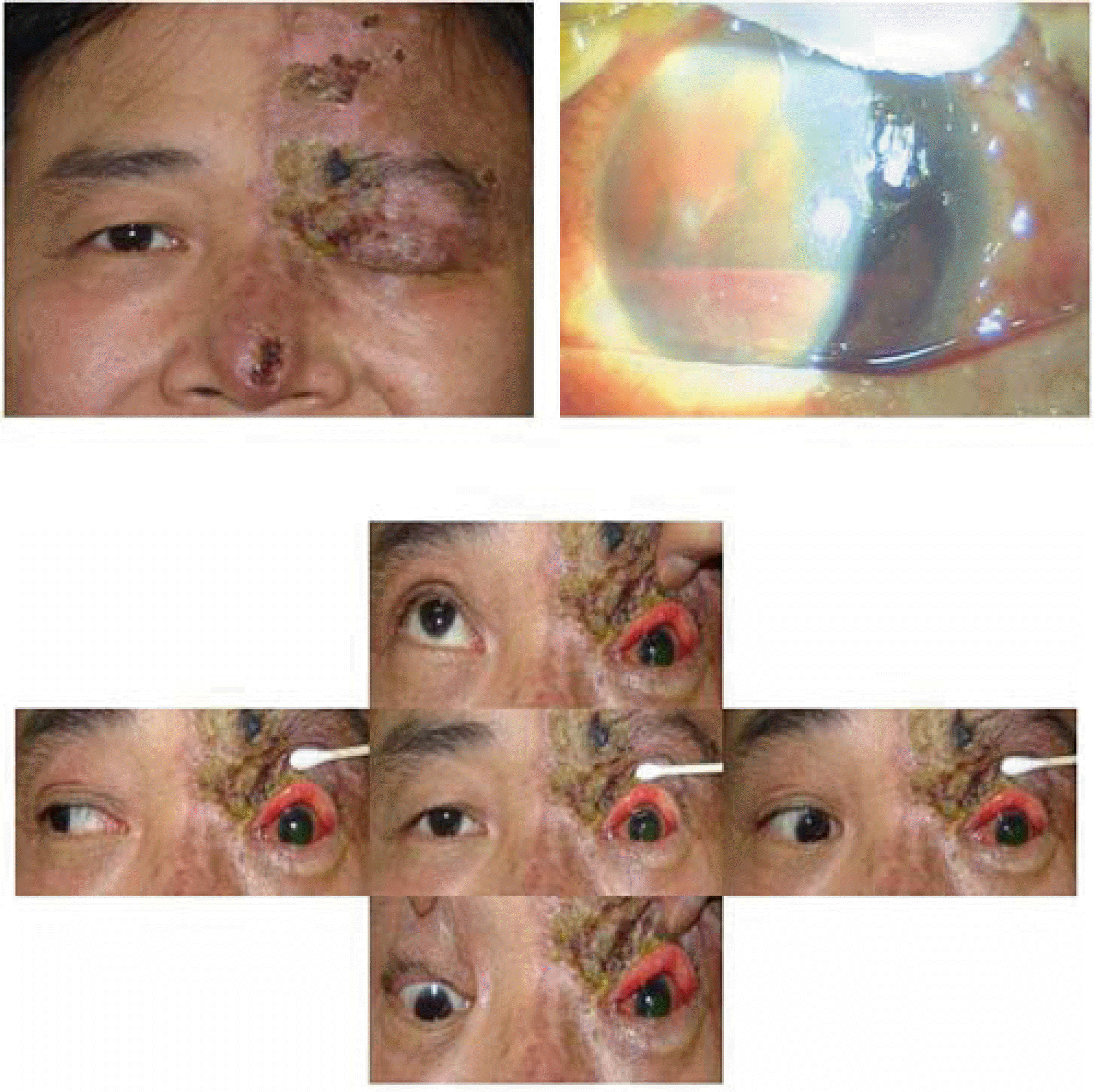
Figure 2.
Photographs of the patient at 4 months after disease onset. (Upper left) The skin lesion turns into scars and lid drooping is somewhat recovered. (Upper right) Left eye shows a hazy and edematous cornea with neovascularization, posterior synechia and iris atrophy. Conjunctival and episcleral vessels are engorged. Left eyeball is soft and perception of light is never recovered. (Lower) Limitation of ocular movement is recovered nearly to normal range.

 XML Download